Introduction: Various bioresorbable materials are proposed for clinical use including medical devices and tissue engineering scaffolds. Especially, polylactic acid (PLA), polyglycolic acid (PGA) and their copolymer have been widely used for scaffolds in virtue of their good mechanical properties and controllable bioresorption. However, their applications are limited due to their brittleness and little flexibility. Therefore, more flexible bioabsorbable materials are highly desired for the applications to soft tissue. In this study, we focused on poly ((R)-3-hydroxybutyrate-co-(R)-3-hydroxyhexanoate) (PHBH), which is a biodegradable polymer produced by bacteria[1]) and should be applicable to agricultural use such as a mulch sheet. PHBH is expected to be a good candidate of scaffolds for soft tissue because it has flexibility at physiological temperature due to its lower glass transition temperature than PLA and PGA. Although reports on the availability of PHBH for scaffold use are increasing[2],[3]), the degradation and resulting inflammatory responses of PHBH in a body have been still poorly understood. Here, to investigate the potency of PHBH as a bioresorbable material, we have examined the biodegradability and biocompatibility of PHBH with two different forms such as fibers and films, by comparing changes in polymer chains after the degradation in vitro and in vivo with poly-L-lactic acid (PLLA).
Experimental: Nonwoven fiber sheets of PHBH and PLLA were fabricated by the electrospinning method and thin films were prepared by a casting method. For the in vitro degradation study, the samples were incubated in PBS at 37 ⁰C in dialysis tubes and their molecular weight distributions and crystallinity after a 1- to 6-month immersion were measured by gel permeation chromatography (GPC) and wide angle X-ray diffraction (WAXD), respectively. For the in vivo degradation study, samples were implanted into a subcutaneous space on the back of rats. After a week, samples were excised for the analysis of GPC, WAXD and histological analysis.
Result and Discussion: From the results of the in vitro degradation study, the molecular distribution of PHBH films and fibers were shifted into low in the early stage, although the top peak of molecular weight of PLLA films and fibers did not show a shift even after 6 months immersion under physiological conditions. (Fig. 1) In addition, PHBH kept approximately a 60% crystallinity in the WAXD analysis after 6 months immersion. These results imply the polymer chains of PHBH were cleaved in a middle (endo) at the amorphous region, which is considered to be caused by a different degradation mechanism from PLLA. In the implantation experiments, PHBH fibers and films also showed fast degradation by GPC analysis. As the result of immunohistochemical staining, initial inflammatory response were suppressed in a PHBH-implanted tissue after two weeks of surgery, but observed in a PLLA-implanted tissue. (Fig. 2) These results suggest that the degradation products of PHBH might be milder for tissue than PLLA because of weak acidity and have potency to alternative materials with flexibility for tissue engineering scaffolds.

Fig.1 Molecular weight distribution of PLLA and PHBH after in vitro degradation test at different months.
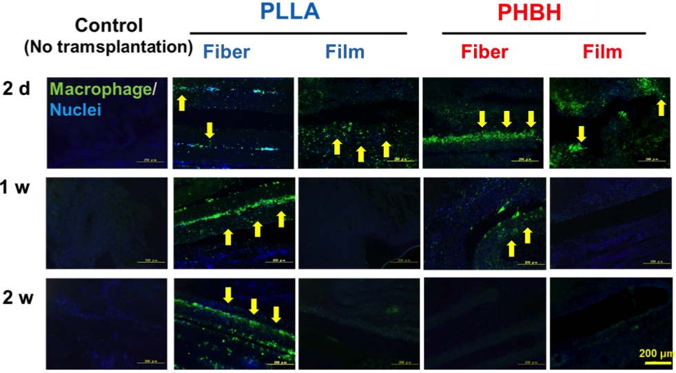
Fig.2 Immunostaining of PLLA and PHBH fibers at different period after transplantation.
This work was supported in part by Kaneka Corporation.
References:
[1] Y. Doi, S. Kitamura, H. Abe, “Microbial Synthesis and Characterization of Poly ( 3-hydroxybutyrate-co-3-hydroxyhexanoate)”, Macromolecules, 28, 4822–4828, 1995.
[2] T.H. Ying, D. Ishii, A. Mahara et al., “Scaffolds from electrospun polyhydroxyalkanoate copolymers: Fabrication, characterization, bioabsorption and tissue response”, Biomaterials, 29, 1307–1317, 2008.
[3] W.R. Webb, T.P. Dale, A.J. Lomas et al., “The application of poly(3-hydroxybutyrate-co-3- hydroxyhexanoate) scaffolds for tendon repair in the rat model”, Biomaterials, 34, 6683–6694, 2013.