Introduction: Freeze-dried chitosan formulations can be solubilised in platelet-rich plasma (PRP) to produce injectable mixtures that coagulate to form implants that resist platelet-mediated clot retraction and are resident for at least 2 weeks in vivo[1],[2]. The purpose of this study was to 1) investigate the mechanisms by which clot retraction is inhibited in chitosan-PRP hybrid clots, 2) to investigate release of platelet-derived growth factors from chitosan-PRP hybrid clots in culture medium, and 3) to inject freeze-dried chitosan-PRP formulations subcutaneously in rabbits to assess long-term implant retention and biodegradability.
Materials and Methods: Freeze-dried chitosan cakes were prepared using 0.56% or 1% w/v chitosan (80% DDA and Mn 40kDa), 1% or 6% w/v trehalose concentration as lyoprotectant and 42.2mM calcium chloride (CaCl2) as clot activator. Cakes were solubilised in PRP obtained from a human donor and dispensed in either 1) Glass tubes for confocal and scanning electron microscopy imaging or 2) 24-well plates to monitor release of PDGF-AB over 7 days. Formulations containing 1% (w/v) chitosan of Mn 40kDa and different DDA (80%, 85%, 90%), 1% (w/v) trehalose and 42.2mM CaCl2 were solubilised with autologous PRP and injected subcutaneously in the back of five NZW rabbits (150µL/implant) to investigate implant retention and biodegradability at 2 weeks; 4 weeks and 6 weeks.
Results: Platelet aggregates were smaller in chitosan-PRP hybrid clots compared to PRP only controls and increasing trehalose concentration further decreased the size of platelet aggregates (Fig 1 left). Platelets, cells and fibrin fibers were coated with chitosan in chitosan-PRP hybrid clots, while fibrin fibers were readily visible in PRP only controls (Fig 1 right). PDGF-AB release profiles were similar for the different chitosan-PRP hybrid clots and for PRP controls, but all were higher than whole blood alone (Fig 2 left). In vivo, PRP controls were completely degraded by 2 weeks, while all chitosan-PRP formulations tested (80%, 85% & 90% DDA) were retained up to 6 weeks post-implantation, where they induced cell recruitment of host-derived cells (Fig 2 right).
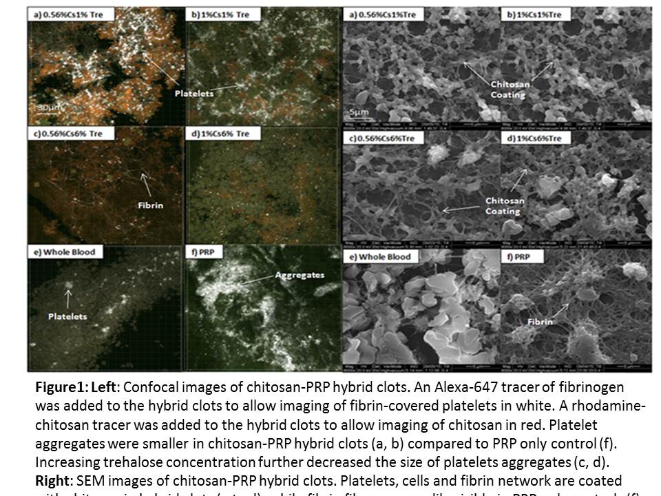
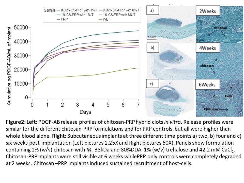
Discussion: Chitosan inhibited platelet aggregation, partially through the physical mechanism of coating the cells and fibrin fibers in the hybrid clots. Increasing trehalose concentration further decreased the size of platelet aggregates through a currently unknown mechanism. Strong platelet aggregation is required for platelet-mediated clot retraction, so this is consistent with our previous results (1). PDGF-AB release was similar in chitosan-PRP hybrid clots and in PRP controls, which is unsurprising considering that PDGF-AB has an isoelectric point of 9.8 and solubilised chitosan-PRP formulations have a pH between 6.8-7.1. Chitosan-PRP hybrids resided up to 6 weeks post-implantation in vivo, while PRP controls were quickly degraded, suggesting that prolonged residency of chitosan itself leads to sustained recruitment of host cells.
Conclusion: Chitosan and trehalose decreased the size of platelet aggregates, which is consistent with their inhibition of clot retraction. Chitosan-PRP does not provide sustained release of PDGF-AB but may do so for other growth factors with different charge properties. Prolonged residency of chitosan-PRP in vivo leads to increased biological activity compared to PRP alone.
We acknowledge the technical contributions of Geneviève Picard, the funding sources (CIHR, CFI, GRSTB, NSERC) and the assistance of Ortho Regenerative Technologies Inc.
References:
[1] Chevrier A et al 2015a; Freeze-dried chitosan formulations for mixing with platelet-rich plasma to form implants for tissue repair. Trans ICRS Chicago, USA.
[2] Chevrier A et al 2015b; Chitosan-platelet-rich plasma implants have in situ tissue building capacity and can be injected into meniscus defects to improve repair. Trans ICRS Chicago, USA.