Introduction: Scaffolds developed for bone tissue engineering (TE) must possess specific structural properties to allow neo-tissue formation and integration within the material[1]. Several polymeric systems and processing methodologies have been proposed to develop bone TE scaffolds. Nevertheless, the so far proposed strategies do not fulfil all the requirements for effective bone regeneration. Textile technologies have recently emerged as an industrial route for producing more complex fibre-based porous scaffolds[2]. Silk fibroin (SF) from Bombyx mori has already proved to be a good biomaterial for bone TE[3]. SF-based structures are known for the impressive mechanical properties and biocompatibility, which meet the basic requirements for developing bone TE scaffolds[4],[5].
Materials and Methods: The present work proposes three-dimensional (3D) spacer structures processed using weft-knitting technology. Two knitted silk layers were assembled/spaced by a monofilament of polyethylene terephthalate (PET). A 3D spacer structure made entirely of PET was used for comparative purposes. The obtained scaffolds were described in terms of morphology and mechanical properties. The in vitro osteogenic differentiation of human adipose-derived stem cells (hASCs) was also investigated. Cells were cultured for 28 days in basal and osteogenic conditions and evaluated through quantitative (ALP, Ca2+, RT-PCR) and qualitative (SEM, Alizarin Red, immunocytochemistry) assays. The in vivo biocompatibility of biotextiles was assessed by subcutaneous implantation in mice for 2 and 4 weeks. Inflammatory response of the collected explants was analyzed by hematoxylin & eosin (H&E) staining. An immunohistochemical analysis of the angiogenic marker SNA-lectin was also performed.
Results and Discussion: The developed spacer textile scaffolds revealed a significant increase of the three-dimensionality induced by the PET monofilament (Fig.1a,b). HASCs cultured for 28 days on the spacer scaffolds were able to cover the surface fibres and colonize scaffolds interior in both culture conditions (Fig.1a). A higher degree of hASCs differentiation and evidences of extracellular matrix mineralization were observed in osteogenic conditions. In vivo results showed that the implanted scaffolds allow tissue ingrowth’s, without inducing any acute inflammatory reaction. In addition blood vessels formation was also observed (Fig.1c).
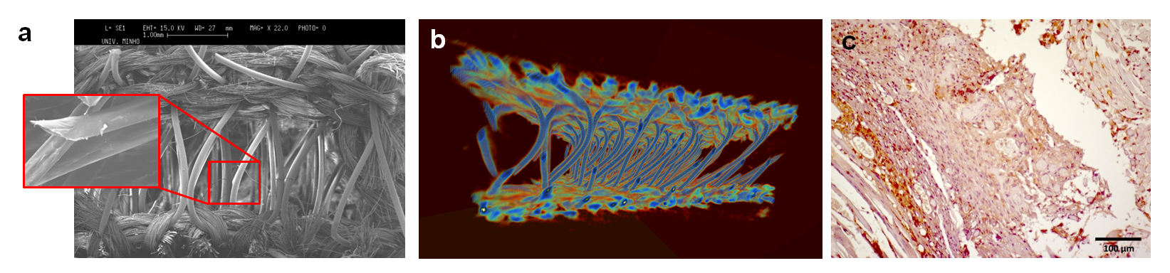
Figure 1. (a) SEM micrographs of SF biotextile cultured with hASCs; (b) Micro-CT image of SF biotextile; (c) SNA-lectin immunostaining of SF textile explant.
Conclusion: In this study, innovative 3D biotextiles were successfully developed. These scaffolds were able to support cell adhesion, proliferation and osteogenic differentiation. New tissue formation and angiogenesis within the spacer scaffolds was also observed after subcutaneous implantation in mice. Thus, the proposed finely tuned and reproducible textile-based scaffolds can be promising candidates for bone TE applications.
Portuguese Foundation for Science and Technology (FCT) for the project TISSUE2TISSUE (PTDC/CTM/105703/2008); Investigator FCT program IF/00423/2012 and IF/00411/2013
References:
[1] Burg, K.J.L., S. Porter, and J.F. Kellam, Biomaterial developments for bone tissue engineering. Biomaterials, 2000. 21(23): p. 2347-2359.
[2] Sumanasinghe, R. and M.W. King, The applications of biotextiles in tissue engineering. Research Journal of Textile and Apparel, 2005. 9(3): p. 80-90.
[3] Kundu, B., R. Rajkhowa, S.C. Kundu, and X.G. Wang, Silk fibroin biomaterials for tissue regenerations. Advanced Drug Delivery Reviews, 2013. 65(4): p. 457-470.
[4] Yan, L.P., A.J. Salgado, J.M. Oliveira, A.L. Oliveira, and R.L. Reis, De novo bone formation on macro/microporous silk and silk/nano-sized calcium phosphate scaffolds. Journal of Bioactive and Compatible Polymers, 2013. 28(5): p. 439-452.
[5] Liu, Y., J. Lim, and S.-H. Teoh, Review: development of clinically relevant scaffolds for vascularised bone tissue engineering. Biotechnology advances, 2013. 31(5): p. 688-705.