Centro de Biología Molecular “Severo Ochoa” (CSIC-UAM), Universidad Autónoma de Madrid, Madrid, Spain
In this short review, I will focus on how a unique tau gene may produce many tau isoforms through alternative splicing and how the phosphorylation of these isoforms by different kinases may affect their activity and behaviour. Indeed, each of the different tau isoforms may play a distinct role under both physiological and pathological conditions. Thus, I will discuss whether a tau code exists that might explain the involvement of different tau isoforms in different cellular functions.
Biological complexity is not only the consequence of the number of genes expressed in an organism but it is also the consequence of further modifications to the gene products. Once a gene is transcribed into nuclear RNA, this RNA can be modified by alternative splicing to yield distinct mRNAs, which may subsequently be translated into different protein isoforms. Post-translational modification of each of these isoforms by phosphorylation (or other processes) can affect the behaviour of each isoform, producing a wide variation in the activity of the different isoforms generated from an unique gene such as tau. It has been suggested that the post-translational modifications that direct the diverse protein functions of a protein may reflect a code and for example, a histone code exists that directs the different functions of chromatin (Jenuwein and Allis, 2001
; Strahl and Allis, 2000
).
In this review, I shall look at how a single tau gene can generate many tau isoforms, first through alternative RNA splicing and afterwards, by different post-translational modifications, phosphorylation being the most important. Finally, I will discuss whether these modifications could define a tau code that might explain the role of different tau isoforms in distinct cellular functions, or in the different neurodegenerative disorders known as tauopathies.
Tau plays a role in different cellular events and as a microtubule associated protein, tau is principally involved in microtubule dynamics, stabilizing the microtubule polymers (Drubin and Kirschner, 1986
; Feinstein and Wilson, 2005
; Goode et al., 1997
). However, it has also been proposed that the tau protein is a key component of axonal transport (Dixit et al., 2008
; Ebneth et al., 1998
; Yuan et al., 2008
) and more recently, tau was found to bind to and inhibit histone deacetylase HDAC6 (Ding and Johnson, 2008
; Perez et al., 2009
), impairing its activity and facilitating tubulin acetylation. In addition, tau can also bind to other proteins apart from tubulin, perhaps influencing their activity (Avila et al., 2004
). In neurodegenerative diseases, modifications to tau are important for the development of tauopathies (Lee et al., 2001
) and the presence of extracellular tau, as a consequence of neuron death, could result from its interaction with muscarinic receptors on surrounding neurons, provoking toxicity in those neurons (Gomez-Ramos et al., 2006
, 2008
). Different functional tau isoforms arise through alternative splicing and their behaviour can be modified by different phosphorylation events Tau is also subject to ubiquitination, glycation, nitration, truncation and other posttranslational modifications that could result in the appearance of additional tau isoforms (Avila et al., 2004
). Thus, these different isoforms could fulfil a different role in all of the processes indicated above. Although in this review I will focus on those tau isoforms arising from alternative splicing or phosphorylation.
Tau is a microtubule associated protein found in the brain that promotes microtubule polymerisation in vitro (Weingarten et al., 1975
). Tau binds to the carboxy terminal region of tubulin (Serrano et al., 1984
), the main component of microtubules, through a region known as the microtubule-binding domain (Goedert et al., 1989
; Lee et al., 1988
). This region of tau contains three or four (depending on the specific tau isoform) similar but not identical motifs of 31 or 32 residues, known as the microtubule binding repeats. Those tau isoforms with three repeats are described as 3Rtau, whereas those with four repeats are defined as 4Rtau.
The unique human tau gene is located on chromosome 17q21 (Andreadis et al., 1992
; Neve et al., 1986
) and it can be transcribed into a nuclear RNA with 16 exons that can yield different tau isoforms through alternative splicing (Goedert et al., 1989
). Six different protein isoforms of tau have been described in the central nervous system (CNS), three 3Rtau and three 4Rtau isoforms, the fourth microtubule binding repeat reflecting the alternative splicing of exon 10.
Alternative splicing of exon 10 in tau, results in isoforms that contain either three (3R tau) or four (4R tau) microtubule binding repeats. The ratio of Tau 4R to Tau 3R approximates to one in the normal brain, both in terms of mRNA and protein, although in some tauopathies this ratio is affected and this might be sufficient to cause neurodegeneration. In Frontotemporal dementia with Parkinson’s linked to chromosome 17 (FTDP-17) and corticobasal degeneration (CBD), there is an increase in the 4R tau/3R tau ratio, while in Pick’s disease there are no phosphorylated tau isoforms containing exon 10 (Buee et al., 2000
). Furthermore, 3Rtau is preferentially expressed in myotonic dystrophy type 1 (DM1), the main isoforms that are expressed in foetal neurons (Ghanem et al., 2009
). By contrast, exonic and intronic mutations in tau associated with FTDP-17 may result in the forced expression of exon 10 (Grover et al., 1999
; Hutton et al., 1998
; Spillantini et al., 1998
; Varani et al., 1999
).
Curiously, some tau mutations present in FTDP-17 patients could produce a change in tau phosphorylation. The K257T mutation generates a new threonine residue although it remains unclear whether or not it may be modified. Alternatively, the P301S mutation results in the appearance of a serine (underlined) in the sequence VSGGGS, generating a SXXXS motif needed for GSK3 phosphorylation. Finally, the S320Y mutation prevents the phosphorylation of a serine that can be phosphorylated by PKA or by MAR kinase in vitro (Schneider et al., 1999
).
The putative enzymes that could participate in establishing a tau code are the kinases and phosphatases that act on tau. There are 45 serine, 35 threonine and 5 tyrosine residues that can be phosphorylated in the longest CNS tau isoform of 441 residues (Goedert et al., 1989
). In the most prevalent tauopathy, Alzheimer’s disease (AD), 40 phosphorylation sites have been reported (Hanger et al., 2009
; Wang and Liu, 2008
), two of which are tyrosine residues (Lee et al., 2004
; Noble, 2004
) and the rest are either serine or threonine residues. These serine and threonine residues can be classified into two groups: those that can be modified by proline directed kinases like GSK3, cdk5, p38MAP kinase or JNK; and those that can be phosphorylated by non-proline directed kinases like PKA, MAR(PAR-1) kinase, CKII, PKC or calcium-calmodulin kinase (Avila et al., 2004
). Among all of these kinases, GSK3 is that which can modify most sites in the tau molecule, although in some cases GSK3 modification requires prior phosphorylation of tau by another kinase (Cho and Johnson, 2003
). On the other hand, several tau phosphatases have been described such as PP1, PP2A and PP2B (Gong et al., 1994a
,b
), of which PP2A is capable of dephosphorylating most phosphorylated sites in the tau molecule (Goedert et al., 1995
; Gong et al., 2000
).
Tau phosphorylation is developmentally regulated, and it is more prevalent in foetal neurons and less intense in adult neurons (Morishima-Kawashima et al., 1995
). Moreover, different tau isoforms may be phosphorylated at different sites (Hernandez et al., 2003
), which could be the origin of the tau code. In addition, tau phosphorylation may facilitate the binding of the protein to other proteins that might regulate its activity, for example tau binding to Pin-1, a chaperone of phosphorylated proteins, (Lu et al., 1999
).
The activity of kinases on tau may also be modulated by different mechanisms, for example beta amyloid peptide can regulate GSK3 activity through the insulin and wnt signalling pathways (Barth et al., 1997
). Moreover, extracellular matrix components, like laminin, can also regulate Akt/GSK3 signalling (Chen et al., 2009
). In addition, p73 (a member of the p53 family) modulates JNK activity to regulate tau phosphorylation (Wetzel et al., 2008
), while cdk5 may increase its activity after an intracellular increase of calcium (Patrick et al., 1999
).
All the previously indicated functions of tau can be regulated by phosphorylation, and since phosphorylation at different sites could have different consequences for each of these functions, a tau code could exist. For example, tau can interact with the neuronal plasma membrane through the amino terminal region of the protein and phosphorylation of that region could prevent such interactions (Arrasate et al., 2000
; Brandt et al., 1995
). The interaction of tau with microtubules is also regulated by phosphorylation whereby phosphorylation generally reduces the affinity of tau for microtubules (Avila et al., 2004
), although some exceptions have been reported (Schneider et al., 1999
). Phosphorylation of the microtubule binding region (residues 244-368 of the tau molecule), mainly at serine 262 and 356, decreases the interaction of tau with microtubules (Buee et al., 2000
). Moreover, phosphorylation in the regions adjacent to the microtubule binding domain may decrease the interaction of the tau protein with microtubules, for example phosphorylation at serine 214 or threonine 231. This phosphothreonine is the binding site for Pin-1, a chaperone that facilitates dephosphorylation of tau at that site by the PP2A phosphatase (Lu et al., 1999
). Phosphorylation of tau regulates its axonal transport (Cuchillo-Ibanez et al., 2008
), as well as facilitating its interaction with other proteins that could regulate some of its activities, like 14-3-3 (Sadik et al., 2009
). In addition, the reported interaction of tau with muscarinic receptors appears to be regulated (decreased) by phosphorylation, probably at the C-terminal of tau (ser 396-404) (Gómez-Ramos, A. et al. unpublished). Finally, phosphorylation at residues 396/404 of the C-terminal region favours the formation of a heterotrimeric complex between phosphotau, α-synuclein and GSK3β (Duka et al., 2009
).
Tau is also known to form self aggregates and this process is also modulated by phosphorylation (Santa-Maria et al., 2004
). Soluble hyperphosphorylated tau (p-tau), associated with AD, can sequester normal tau, MAP1 and MAP2, causing the inhibition of microtubule assembly and the disassembly of already formed microtubules (Alonso et al., 1997
). It was suggested that P-tau but not unmodified tau can interact with MAPs, although the nature of the phosphorylated residues involved in this interaction remains unclear. In AD, another post-translational modification of tau could also be regulated by phosphorylation, its truncation (Mondragon-Rodriguez et al., 2008
; Wischik et al., 1997
). Thus, phosphorylation of residues in different regions of tau produces different effects on tau function or dysfunction.
Up to 40 residues have been seen to be modified in tau protein isolated from the brain of AD patients (Hanger et al., 2009
), with GSK3 being identified as the kinase that could phosphorylate more sites in the tau molecule (Figure 1
). However, tau phosphorylation in AD could result from the phosphorylation of different tau isoforms modified at different sites, rather than the modification of the 40 sites in a single tau molecule (Hernandez et al., 2003
).
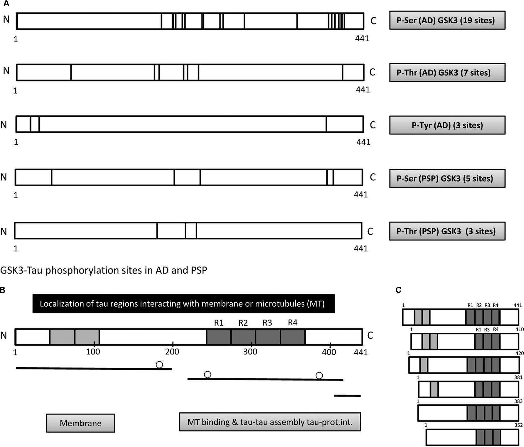
Figure 1. Tau phosphorylation in tauopathies. (A) More than 40 residues of the different tau isoforms can be modified in Alzheimer disease (AD). Some of these sites are modified by GSK3, and the modified serine residues (184, 198, 199, 202, 210, 214, 235, 237, 258, 262, 289, 356, 396, 400, 404, 409, 412, 413, 416) or threonine residues (69, 175, 181, 212, 217, 231, 414) have been localized within the tau molecule (Hanger et al., 2009
). In addition, there are three possible tyrosine residues (18, 29, 394) that could be modified in AD, the main candidates each located in the amino terminal domain. Fewer residues modified by GSK3 have been identified in tau from progressive supracellular palsy (PSP). (B) These and other phosphorylation events can regulate the interaction of tau protein with the cell membrane, microtubules, other proteins or its self association. The circles indicate the localization of the phosphorylated residues that prevent interactions (Ser 199-202) with the cell membrane, (Ser 262) with microtubules, or (Ser 396-404) with muscarine receptors. Also, when the serines (396-404) are phosphorylated the interaction of phospho tau with α-synuclein and GSK3β is favoured. (C) Scheme showing the six CNS tau isoforms. Three 3R and three 4R tau isoforms.)
In other tauopathies, like progressive supranuclear palsy (PSP), only eight phosphorylation sites have been reported (Wray et al., 2008
), although all of these sites (Ser 46, Thr181, Ser202, Thr217, Thr231, Ser235, Ser396 or Ser400, and Thr403 or Ser404) are also modified in AD and they might be modified by GSK3. It is not known whether these differences in the number of phosphorylated residues in tauopathies like AD or PSP could provoke the appearance of different filamentous tau polymers in these conditions (Arrasate et al., 1997
), in addition to other reported factors that could also influence the morphology of these filaments (Arrasate et al., 1997
).
In AD, paired helical (PHF) and straight (SF) filaments can be detected in the patients’ brain (Kidd, 1963
), whereas mainly straight filaments are observed in PSP (Perez et al., 1998
). Alternatively, tau can be proteolyzed (truncated) in different ways in tauopathies like AD or PSP, and truncated tau can be phosphorylated by kinases like GSK3 (Rissman et al., 2004
) and indeed, it has been suggested that tau cleavage may precede its hyperphosphorylation (Cribbs et al., 2004
). In AD, tau fragments of different sizes have been described that lack C-terminal residues (up to 17 kDa) (Binder et al., 2005
; Gamblin et al., 2003
; Park and Ferreira, 2005
). However, in PSP (Wray et al., 2008
) a 35 kDa tau fragments lacking the N-Terminal region has been identified. Curiously, this tau fragment is less evident in the CSF of PSP patients (Borroni et al., 2008
), perhaps because it forms aggregates that are not secreted into the CSF. On the other hand, tau truncated at asp 421 is toxic since it induces mitochondrial fragmentation and elevated oxidative stress, which could in turn result in neuron death (Quintanilla et al., 2009
).
In summary, many tau protein isoforms can be generated from a single tau gene and some of these isoforms may undergo different modifications, such as phosphorylation. These differences in phosphorylation could reflect a tau code that could explain the different activities fulfilled by tau (Figure 1
B). Indeed, phosphorylation of the N-terminal half of the tau molecule could affect its interaction with the cell membrane, while phosphorylation in the microtubule binding domain and its adjacent regions will impair the interaction of tau with microtubules, as well as influencing tau-tau self assembly. By contrast, phosphorylation at the C-terminal region affects the interaction of tau with other proteins, like muscarinic receptors, or the formation of the α-synuclein-GSK3β-tau complex.
The author declare that the research was conducted in the absence of any commercial or financial relationships that could be construed as a potential conflict of interest. On the other hand, J. Avila is a consultant for Noscira.
This work was supported by Spanish Plan Nacional, CIBERNED, Comunidad de Madrid, Fundación Marcelino Botín and an Institutional grant from Fundación Ramón Areces. I will like to acknowledge the excellent technical assistance of Ms Nuria de la Torre.
Alonso, A. D., Grundke-Iqbal, I., Barra, H. S., and Iqbal, K. (1997). Abnormal phosphorylation of tau and the mechanism of Alzheimer neurofibrillary degeneration: sequestration of microtubule-associated proteins 1 and 2 and the disassembly of microtubules by the abnormal tau. Proc. Natl. Acad. Sci. U.S.A. 94, 298–303.
Gamblin, T. C., Chen, F., Zambrano, A., Abraha, A., Lagalwar, S., Guillozet, A. L., Lu, M., Fu, Y., Garcia-Sierra, F., LaPointe, N., Miller, R., Berry, R. W., Binder, L. I., and Cryns, V. L. (2003). Caspase cleavage of tau: linking amyloid and neurofibrillary tangles in Alzheimer’s disease. Proc. Natl. Acad. Sci. U.S.A. 100, 10032–10037.
Gong, C. X., Lidsky, T., Wegiel, J., Zuck, L., Grundke-Iqbal, I., and Iqbal, K. (2000). Phosphorylation of microtubule-associated protein tau is regulated by protein phosphatase 2A in mammalian brain. Implications for neurofibrillary degeneration in Alzheimer’s disease. J. Biol. Chem. 275, 5535–5544.
Grover, A., Houlden, H., Baker, M., Adamson, J., Lewis, J., Prihar, G., Pickering-Brown, S., Duff, K., and Hutton, M. (1999). 5’ splice site mutations in tau associated with the inherited dementia FTDP-17 affect a stem-loop structure that regulates alternative splicing of exon 10. J. Biol. Chem. 274, 15134–15143.
Hutton, M., Lendon, C. L., Rizzu, P., Baker, M., Froelich, S., Houlden, H., Pickering-Brown, S., Chakraverty, S., Isaacs, A., Grover, A., Hackett, J., Adamson, J., Lincoln, S., Dickson, D., Davies, P., Petersen, R. C., Stevens, M., de Graaff, E., Wauters, E., van Baren, J., Hillebrand, M., Joosse, M., Kwon, J. M., Nowotny, P., Che, L. K., Norton, J., Morris, J. C., Reed, L. A., Trojanowski, J., Basun, H., Lannfelt, L., Neystat, M., Fahn, S., Dark, F., Tannenberg, T., Dodd, P. R., Hayward, N., Kwok, J. B., Schofield, P. R., Andreadis, A., Snowden, J., Craufurd, D., Neary, D., Owen, F., Oostra, B. A., Hardy, J., Goate, A., van Swieten, J., Mann, D., Lynch, T., and Heutink, P. (1998). Association of missense and 5’-splice-site mutations in tau with the inherited dementia FTDP-17. Nature 393, 702–705.
Varani, L., Hasegawa, M., Spillantini, M. G., Smith, M. J., Murrell, J. R., Ghetti, B., Klug, A., Goedert, M., and Varani, G. (1999). Structure of tau exon 10 splicing regulatory element RNA and destabilization by mutations of frontotemporal dementia and parkinsonism linked to chromosome 17. Proc. Natl. Acad. Sci. U.S.A. 96, 8229–8234.
Wetzel, M. K., Naska, S., Laliberte, C. L., Rymar, V. V., Fujitani, M., Biernaskie, J. A., Cole, C. J., Lerch, J. P., Spring, S., Wang, S. H., Frankland, P. W., Henkelman, R. M., Josselyn, S. A., Sadikot, A. F., Miller, F. D., and Kaplan, D. R. (2008). p73 regulates neurodegeneration and phospho-tau accumulation during aging and Alzheimer’s disease. Neuron 59, 708–721.