Microtissue array to screen the impact of carbon nanotube on lung cellular and tissue biomechanics
-
1
State University of New York at Buffalo, Biomedical Engineering, United States
Introduction: Multi-wall carbon nanotubes (MWCNT) are widely used in many biomedical applications such as drug delivery and medical imaging. However, the application of MWCNT also raises health concerns. In vivo animal studies have shown that certain types of MWCNT cause cellular apoptosis and inflammation in the lung, which lead to the thickening and stiffening of the lung tissue. Such elevation of the stiffness and contractility of the tissue is a known hallmark of tissue fibrosis. Therefore, identifying the biomechanical impacts of specific types of MWCNT at the cellular and tissue levels are important to the health safety in applications involving carbon nanotube. The recent emergence of bio-microelectromechanical systems (bio-MEMS) technology allows realistic modeling of 3D human tissue and high throughput testing of many samples in a single device, thus provides a potential viable solution to screen the pathogenic factors. Here we report the development of a 3D human lung microtissue array device for the screening of the biomechanical impact of two different types of MWCNT at different concentrations.
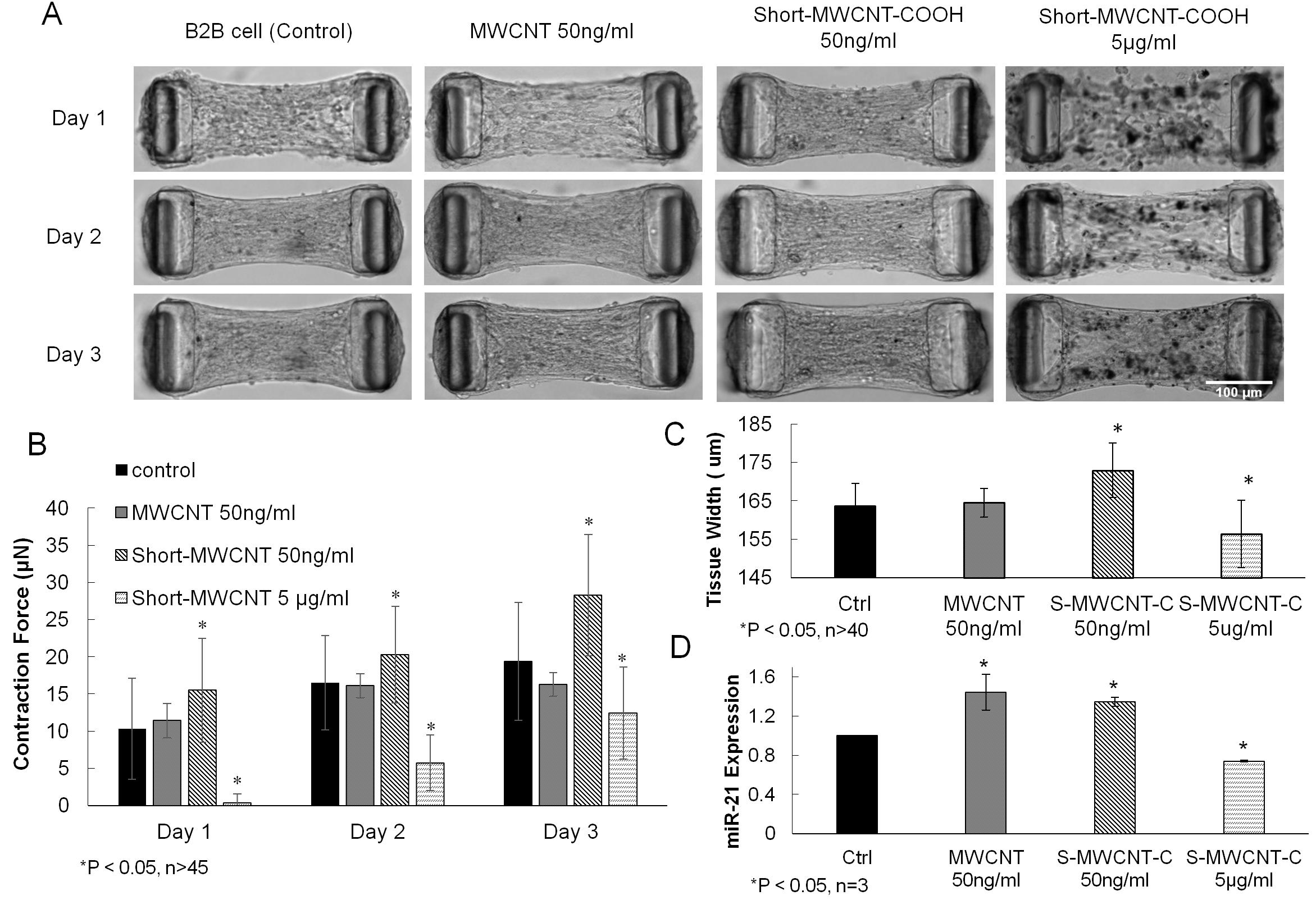
We fabricated microtissue array device by PDMS replica molding from masters made using a multilayer microlithography technique. Each device contains an array of 10 x 13 microwells (Fig. 1A, B). The contraction force F generated by the microtissues is calculated according to the cantilever bending theory as F = kδ, where δ is the cantilever deflection and k is the spring constant (Fig. 1C). During cell seeding, suspensions of human lung bronchial epithelial cells (B2B) in unpolymerized type-I collagen were centrifuged into microwells and the collagen was polymerized under 37°C. Within 24 hours, the contractile action of the cells caused the compaction of the matrix hydrogel and formation of a dog-bone shaped microtissue between the two pillars (Fig. 1D). To study the biomechanical impacts of MWCNT on B2B cell-populated microtissue, three different conditions (MWCNT 50ng/ml, short-MWCNT 50ng/ml and short-MWCNT 5ug/ml) were used to treat the microtissue for three continuous days. The changes in tissue contraction force and tissue width under different conditions were measured daily (Fig. 2A). We also used qRT-PCR to measure fibrosis-related biomarkers in the microtissue after MWCNT treatments.
The results showed that contraction forces continued to increase for all the groups over a 3-day culture period with the short-MWCNT 50ng/ml group producing the largest contraction force (Fig. 2B). The elevated contraction force correlated with increased tissue width (Fig. 2C). This is because the squeezing of microtissue in the longitudinal direction under high contraction force caused increase in the cross-section area. Results of qRT-PCR showed significant up-regulation of miR-21 (a known marker for fibrosis) in short-MWCNT 50ng/ml group (Fig. 2D). Together, the above results suggested that short-MWCNT 50ng/ml is a potent stimulant for tissue contractility and a potential pathogenic risk of lung fibrosis. In this study, the integrated 3D biomechanical capacity of the device allowed in situ collection of mechanical properties of the model tissue under different treatment conditions. Such information was then used as a phonotypical marker to compare with the results obtained through qRT-PCR test. Together, the cross examination of the results obtained from this novel system enabled identification of the potential pathogenic factors for lung fibrosis.
Keywords:
Bio-MEMS,
array,
microstructure,
mechanical property
Conference:
10th World Biomaterials Congress, Montréal, Canada, 17 May - 22 May, 2016.
Presentation Type:
New Frontier Oral
Topic:
Microdevices: reproducing physiology at microscale
Citation:
Chen
Z,
Wang
Q,
Asmani
M,
Li
Y,
Liu
C,
Wu
Y and
Zhao
R
(2016). Microtissue array to screen the impact of carbon nanotube on lung cellular and tissue biomechanics.
Front. Bioeng. Biotechnol.
Conference Abstract:
10th World Biomaterials Congress.
doi: 10.3389/conf.FBIOE.2016.01.00692
Copyright:
The abstracts in this collection have not been subject to any Frontiers peer review or checks, and are not endorsed by Frontiers.
They are made available through the Frontiers publishing platform as a service to conference organizers and presenters.
The copyright in the individual abstracts is owned by the author of each abstract or his/her employer unless otherwise stated.
Each abstract, as well as the collection of abstracts, are published under a Creative Commons CC-BY 4.0 (attribution) licence (https://creativecommons.org/licenses/by/4.0/) and may thus be reproduced, translated, adapted and be the subject of derivative works provided the authors and Frontiers are attributed.
For Frontiers’ terms and conditions please see https://www.frontiersin.org/legal/terms-and-conditions.
Received:
27 Mar 2016;
Published Online:
30 Mar 2016.