New Information about Albert Einstein's brain

A commentary on
New information about Albert Einstein’s brain
by Dean Falk. Front. Evol. Neurosci. 1:3.
Scientists continue to look to Einstein’s brain in hopes of discovering the biology of intelligence. At the time of Einstein’s death, statistical data on individuals of normal intelligence was limited (Herskovitz, 2000). However, our bank of statistical data on individuals in the “normal intelligence” range has since grown; a comparison of this new data to Einstein’s brain may provide us with a greater context for understanding the relationship between the brain and intelligence. New Information about Albert Einstein’s brain asks new questions about the famous mathematician’s brain from a form and function point of view. In-depth studies of cortical surfaces, weight, and sulcal patterns serve as the foundation for this research, but Dr. Dean Falk at Florida State University has made a new discovery by closely examining Einstein’s cortical sulci. Using existing neuroanatomy and functional neuroimaging studies, Falk identifies two significant differences on Einstein’s brain that shed light on both his superior intelligence and his developmental struggles.
Falk closely analyzed photographs of Einstein’s brain that were taken within the first few hours of his death. These calibrated photographs, along with direct caliper measurements were the primary materials Falk used to reanalyze Einstein’s brain. Two identifications, a long, unnamed sulcus (u), and a knob (K), are the emphasis of Falk’s research because they are both unusual and uncommon (see Figure 1).
FIGURE 1
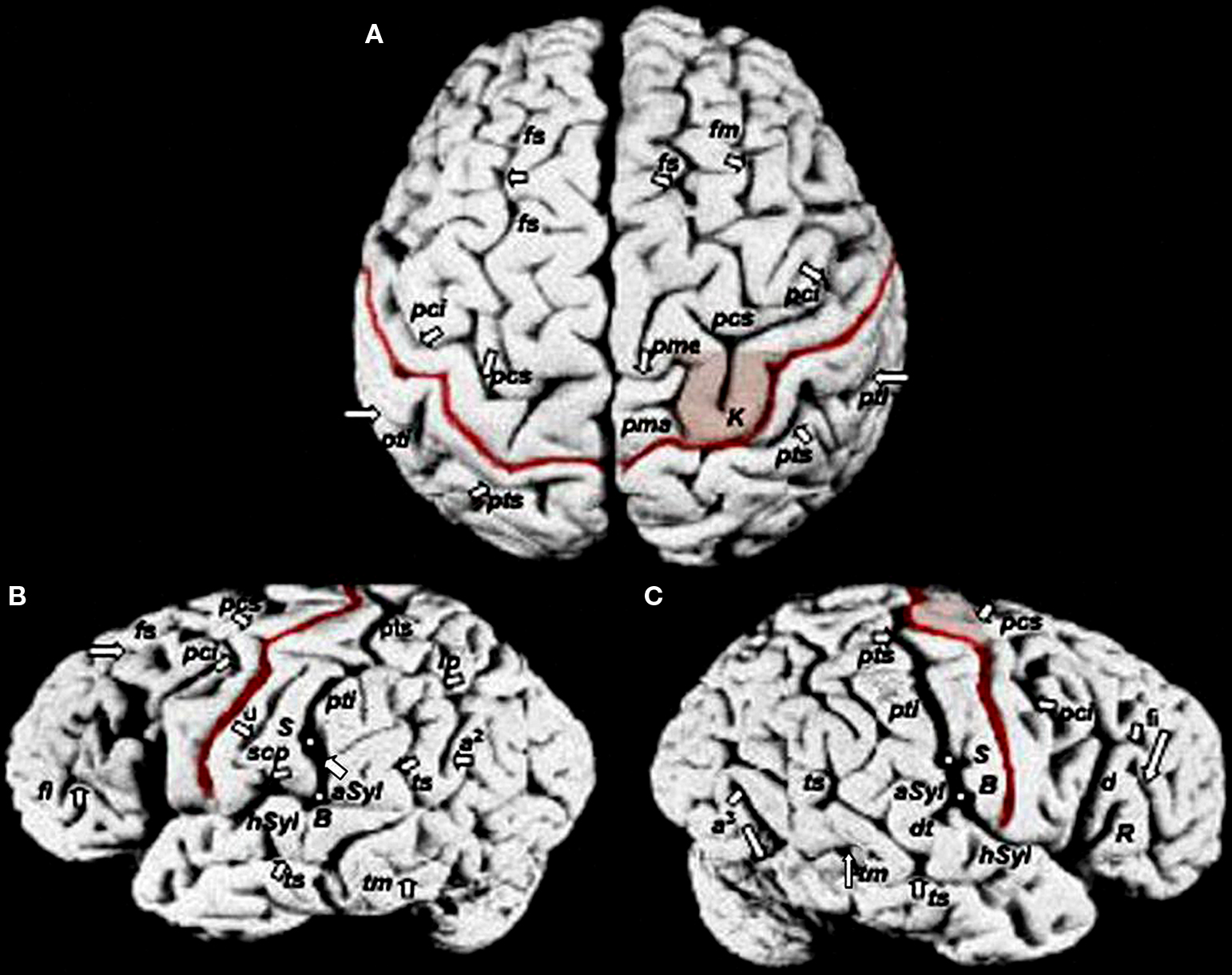
Figure 1. Photographs of Einstein’s brain that were taken in 1955, adapted from Witelson et al. (1999b) with identifications added here. (A) Dorsal view, (B) left lateral view, (C) right lateral view. Sulci: angular (a2), anterior occipital (a3), ascending limb of the posterior Sylvian fissure (aSyl), central fissure (red lines), diagonal (d), descending terminal portion of aSyl (dt), inferior frontal (fi), middle frontal (fm), superior frontal (fs), horizontal limb of the posterior Sylvian fissure (hSyl), intraparietal (ip), precentral inferior and superior (pci, pcs), marginal precentral (pma), medial precentral (pme), postcentral inferior and superior (pti, pts), ascending ramus of Sylvian fissure (R), subcentral posterior sulcus (scp), middle temporal (tm), superior temporal sulcus (ts), unnamed sulcus in postcentral gyrus (u). Other features: branching point between hSyl and aSyl (white dots, B), hand motor cortex knob (K, shaded in A, C), termination of aSyl (white dots, S). (adopted from Falk D (2009) New Information about Albert Einstein’s brain. Front. Evol. Neurosci. 1:3. doi:10.3389/neuro.18.003.2009)
The u located in the post-central gyrus suggests greater development in areas of the brain that correspond to the face and tongue (Falk, 2009). As the post-central gyrus has been shown to correlate with auditory “what” and “where” pathways, this unnamed sulcus might similarly correspond to Einstein’s musical ability and aural discrimination (Alain et al., 2008).
One remarkable feature of Einstein’s brain is a K, an omega-shaped curve in the perirolandic region of the right hemisphere. Falk suggests this may be so pronounced in Einstein because of his extensive musical training on the violin. Gromko et al. (2009) reported that children with good aural discrimination skills had similar number recall accuracy. It is not a surprise that Einstein was both gifted in music and a genius in mathematics; the pronounced K may be the testament to his superior abilities in these subjects.
In addition to the u and the K, Falk discovered noticeably wider post-central gyri at their lateral ends. This subtle, but important variation is suggested to partially account for Einstein’s known language difficulties.
Another significant component of Einstein’s brain was an unusual symmetry particularly with the post-central inferior sulcus (pti). Violin players often develop ambidexterity (e.g., equal use of both hands) in order to successfully play their instrument, and this may have been the case with Einstein; the symmetry seen in his brain reinforces the long-time argument of Einstein’s ambidexterity.
Falk’s analysis of Einstein’s sulci is a new piece of the puzzle that addresses key features that separate Einstein from the mean. One of Falk’s most groundbreaking findings is the presence of continuous precentral superior and inferior sulci (pcs and pci). These are present on both right and left hemispheres of Einstein’s brain; symmetry not found in 98% of the 50 hemispheres scored by Ono et al. (1990:43; as cited in Falk, 2009).
Falk’s analysis of Einstein’s brain sheds light on the relationship between his biology and his success. The most notable differences seen in his brain appear to have more to do with his training in the violin as a child, and less to do with his genetics. Interestingly, recent findings suggest that early musical training may indeed increase one’s aptitude for math; Einstein is testament to this idea. These findings help us not only understand the genius of Albert Einstein, but also lend to understanding individual variation in mental aptitude and the effects of development on neuroanatomy and function. This study is similar to existing studies of patient populations (e.g., mental illness, developmental disorders, autism, and learning disorders) where functional neuroimaging researchers are employing voxel based morphometry to quantify individual variation in neuroanatomy related to illness-related deficits in cognition, affect, and sociality.
References
Alain, C., He, Y., and Grady, C. (2008). The contribution of the inferior parietal lobe to auditory spatial working memory. J. Cogn. Neurosci. 20, 285–295.
Falk, D. (2009). New information about Albert Einstein’s brain. Front. Evol. Neurosci. 1. doi:10.3389/neuro.18.003.2009.
Citation: Spicer KR and Platek SM (2010) An old brain with new tricks. Front. Neurosci. 4:40. doi: 10.3389/fnins.2010.00040
Received: 02 February 2010;
Paper pending published: 26 February 2010;
Accepted: 27 May 2010;
Published online: 15 September 2010
Copyright: © 2010 Spicer and Platek. This is an open-access article subject to an exclusive license agreement between the authors and the Frontiers Research Foundation, which permits unrestricted use, distribution, and reproduction in any medium, provided the original authors and source are credited.
*Correspondence: splatek@gmail.com