Role of Microglia TLRs in Neurodegeneration
 Bernd L. Fiebich1*
  Carla Ribeiro Alvares Batista2
  Soraya Wilke Saliba1,3
  Nizar M. Yousif1,3
  Antonio Carlos Pinheiro de Oliveira2*
- 1Neuroimmunology and Neurochemistry Research Group, Department of Psychiatry and Psychotherapy, Medical Center—University of Freiburg, Faculty of Medicine, University of Freiburg, Freiburg, Germany
- 2Department of Pharmacology, Universidade Federal de Minas Gerais, Belo Horizonte, Brazil
- 3Faculty of Biology, University of Freiburg, Freiburg, Germany
Toll-like receptors (TLRs) are a group of receptors widely distributed in the organism. In the central nervous system, they are expressed in neurons, astrocytes and microglia. Although their involvement in immunity is notorious, different articles have demonstrated their roles in physiological and pathological conditions, including neurodegeneration. There is increasing evidence of an involvement of TLRs, especially TLR2, 4 and 9 in neurodegenerative diseases such as Alzheimer’s disease (AD), Parkinson’s disease (PD), and amyotrophic lateral sclerosis (ALS). In this sense, their expression in microglia might modulate the activity of these cells, which in turn, lead to protective or deleterious effects over neurons and other cells. Therefore, TLRs might mediate the link between inflammation and neurodegenerative diseases. However, further studies have to be performed to elucidate the role of the other TLRs in these diseases and to further prove and confirm the pathophysiological role of all TLRs in neurodegeneration. In this article, we revise and summarize the current knowledge regarding the role of TLRs in neurodegeneration with the focus on the possible functions of these receptors in microglia.
General Aspects of Toll-Like Receptors
Toll-like receptors (TLRs) are a growing family that plays critical roles in innate immune responses. Several TLRs have already been identified in human and murine, such as TLR1–5 (Chaudhary et al., 1998; Rock et al., 1998), TLR6 (Takeuchi et al., 1999), TLR7–9 (Du et al., 2000). TLRs are located on the plasma membrane with the exception of TLR3, TLR7, TLR9 that are presented intracellularly in vesicles such as the endoplasmic reticulum, endosomes or lysosomes (Kawai and Akira, 2010).
Signaling
TLRs are activated by different pathogen-associated molecular patterns (PAMPs), including compounds derived from bacteria, virus, or fungi (Takeuchi et al., 2002; Li et al., 2013). TLR signaling pathways depend on receptor dimerization and recruitment of adapter proteins that mediate other protein-protein interactions, such as myeloid differentiation primary-response protein 88 (MyD88) and Toll/interleukin 1 receptor (TIR) domain-containing adaptor interferon-β (TRIF; Takeda et al., 2003; Yamamoto et al., 2003a). Importantly, all TLR signaling pathways are dependent on MyD88, except TLR3, which initiates signaling via the TRIF adapter. In addition, TLR4 is activated by both MyD88-associated and TRIF-related adapter molecule (TRAM)-associated pathways (Fitzgerald et al., 2003a; Ruckdeschel et al., 2004).
The MyD88-dependent pathway initiates with the recruitment of tumor-necrosis-factor-receptor-associated factor 6 (TRAF6) and members of the IL-1R-associated kinases (IRAK) family. The activation of TRAF6 allows the translocation of nuclear factor kappa B (NF-κB) to the nucleus, which results in the production of different pro-inflammatory mediators such as cytokines and cyclooxygenase-2 (COX-2; Zhang et al., 1999; Takeda and Akira, 2004; Broad et al., 2007; Kawai and Akira, 2010).
While the MyD88-dependent pathway promotes the production of pro-inflammatory cytokines, the MyD88-independent pathway is associated with the induction of interferon (IFN)-inducible genes and maturation of dendritic cells (Yamamoto et al., 2003b). The MyD88-independent pathway is mediated by TRAM and TRIF. Inhibitor of NF-κB kinase subunit epsilon (IKKε) and TANK-binding kinase (TBK) 1 interact with TRIF or TRAM and activate interferon regulatory factor 3 (IRF-3) and NF-κB-dependent signaling pathways (Fitzgerald et al., 2003b; Palsson-McDermott and O’Neill, 2004). IRF3 mediates the expression of IFN type I, e.g., IFN-β and chemokines, such as C-C motif chemokine ligand 5 (CCL5) and C-X-C motif chemokine ligand 10 (CXCL10; Schafer et al., 1998; Melchjorsen and Paludan, 2003).
TLR Expression in the CNS
TLRs are widely expressed in the CNS and play different roles in cell survival or death (Okun et al., 2011). Studies have demonstrated that TLRs are expressed in neurons (Tang et al., 2007), microglia (Olson and Miller, 2004), astrocytes (Bowman et al., 2003), oligodendrocytes (Lehnardt et al., 2006) and neural stem cells (NSC; Okun et al., 2009; van Noort and Bsibsi, 2009). It has been demonstrated that NSC transplantation in APP/PS1 mice decreased inflammatory injury through suppression of glial and TLR4-mediated inflammatory pathway activation (Zhang et al., 2016). The expression of TLRs is affected by aging, which is a risk factor of neurodegenerative diseases, such as Alzheimer’s disease (AD). Aged cultures of rat hippocampal neurons presented an increase of TLR4 expression (Calvo-Rodríguez et al., 2017) while the expression levels of TLR2 and TLR4 increased during the suckling period and then decreased once the infants reached 1 year of age (Savino et al., 2018).
Microglia are the resident mononuclear immune cells of the CNS and play important roles in the maintenance of homeostasis and neuroinflammation (Ransohoff and Perry, 2009; Perry et al., 2010; Boche et al., 2013). Microglia express all TLRs and their adapter proteins as shown in mice, rats, and humans (Bsibsi et al., 2002; Olson and Miller, 2004; Zhang et al., 2013), such as TLR1 and TLR6 (Bsibsi et al., 2002), TLR2 (Laflamme et al., 2001), TLR3, TLR5, TLR7, TLR8 (Bsibsi et al., 2002), TLR4 (Laflamme and Rivest, 2001) and TLR9 (Dalpke et al., 2002). Constitutive expression of TLRs is primarily in microglia and largely restricted to the circumventricular organs (CVOs) and meninges, areas with direct access to the circulation, although they may also be expressed at lower levels in other regions (Laflamme et al., 2001; Laflamme and Rivest, 2001; Chakravarty and Herkenham, 2005).
Different stimuli may lead to increased expression of microglia TLRs, such as hypoxia (Smith et al., 2013; Yao et al., 2013), LPS (Kielian et al., 2005; Yousif et al., 2018), Poly(I:C; Yousif et al., 2018), kainic acid (Wang et al., 2015; Hu and Mao, 2016), α-synuclein (Béraud et al., 2011; Daniele et al., 2015) and amyloid beta (Aβ; Jana et al., 2008; Richard et al., 2008; Caldeira et al., 2017).
TLRs in Neurodegeneration
TLR activation can be beneficial or detrimental to the host. In addition to the known role of being pro-inflammatory in the protective response against pathogens in the CNS, TLRs may be activated in the absence of microbial invaders. It has been shown that TLRs are upregulated in brains of AD patients (Liu et al., 2005) and other neurodegenerative diseases (Bsibsi et al., 2002; Walter et al., 2006), such as amyotrophic lateral sclerosis (ALS) and Parkinson’s disease (PD; van Noort and Bsibsi, 2009; De Paola et al., 2012; Lee et al., 2015). An increased expression of TLR2, TLR4, TLR5, TLR7, TLR9 and the TLR co-receptor CD14 has been shown in microglia cells situated around senile plaques in post-mortem brain of AD patients and related mouse models (Liu et al., 2005; Walter et al., 2007; Jana et al., 2008; Letiembre et al., 2009). Another study demonstrated a significant alteration of cognitive functions in TLR2-deficient APP/PS1 mice and synaptic plasticity associated with increased levels of toxic Aβ1–42 (Richard et al., 2008). Further studies associated TLRs with ALS in mouse models, suggesting their crucial role in the neurodegenerative process of this pathological condition (De Paola et al., 2012; Lee et al., 2015).
Finally, a crosstalk between TLR and cell stressors has been observed in immune cells, such as the unfolded protein response (UPR) and oxidative stress, which are present in different neurodegenerative diseases (Qin et al., 2005; Gill et al., 2010; Gough, 2010; Coope et al., 2012; Grootjans et al., 2016; Sprenkle et al., 2017; Wang et al., 2017). Besides, various components present in neurodegenerative diseases, which will be discussed below, activate the TLRs present in microglia. In this sense, many stimuli modulate the activity of these receptors and alter microglia phenotype and activation. Thus, TLRs present in microglia may partially mediate the development of these pathological conditions.
TLR2
TLR2 is one of the most studied TLR in respect to neurodegenerative diseases (Guillot-Sestier and Town, 2018). In microglia, the coupling of CD14 to TLR2 and TLR4 is necessary for the induction of immune response associated with fibrillar Aβ phagocytosis (Reed-Geaghan et al., 2009). Moreover, it was reported that activation of TLR2 in microglia enhances pathological Aβ uptake (Chen et al., 2006). However, others showed that inhibition of TLR2 activation decreased glial cell reactivity, leading to reduced Aβ deposits and improved cognitive performance in APP/PS1 mice (McDonald et al., 2016). Likewise, knocking out TLR2 in mice prevented memory impairment occurring after Aβ1–42 immunization (Vollmar et al., 2010). This is supported by results from cultured microglia showing that TLR2 deficiency reduced Aβ42-induced inflammation and enhanced Aβ clearance (Jana et al., 2008; Liu et al., 2012). In accordance, TLR2-dependent JNK/NF-κB signaling partially contributed to the inflammatory response mediated by Aβ1–42 (Lin et al., 2013). Immunohistochemistry data demonstrated the TLR2 localization on microglia associated with Aβ plaques in old APP23 transgenic mice (Frank et al., 2009). Genetic studies identified TLR2 as one of the risk factors associated with Late-Onset AD (LOAD) in Azeri Turk ancestry and Han Chinese populations (Wang et al., 2011; Yu et al., 2011a; Rezazadeh et al., 2016). However, results from another report argue against the relationship between TLR2 polymorphism and LOAD (Yu et al., 2011b).
The potential role of TLR2 in PD is supported by the existing link between TLR2 polymorphism and increased risk of PD (Kalinderi et al., 2013). α-synuclein released by neurons extracellularly acts as an endogenous ligand for TLR2 on microglia, which in turn produce toxic molecules leading to neurodegeneration (Kim et al., 2013, 2016). In mice, overexpression of α-synuclein increased TLR2 expression and led to increased microglial activation (Drouin-Ouellet et al., 2015). Disease stage and region-dependent expression of TLR2 consistent with region-specific phenotype and activation of microglia shown in different groups of PD patients suggest a critical role of TLR2 in the microglia-related responses observed in PD (Doorn et al., 2014). Upon treatment with α-synuclein, microglia exhibit pro-inflammatory phenotype by forming a heterodimer complex that engage TLR1 and TLR2, leading to a chronic neuroinflammatory milieu. In this context, candesartan cilexetil, a drug used for treatment of hypertension also known to suppress the expression of TLR2, reversed the pro-inflammatory microglia phenotype activated by α-synuclein exposure (Daniele et al., 2015). Interestingly, TLR2 and its downstream signaling were shown to play a key role in the neuroprotective effects achieved through exercise approaches in 1-methyl-4-phenyl-1,2,3,6-tetrahydropyridine (MPTP) mouse model of PD (Jang et al., 2017; Koo et al., 2017).
Furthermore, induced expression of TLR2 was detected strictly within microglia in the chronically LPS-stimulated SOD1 transgenic mouse model of ALS. The increased TLR2 expression was strongly correlated with robust immune response and degeneration of motor neurons and motor axons (Nguyen et al., 2004). Another report demonstrated that overexpression of ALS-related SOD1 mutant promotes microglia neurotoxic effects via TLR2 (Liu et al., 2009). The fact that marked upregulation of TLR2 has already been observed in a previous study implemented in the ALS mouse model is in line with these findings (Nguyen et al., 2001). In two culture models of mSOD1-linked ALS, microglia TLR2 dependent activation was required to induce toxic effects and injury of motoneurons (Zhao et al., 2010). Increased expression of TLR2 in reactive glial cells was detected in spinal cord of ALS patients. Interestingly, TLR2 was mainly detected in myeloid cells (microglia, macrophages; Casula et al., 2011). Taken together, the role of microglial TLR2 in ALS seems to be consistent, since data from various reports are in agreement. This fact also suggests that modulation of TLR2 in ALS might be a potential target for effective therapy.
TLR3
TLR3 is less studied in respect to neurodegenerative diseases, although it may be involved in neuroinflammation. For example, incubation of microglia with Poly(I:C), a TLR3 agonist, increases the synthesis of COX-2, mPGES-1 and prostaglandin E2 release (de Oliveira et al., 2016), and increased the expression of TLR3 (Yousif et al., 2018). The production of these inflammatory mediators might contribute to neurodegenerative processes observed in some conditions. Indeed, a previous study shows that peripheral challenge with Poly(I:C) increased Aβ levels in the hippocampus linked to cognitive dysfunction in non-transgenic mice (Weintraub et al., 2014). Moreover, systemic challenge with Poly(I:C) amplified CNS inflammation and exacerbated chronic neurodegeneration in the ME7 model of prion disease (Field et al., 2010). Importantly, a very recent study in human AD brains identified increased expression of TLR3 in microglia associated with Aβ plaques (Walker et al., 2018). In addition, TLR3 is an essential receptor for viral detection (Hanke and Kielian, 2011). It has already been shown that influenza virus infection may lead to neurodegeneration (Jang et al., 2009). Other viruses such as hepatitis C virus, Eppstein-Barr virus and human immunodeficiency virus (HIV) have been also linked to PD (Woulfe et al., 2014; Moulignier et al., 2015; Wu et al., 2015).
TLR4
An elegant and recent study demonstrated that microglia immune memory shapes neuropathology and provided potential mechanism for LPS-triggered immune memory in the brain, suggesting a role of TLR4 in this phenomenon (Wendeln et al., 2018). Interestingly, both, beneficial and detrimental roles of TLR4 in the context of AD are reported. Transgenic AD mice deficient in TLR4 exhibit reduced microglial activation, increased Aβ deposition, and reduced cognitive function (Song et al., 2011). In the transgenic mouse model of tau pathology P301S, chronic mild stimulation of TLR4 with LPS reduced cerebral p-tau proteins and improved memory impairment through microglia-dependent activation of autophagy (Qin et al., 2016). In contrast, activation of TLR4 but not TLR2 reduced microglial Aβ42-clearance through modulation of the scavenger receptor CD36 activity (Li et al., 2015). Pronounced TLR4 expression in APP mice and an increased expression of TLR4 in brains of AD patients associated with Aβ plaques have been identified (Walter et al., 2007). TLR4 was shown to be necessary for glial cell activation that resulted in impairment in memory in a mouse model of AD based on acute Aβ injection (Balducci et al., 2017). A recent report suggests that alteration in microglial TLR4 signaling contribute to AD pathogenesis in APP/PS1 mice (Go et al., 2016).
Collectively, the role of TLR4, likewise TLR2, in AD is perhaps disease-stage dependent and this could be at least one explanation for the controversy found in the literature about the impact of these receptors in AD pathology. In this regard, activation of TLR4 in the initial stage has beneficial effects on Aβ-clearance, while chronic activation contributes to Aβ aggregation (Huang et al., 2017).
Previous studies suggest that TLR4 deregulation may play an important role in α-synucleinopathies (Caputi and Giron, 2018). TLR4 was shown to be essential for microglia to induce responses against α-synuclein in vitro such as uptake of this protein, pro-inflammatory cytokine release, and reactive oxygen species production (ROS; Fellner et al., 2013). TLR4 deficiency in the MPTP induced PD model resulted in reduced cell death and decreased number of reactive microglia (Noelker et al., 2013). Impaired microglial phagocytosis, enhanced neuronal loss and exacerbated motor dysfunction upon TLR4 ablation has been shown in a transgenic mouse model of multiple system atrophy with oligodendroglial α-synuclein overexpression (Stefanova et al., 2011). Moreover, stimulation of TLR4 with monophosphoryl lipid A in mice overexpressing α-synuclein increased microglia phagocytosis of this protein, improved motor deficits, and prevented neuronal degeneration (Venezia et al., 2017). In BV2 microglia-like cells, TLR4-dependent immune response regulated Nurr1 and NF-κB signals, which were shown to be vital during neuroinflammation stimulated by α-synuclein (Shao et al., 2018). In addition, TLR4 signaling was shown to exert a crucial function in the activation of BV2 cells and the promotion of inflammation induced by 1-methyl-4-phenylpyridinium (MPP(+); Zhou et al., 2016). In an in vitro model of PD, neuron-derived IgG mediates dopaminergic neurons survival by reducing apoptosis induced by 6-hydroxydopamine toxicity through a mechanism that involved the activation of microglial TLR4 (Zhang et al., 2013). Taken together, TLR4 seems to exert different roles in relation to PD. Nevertheless, future investigations aiming to target TLR4 in a specific cell subset in the brain will provide a better understanding concerning the role of this receptor in the context of PD.
Recently, the impact of TLRs in respect to ALS has been given some attention. For instance, in the hSOD1 G93A mouse model of ALS, increased TLR4 expression has been detected in microglia and astrocytes. In the same model, TLR4 deficiency improved motor function and extended life expectancy (Lee et al., 2015). In vitro and in vivo approaches of motor neuron degeneration, treatment with TLR4 antagonist decreased microglial activation, exerted neuroprotective effects, and improved behavior performance (De Paola et al., 2012). In line with these findings, antagonizing TLR4 in motoneurons/glia co-cultures isolated from SOD1 G93A mice prevented cell death induced by SOD1mut glia (De Paola et al., 2016).
The role of TLRs in Huntington’s disease (HD) is not covered. There is only one study that directly addressed the importance of TLR2, TLR3 and TLR4 in the N171–82Q mouse model of HD. In this study, when TLR2, TLR3 and TLR4 were lacking, the survival rate of HD mice was increased (Griffioen et al., 2018).
TLR5
In the context of neurodegenerative diseases, Bsibsi et al. (2002) have demonstrated that human microglial cell cultures from donors with AD, olivopontocerebellar atrophy (OPCA), and Pick’s disease (PID) expressed increasing levels of TLR5 but differing between the donors. Additionally, overexpression of TLR5 in the substantia nigra, striatum, cerebral cortex and nucleus dentatus areas, was observed in multiple systems atrophy patients (Brudek et al., 2013) and in the brain stem of a genetic PD mouse model (Letiembre et al., 2009). In contrast, down-regulation of TLR5 expression was observed in the frontal cortex of a PD rat model, using intranasal administration of MPTP (Viana et al., 2017). However, additional studies are needed to elucidate the effects of TLR5 in neurodegenerative diseases and to comprehend its role in microglia.
TLR7
The expression of TLR7 has been demonstrated in different neurodegenerative diseases (AD, PD, OPCA and PID) and in disease models including human microglial cell culture (Bsibsi et al., 2002) or murine microglial cells (Béraud et al., 2011; Béraud and Maguire-Zeiss, 2012). Furthermore, the expression of TLR7 has also been demonstrated in some areas of the CNS of genetic mice models of AD (brain cortex) and ALS (spinal cord; Letiembre et al., 2009; Liu et al., 2017). Other studies have demonstrated that TLR7 activation induced neurodegeneration. Rosenberger et al. (2014) have shown that activation of TLR7 in neurons, but not in microglia, significantly enhanced apoptosis in co-cultures and the TLR7 agonist (loxoribine) induced loss of neurons, axonal injury in the cerebral cortex and pronounced microglia activation in the mouse brain. Another study has verified the activation of TLR7 by microRNA let-7, a regulator of gene expression in the CNS, which induced neurodegeneration independent of microglia (Lehmann et al., 2012a). In contrast, the same group has shown TLR7 activation by ssRNA40-induced neuronal cell death through microglia (Lehmann et al., 2012b).
In a transgenic mouse model of AD, deletion of TLR7 ameliorates spatial learning, but did not affect microglia activation, cytokine expression, or Aβ deposition (Liu et al., 2017).
Nevertheless, the activation of TLR7 has been described to induce autophagy (Delgado et al., 2008) and has been proposed to be involved in the Aβ clearance by microglia (Gambuzza et al., 2014). Further studies have to be performed to elucidate the function of TLR7 in autophagy and its impact in neurodegenerative diseases.
At present, the role of microglial TLR7 in neurodegenerative diseases remains elusive. However, one potential strategy to address this issue would be to use a TLR7 knockdown model specifically in microglia and associate it with models of neurodegeneration.
TLR9
TLR9 is overexpressed in brain regions such as substantia nigra and putamen from PD patients (Ros-Bernal et al., 2011), in striatum of a PD mouse model (Ros-Bernal et al., 2011), and in the spinal cord in an ALS mouse model (Letiembre et al., 2009).
Some studies have demonstrated that the activation of TLR9 signaling exacerbate neurodegeneration by inducing oxidative stress and inflammation. CpG-DNA activated microglia and induced TNF-α and nitric oxide (Dalpke et al., 2002; Ebert et al., 2005). In a co-culture containing microglia and neurons, the activation of microglial cells by CpG-DNA via TLR9 induced neuronal toxicity mediated partially through TNF-α (Iliev et al., 2004). Intracerebroventricular infusions of CpG-DNA caused impairment in spatial memory, microglia activation and acute axonal damage (Tauber et al., 2009). Furthermore, intrathecal injection of CpG oligodeoxynucleotide (ODN) induced loss of neurons, axonal injury in the cerebral cortex, and pronounced microglia activation (Rosenberger et al., 2014). TLR9-deficient mice demonstrated reduction of inflammation, demyelination and axonal damage during the late phase of an MS mouse model (Prinz et al., 2006). It was also hypothesized that a reduced glucocorticoid receptor activity in PD due to chronically high cortisol levels could lead to TLR9 activation by components released by dopaminergic neurons, which could, in turn, exacerbate the death of these cells (Maatouk et al., 2018).
On the other hand, a neuroprotective role of microglia through activation of TLR9 has been suggested. In an AD mouse model, activation of TLR9 by CpG ODN appears to be involved in the enhanced Aβ ingestion by microglia, which reduces Aβ aggregation and improves memory impairment (Iribarren et al., 2005; Tahara et al., 2006; Doi et al., 2009; Scholtzova et al., 2009). In a mouse model of epilepsy, activation of TLR9 in microglia by DNA from degenerating neurons induced TNF-α expression. Moreover, this TLR9 activation in microglia resulted in the attenuation of aberrant neurogenesis in the hippocampus and reduced seizure mediated by kainic acid (Matsuda et al., 2015). These studies suggest that microglial TLR9 could be involved in neuroprotection in various neurodegenerative diseases.
Conclusion and Future Perspectives
TLRs are involved in a plethora of physiological and pathological mechanisms. Activation of both endosomal and plasma membrane receptors control the microglial activity and may alter its phenotypes, which could control the evolution of neurodegenerative processes (Table 1). Thus, TLRs could represent potential pharmacological targets for the development of neuroprotective drugs. However, the current knowledge on the effects and pathways modulated by them in microglia is still modest and further studies are necessary to establish their exact roles neuropathological events. Finally, studies that specifically delete these receptors in microglia using models of neurodegeneration could contribute to clarify their roles in these pathological conditions.
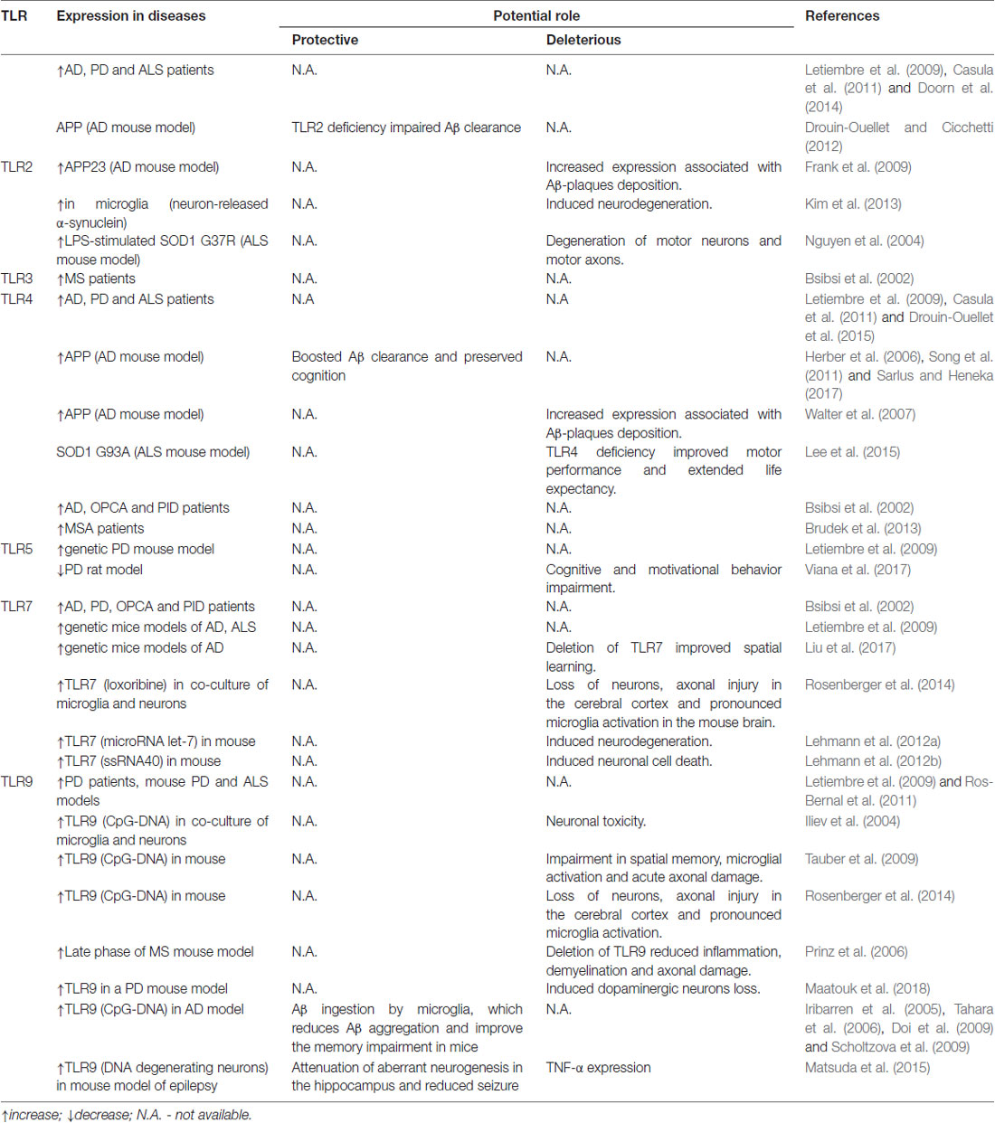
Table 1. Expression and potential roles of TLR in neurodegenerative conditions.
Author Contributions
AO reviewed the literature, designed, wrote part of and revised the manuscript. CB, SS and NY reviewed the literature, wrote part of the manuscript. BF reviewed the literature, designed the manuscript and revised it.
Funding
We thank Fundação de Amparo à Pesquisa do Estado de Minas Gerais (FAPEMIG; #CBB-APQ-02044-15) and Conselho Nacional de Desenvolvimento Científico e Tecnológico (CNPq; #424588-2016-1) for the financial support. SS acknowledges CNPq/CSF (Brasília/Brazil) and DAAD (Germany) for the fellowship. AO acknowledges CNPq for the Research Productivity Fellowship (#309718/2015-5). NY was supported by a scholarship as per the State Law on Graduate Funding (LGFG-IGA, Freiburg). The article processing charge was funded by the University of Freiburg in the funding program Open Access Publishing.
Conflict of Interest Statement
The authors declare that the research was conducted in the absence of any commercial or financial relationships that could be construed as a potential conflict of interest.
References
Balducci, C., Frasca, A., Zotti, M., La Vitola, P., Mhillaj, E., Grigoli, E., et al. (2017). Toll-like receptor 4-dependent glial cell activation mediates the impairment in memory establishment induced by β-amyloid oligomers in an acute mouse model of Alzheimer’s disease. Brain Behav. Immun. 60, 188–197. doi: 10.1016/j.bbi.2016.10.012
Béraud, D., and Maguire-Zeiss, K. A. (2012). Misfolded α-synuclein and toll-like receptors: therapeutic targets for Parkinson’s disease. Parkinsonism Relat. Disord. 18, S17–S20. doi: 10.1016/S1353-8020(11)70008-6
Béraud, D., Twomey, M., Bloom, B., Mittereder, A., Ton, V., Neitzke, K., et al. (2011). α-synuclein alters toll-like receptor expression. Front. Neurosci. 5:80. doi: 10.3389/fnins.2011.00080
Boche, D., Perry, V. H., and Nicoll, J. A. R. (2013). Review: activation patterns of microglia and their identification in the human brain. Neuropathol. Appl. Neurobiol. 39, 3–18. doi: 10.1111/nan.12011
Bowman, C. C., Rasley, A., Tranguch, S. L., and Marriott, I. (2003). Cultured astrocytes express toll-like receptors for bacterial products. Glia 43, 281–291. doi: 10.1002/glia.10256
Broad, A., Kirby, J. A., Jones, D. E. J., and Applied Immunology and Transplantation Research Group. (2007). Toll-like receptor interactions: tolerance of MyD88-dependent cytokines but enhancement of MyD88-independent interferon-β production. Immunology 120, 103–111. doi: 10.1111/j.1365-2567.2006.02485.x
Brudek, T., Winge, K., Agander, T. K., and Pakkenberg, B. (2013). Screening of toll-like receptors expression in multiple system atrophy brains. Neurochem. Res. 38, 1252–1259. doi: 10.1007/s11064-013-1020-5
Bsibsi, M., Ravid, R., Gveric, D., and van Noort, J. M. (2002). Broad expression of toll-like receptors in the human central nervous system. J. Neuropathol. Exp. Neurol. 61, 1013–1021. doi: 10.1093/jnen/61.11.1013
Caldeira, C., Cunha, C., Vaz, A. R., Falcão, A. S., Barateiro, A., Seixas, E., et al. (2017). Key aging-associated alterations in primary microglia response to β-amyloid stimulation. Front. Aging Neurosci. 9:277. doi: 10.3389/fnagi.2017.00277
Calvo-Rodríguez, M., de la Fuente, C., García-Durillo, M., García-Rodríguez, C., Villalobos, C., and Núñez, L. (2017). Aging and amyloid β oligomers enhance TLR4 expression, LPS-induced Ca2+ responses, and neuron cell death in cultured rat hippocampal neurons. J. Neuroinflammation 14:24. doi: 10.1186/s12974-017-0802-0
Caputi, V., and Giron, M. C. (2018). Microbiome-gut-brain axis and toll-like receptors in Parkinson’s disease. Int. J. Mol. Sci. 19:E1689. doi: 10.3390/ijms19061689
Casula, M., Iyer, A. M., Spliet, W. G. M., Anink, J. J., Steentjes, K., Sta, M., et al. (2011). Toll-like receptor signaling in amyotrophic lateral sclerosis spinal cord tissue. Neuroscience 179, 233–243. doi: 10.1016/j.neuroscience.2011.02.001
Chakravarty, S., and Herkenham, M. (2005). Toll-like receptor 4 on nonhematopoietic cells sustains CNS inflammation during endotoxemia, independent of systemic cytokines. J. Neurosci. 25, 1788–1796. doi: 10.1523/JNEUROSCI.4268-04.2005
Chaudhary, P. M., Ferguson, C., Nguyen, V., Nguyen, O., Massa, H. F., Eby, M., et al. (1998). Cloning and characterization of two Toll/Interleukin-1 receptor-like genes TIL3 and TIL4: evidence for a multi-gene receptor family in humans. Blood 91, 4020–4027.
Chen, K., Iribarren, P., Hu, J., Chen, J., Gong, W., Cho, E. H., et al. (2006). Activation of toll-like receptor 2 on microglia promotes cell uptake of Alzheimer disease-associated amyloid β peptide. J. Biol. Chem. 281, 3651–3659. doi: 10.1074/jbc.M508125200
Coope, A., Milanski, M., Arruda, A. P., Ignacio-Souza, L. M., Saad, M. J., Anhê, G. F., et al. (2012). Chaperone insufficiency links TLR4 protein signaling to endoplasmic reticulum stress. J. Biol. Chem. 287, 15580–15589. doi: 10.1074/jbc.M111.315218
Dalpke, A. H., Schafer, M. K.-H., Frey, M., Zimmermann, S., Tebbe, J., Weihe, E., et al. (2002). Immunostimulatory CpG-DNA activates murine microglia. J. Immunol. 168, 4854–4863. doi: 10.4049/jimmunol.168.10.4854
Daniele, S. G., Béraud, D., Davenport, C., Cheng, K., Yin, H., and Maguire-Zeiss, K. A. (2015). Activation of MyD88-dependent TLR1/2 signaling by misfolded α-synuclein, a protein linked to neurodegenerative disorders. Sci. Signal. 8:ra45. doi: 10.1126/scisignal.2005965
de Oliveira, A. C. P., Yousif, N. M., Bhatia, H. S., Hermanek, J., Huell, M., and Fiebich, B. L. (2016). Poly(I:C) increases the expression of mPGES-1 and COX-2 in rat primary microglia. J. Neuroinflammation 13:11. doi: 10.1186/s12974-015-0473-7
De Paola, M., Mariani, A., Bigini, P., Peviani, M., Ferrara, G., Molteni, M., et al. (2012). Neuroprotective effects of toll-like receptor 4 antagonism in spinal cord cultures and in a mouse model of motor neuron degeneration. Mol. Med. 18, 971–981. doi: 10.2119/molmed.2012.00020
De Paola, M., Sestito, S. E., Mariani, A., Memo, C., Fanelli, R., Freschi, M., et al. (2016). Synthetic and natural small molecule TLR4 antagonists inhibit motoneuron death in cultures from ALS mouse model. Pharmacol. Res. 103, 180–187. doi: 10.1016/j.phrs.2015.11.020
Delgado, M. A., Elmaoued, R. A., Davis, A. S., Kyei, G., and Deretic, V. (2008). Toll-like receptors control autophagy. EMBO J. 27, 1110–1121. doi: 10.1038/emboj.2008.31
Doi, Y., Mizuno, T., Maki, Y., Jin, S., Mizoguchi, H., Ikeyama, M., et al. (2009). Microglia activated with the toll-like receptor 9 ligand CpG attenuate oligomeric amyloid β neurotoxicity in in vitro and in vivo models of Alzheimer’s disease. Am. J. Pathol. 175, 2121–2132. doi: 10.2353/ajpath.2009.090418
Doorn, K. J., Moors, T., Drukarch, B., van de Berg, W. D., Lucassen, P. J., and van Dam, A.-M. (2014). Microglial phenotypes and toll-like receptor 2 in the substantia nigra and hippocampus of incidental Lewy body disease cases and Parkinson’s disease patients. Acta Neuropathol. Commun. 2:90. doi: 10.1186/s40478-014-0090-1
Drouin-Ouellet, J., and Cicchetti, F. (2012). Inflammation and neurodegeneration: the story ‘retolled’. Trends Pharmacol. Sci. 33, 542–551. doi: 10.1016/j.tips.2012.07.002
Drouin-Ouellet, J., St-Amour, I., Saint-Pierre, M., Lamontagne-Proulx, J., Kriz, J., Barker, R. A., et al. (2015). Toll-like receptor expression in the blood and brain of patients and a mouse model of Parkinson’s disease. Int. J. Neuropsychopharmacol. 18:pyu103. doi: 10.1093/ijnp/pyu103
Du, X., Poltorak, A., Wei, Y., and Beutler, B. (2000). Three novel mammalian toll-like receptors: gene structure, expression, and evolution. Eur. Cytokine Netw. 11, 362–371.
Ebert, S., Gerber, J., Bader, S., Mühlhauser, F., Brechtel, K., Mitchell, T. J., et al. (2005). Dose-dependent activation of microglial cells by Toll-like receptor agonists alone and in combination. J. Neuroimmunol. 159, 87–96. doi: 10.1016/j.jneuroim.2004.10.005
Fellner, L., Irschick, R., Schanda, K., Reindl, M., Klimaschewski, L., Poewe, W., et al. (2013). Toll-like receptor 4 is required for α-synuclein dependent activation of microglia and astroglia. Glia 61, 349–360. doi: 10.1002/glia.22437
Field, R., Campion, S., Warren, C., Murray, C., and Cunningham, C. (2010). Systemic challenge with the TLR3 agonist poly I:C induces amplified IFNα/β and IL-1β responses in the diseased brain and exacerbates chronic neurodegeneration. Brain Behav. Immun. 24, 996–1007. doi: 10.1016/j.bbi.2010.04.004
Fitzgerald, K. A., McWhirter, S. M., Faia, K. L., Rowe, D. C., Latz, E., Golenbock, D. T., et al. (2003a). IKKε and TBK1 are essential components of the IRF3 signaling pathway. Nat. Immunol. 4, 491–496. doi: 10.1038/ni921
Fitzgerald, K. A., Rowe, D. C., Barnes, B. J., Caffrey, D. R., Visintin, A., Latz, E., et al. (2003b). LPS-TLR4 signaling to IRF-3/7 and NF-κB involves the toll adapters TRAM and TRIF. J. Exp. Med. 198, 1043–1055. doi: 10.1084/jem.20031023
Frank, S., Copanaki, E., Burbach, G. J., Müller, U. C., and Deller, T. (2009). Differential regulation of toll-like receptor mRNAs in amyloid plaque-associated brain tissue of aged APP23 transgenic mice. Neurosci. Lett. 453, 41–44. doi: 10.1016/j.neulet.2009.01.075
Gambuzza, M., Sofo, V., Salmeri, F., Soraci, L., Marino, S., and Bramanti, P. (2014). Toll-like receptors in Alzheimer’s disease: a therapeutic perspective. CNS Neurol. Disord. Drug Targets 13, 1542–1558. doi: 10.2174/1871527313666140806124850
Gill, R., Tsung, A., and Billiar, T. R. (2010). Linking oxidative stress to inflammation: toll-like receptors. Free Radic. Biol. Med 48, 1121–1132. doi: 10.1016/j.freeradbiomed.2010.01.006
Go, M., Kou, J., Lim, J.-E., Yang, J., and Fukuchi, K. (2016). Microglial response to LPS increases in wild-type mice during aging but diminishes in an Alzheimer’s mouse model: Implication of TLR4 signaling in disease progression. Biochem. Biophys. Res. Commun. 479, 331–337. doi: 10.1016/j.bbrc.2016.09.073
Griffioen, K., Mattson, M. P., and Okun, E. (2018). Deficiency of Toll-like receptors 2, 3 or 4 extends life expectancy in Huntington’s disease mice. Heliyon 4:e00508. doi: 10.1016/j.heliyon.2018.e00508
Grootjans, J., Kaser, A., Kaufman, R. J., and Blumberg, R. S. (2016). The unfolded protein response in immunity and inflammation. Nat. Rev. Immunol. 16, 469–484. doi: 10.1038/nri.2016.62
Guillot-Sestier, M. V., and Town, T. (2018). Let’s make microglia great again in neurodegenerative disorders. J. Neural Transm. 125, 751–770. doi: 10.1007/s00702-017-1792-x
Hanke, M. L., and Kielian, T. (2011). Toll-like receptors in health and disease in the brain: mechanisms and therapeutic potential. Clin. Sci. 121, 367–387. doi: 10.1042/cs20110164
Herber, D. L., Maloney, J. L., Roth, L. M., Freeman, M. J., Morgan, D., and Gordon, M. N. (2006). Diverse microglial responses after intrahippocampal administration of lipopolysaccharide. Glia 53, 382–391. doi: 10.1002/glia.20272
Hu, Q.-P., and Mao, D.-A. (2016). Histone deacetylase inhibitor SAHA attenuates post-seizure hippocampal microglia TLR4/MYD88 signaling and inhibits TLR4 gene expression via histone acetylation. BMC Neurosci. 17:22. doi: 10.1186/s12868-016-0264-9
Huang, N.-Q., Jin, H., Zhou, S., Shi, J., and Jin, F. (2017). TLR4 is a link between diabetes and Alzheimer’s disease. Behav. Brain Res. 316, 234–244. doi: 10.1016/j.bbr.2016.08.047
Iliev, A. I., Stringaris, A. K., Nau, R., and Neumann, H. (2004). Neuronal injury mediated via stimulation of microglial toll-like receptor-9 (TLR9). FASEB J. 18, 412–414. doi: 10.1096/fj.03-0670fje
Iribarren, P., Chen, K., Hu, J., Gong, W., Cho, E. H., Lockett, S., et al. (2005). CpG-containing oligodeoxynucleotide promotes microglial cell uptake of amyloid β 1–42 peptide by up-regulating the expression of the G-protein- coupled receptor mFPR2. FASEB J. 19, 2032–2034. doi: 10.1096/fj.05-4578fje
Jana, M., Palencia, C. A., and Pahan, K. (2008). Fibrillar amyloid-β peptides activate microglia via TLR2: implications for Alzheimer’s disease. J. Immunol. 181, 7254–7262. doi: 10.4049/jimmunol.181.10.7254
Jang, H., Boltz, D., Sturm-Ramirez, K., Shepherd, K. R., Jiao, Y., Webster, R., et al. (2009). Highly pathogenic H5N1 influenza virus can enter the central nervous system and induce neuroinflammation and neurodegeneration. Proc. Natl. Acad. Sci. U S A 106, 14063–14068. doi: 10.1073/pnas.0900096106
Jang, Y., Koo, J.-H., Kwon, I., Kang, E.-B., Um, H.-S., Soya, H., et al. (2017). Neuroprotective effects of endurance exercise against neuroinflammation in MPTP-induced Parkinson’s disease mice. Brain Res. 1655, 186–193. doi: 10.1016/j.brainres.2016.10.029
Kalinderi, K., Bostantjopoulou, S., Katsarou, Z., and Fidani, L. (2013). TLR9 −1237 T/C and TLR2 −194 to −174 del polymorphisms and the risk of Parkinson’s disease in the Greek population: a pilot study. Neurol. Sci. 34, 679–682. doi: 10.1007/s10072-012-1106-x
Kawai, T., and Akira, S. (2010). The role of pattern-recognition receptors in innate immunity: update on Toll-like receptors. Nat. Immunol. 11, 373–384. doi: 10.1038/ni.1863
Kielian, T., Esen, N., and Bearden, E. D. (2005). Toll-like receptor 2 (TLR2) is pivotal for recognition of S. aureus peptidoglycan but not intact bacteria by microglia. Glia 49, 567–576. doi: 10.1002/glia.20144
Kim, C., Ho, D.-H., Suk, J.-E., You, S., Michael, S., Kang, J., et al. (2013). Neuron-released oligomeric α-synuclein is an endogenous agonist of TLR2 for paracrine activation of microglia. Nat. Commun. 4:1562. doi: 10.1038/ncomms2534
Kim, C., Lee, H.-J., Masliah, E., and Lee, S.-J. (2016). Non-cell-autonomous neurotoxicity of α-synuclein through microglial toll-like receptor 2. Exp. Neurobiol. 25, 113–119. doi: 10.5607/en.2016.25.3.113
Koo, J.-H., Jang, Y.-C., Hwang, D.-J., Um, H.-S., Lee, N.-H., Jung, J.-H., et al. (2017). Treadmill exercise produces neuroprotective effects in a murine model of Parkinson’s disease by regulating the TLR2/MyD88/NF-κB signaling pathway. Neuroscience 356, 102–113. doi: 10.1016/j.neuroscience.2017.05.016
Laflamme, N., Soucy, G., and Rivest, S. (2001). Circulating cell wall components derived from gram-negative, not gram-positive, bacteria cause a profound induction of the gene-encoding Toll-like receptor 2 in the CNS: cell wall components and TLR2 in the CNS. J. Neurochem. 79, 648–657. doi: 10.1046/j.1471-4159.2001.00603.x
Laflamme, N., and Rivest, S. (2001). Toll-like receptor 4: the missing link of the cerebral innate immune response triggered by circulating gram-negative bacterial cell wall components. FASEB J. 15, 155–163. doi: 10.1096/fj.00-0339com
Lee, J. Y., Lee, J. D., Phipps, S., Noakes, P. G., and Woodruff, T. M. (2015). Absence of toll-like receptor 4 (TLR4) extends survival in the hSOD1G93A mouse model of amyotrophic lateral sclerosis. J. Neuroinflammation 12:90. doi: 10.1186/s12974-015-0310-z
Lehmann, S. M., Krüger, C., Park, B., Derkow, K., Rosenberger, K., Baumgart, J., et al. (2012a). An unconventional role for miRNA: let-7 activates Toll-like receptor 7 and causes neurodegeneration. Nat. Neurosci. 15, 827–835. doi: 10.1038/nn.3113
Lehmann, S. M., Rosenberger, K., Krüger, C., Habbel, P., Derkow, K., Kaul, D., et al. (2012b). Extracellularly delivered single-stranded viral RNA causes neurodegeneration dependent on TLR7. J. Immunol. 189, 1448–1458. doi: 10.4049/jimmunol.1201078
Lehnardt, S., Henneke, P., Lien, E., Kasper, D. L., Volpe, J. J., Bechmann, I., et al. (2006). A mechanism for neurodegeneration induced by group B streptococci through activation of the TLR2/MyD88 pathway in microglia. J. Immunol. 177, 583–592. doi: 10.4049/jimmunol.177.1.583
Letiembre, M., Liu, Y., Walter, S., Hao, W., Pfander, T., Wrede, A., et al. (2009). Screening of innate immune receptors in neurodegenerative diseases: a similar pattern. Neurobiol. Aging 30, 759–768. doi: 10.1016/j.neurobiolaging.2007.08.018
Li, X., Melief, E., Postupna, N., Montine, K. S., Keene, C. D., and Montine, T. J. (2015). Prostaglandin E2 receptor subtype 2 regulation of scavenger receptor CD36 modulates microglial Aβ42 phagocytosis. Am. J. Pathol. 185, 230–239. doi: 10.1016/j.ajpath.2014.09.016
Li, J., Wang, X., Zhang, F., and Yin, H. (2013). Toll-like receptors as therapeutic targets for autoimmune connective tissue diseases. Pharmacol. Ther. 138, 441–451. doi: 10.1016/j.pharmthera.2013.03.003
Lin, W., Ding, M., Xue, J., and Leng, W. (2013). The role of TLR2/JNK/NF-κB pathway in amyloid β peptide-induced inflammatory response in mouse NG108–15 neural cells. Int. Immunopharmacol. 17, 880–884. doi: 10.1016/j.intimp.2013.09.016
Liu, S., Liu, Y., Hao, W., Wolf, L., Kiliaan, A. J., Penke, B., et al. (2012). TLR2 is a primary receptor for Alzheimer’s amyloid peptide to trigger neuroinflammatory activation. J. Immunol. 188, 1098–1107. doi: 10.4049/jimmunol.1101121
Liu, Y., Hao, W., Dawson, A., Liu, S., and Fassbender, K. (2009). Expression of amyotrophic lateral sclerosis-linked SOD1 mutant increases the neurotoxic potential of microglia via TLR2. J. Biol. Chem. 284, 3691–3699. doi: 10.1074/jbc.m804446200
Liu, H.-Y., Hung, Y.-F., Lin, H.-R., Yen, T.-L., and Hsueh, Y.-P. (2017). Tlr7 deletion selectively ameliorates spatial learning but does not influence β deposition and inflammatory response in an Alzheimers disease mouse model. Neuropsychiatry 7, 509–521. doi: 10.4172/neuropsychiatry.1000243
Liu, Y., Walter, S., Stagi, M., Cherny, D., Letiembre, M., Schulz-Schaeffer, W., et al. (2005). LPS receptor (CD14): a receptor for phagocytosis of Alzheimer’s amyloid peptide. Brain 128, 1778–1789. doi: 10.1093/brain/awh531
Maatouk, L., Compagnion, A.-C., Sauvage, M.-A. C., Bemelmans, A.-P., Leclere-Turbant, S., Cirotteau, V., et al. (2018). TLR9 activation via microglial glucocorticoid receptors contributes to degeneration of midbrain dopamine neurons. Nat. Commun. 9:3109. doi: 10.1038/s41467-018-05680-w
Matsuda, T., Murao, N., Katano, Y., Juliandi, B., Kohyama, J., Akira, S., et al. (2015). TLR9 signalling in microglia attenuates seizure-induced aberrant neurogenesis in the adult hippocampus. Nat. Commun. 6:6514. doi: 10.1038/ncomms7514
McDonald, C. L., Hennessy, E., Rubio-Araiz, A., Keogh, B., McCormack, W., McGuirk, P., et al. (2016). Inhibiting TLR2 activation attenuates amyloid accumulation and glial activation in a mouse model of Alzheimer’s disease. Brain Behav. Immun. 58, 191–200. doi: 10.1016/j.bbi.2016.07.143
Melchjorsen, J., and Paludan, S. R. (2003). Induction of RANTES/CCL5 by herpes simplex virus is regulated by nuclear factor κ B and interferon regulatory factor 3. J. Gen. Virol. 84, 2491–2495. doi: 10.1099/vir.0.19159-0
Moulignier, A., Gueguen, A., Lescure, F.-X., Ziegler, M., Girard, P.-M., Cardon, B., et al. (2015). Does HIV infection alter parkinson disease? J. Acquir. Immune Defic. Syndr. 70, 129–136. doi: 10.1097/qai.0000000000000677
Nguyen, M. D., D’Aigle, T., Gowing, G., Julien, J. P., and Rivest, S. (2004). Exacerbation of motor neuron disease by chronic stimulation of innate immunity in a mouse model of amyotrophic lateral sclerosis. J. Neurosci. 24, 1340–1349. doi: 10.1523/JNEUROSCI.4786-03.2004
Nguyen, M. D., Julien, J. P., and Rivest, S. (2001). Induction of proinflammatory molecules in mice with amyotrophic lateral sclerosis: no requirement for proapoptotic interleukin-1β in neurodegeneration. Ann. Neurol. 50, 630–639. doi: 10.1002/ana.1256
Noelker, C., Morel, L., Lescot, T., Osterloh, A., Alvarez-Fischer, D., Breloer, M., et al. (2013). Toll like receptor 4 mediates cell death in a mouse MPTP model of Parkinson disease. Sci. Rep. 3:1393. doi: 10.1038/srep01393
Okun, E., Griffioen, K. J., and Mattson, M. P. (2011). Toll-like receptor signaling in neural plasticity and disease. Trends Neurosci. 34, 269–281. doi: 10.1016/j.tins.2011.02.005
Okun, E., Griffioen, K. J., Lathia, J. D., Tang, S.-C., Mattson, M. P., and Arumugam, T. V. (2009). Toll-like receptors in neurodegeneration. Brain Res. Rev. 59, 278–292. doi: 10.1016/j.brainresrev.2008.09.001
Olson, J. K., and Miller, S. D. (2004). Microglia initiate central nervous system innate and adaptive immune responses through multiple TLRs. J. Immunol. 173, 3916–3924. doi: 10.4049/jimmunol.173.6.3916
Palsson-McDermott, E. M., and O’Neill, L. A. J. (2004). Signal transduction by the lipopolysaccharide receptor, Toll-like receptor-4. Immunology 113, 153–162. doi: 10.1111/j.1365-2567.2004.01976.x
Perry, V. H., Nicoll, J. A. R., and Holmes, C. (2010). Microglia in neurodegenerative disease. Nat. Rev. Neurol. 6, 193–201. doi: 10.1038/nrneurol.2010.17
Prinz, M., Garbe, F., Schmidt, H., Mildner, A., Gutcher, I., Wolter, K., et al. (2006). Innate immunity mediated by TLR9 modulates pathogenicity in an animal model of multiple sclerosis. J. Clin. Invest. 116, 456–464. doi: 10.1172/jci26078
Qin, L., Li, G., Qian, X., Liu, Y., Wu, X., Liu, B., et al. (2005). Interactive role of the toll-like receptor 4 and reactive oxygen species in LPS-induced microglia activation. Glia 52, 78–84. doi: 10.1002/glia.20225
Qin, Y., Liu, Y., Hao, W., Decker, Y., Tomic, I., Menger, M. D., et al. (2016). Stimulation of TLR4 attenuates Alzheimer’s disease-related symptoms and pathology in tau-transgenic mice. J. Immunol. 197, 3281–3292. doi: 10.4049/jimmunol.1600873
Ransohoff, R. M., and Perry, V. H. (2009). Microglial physiology: unique stimuli, specialized responses. Annu. Rev. Immunol. 27, 119–145. doi: 10.1146/annurev.immunol.021908.132528
Reed-Geaghan, E. G., Savage, J. C., Hise, A. G., and Landreth, G. E. (2009). CD14 and toll-like receptors 2 and 4 are required for fibrillar Aβ-stimulated microglial activation. J. Neurosci. 29, 11982–11992. doi: 10.1523/JNEUROSCI.3158-09.2009
Rezazadeh, M., Khorrami, A., Yeghaneh, T., Talebi, M., Kiani, S. J., Heshmati, Y., et al. (2016). Genetic factors affecting late-onset Alzheimer’s disease susceptibility. Neuromolecular Med. 18, 37–49. doi: 10.1007/s12017-015-8376-4
Richard, K. L., Filali, M., Prefontaine, P., and Rivest, S. (2008). Toll-like receptor 2 acts as a natural innate immune receptor to clear amyloid 1–42 and delay the cognitive decline in a mouse model of Alzheimer’s disease. J. Neurosci. 28, 5784–5793. doi: 10.1523/JNEUROSCI.1146-08.2008
Rock, F. L., Hardiman, G., Timans, J. C., Kastelein, R. A., and Bazan, J. F. (1998). A family of human receptors structurally related to Drosophila Toll. Proc. Natl. Acad. Sci. U S A 95, 588–593. doi: 10.1073/pnas.95.2.588
Ros-Bernal, F., Hunot, S., Herrero, M. T., Parnadeau, S., Corvol, J.-C., Lu, L., et al. (2011). Microglial glucocorticoid receptors play a pivotal role in regulating dopaminergic neurodegeneration in parkinsonism. Proc. Natl. Acad. Sci. U S A 108, 6632–6637. doi: 10.1073/pnas.1017820108
Rosenberger, K., Derkow, K., Dembny, P., Krüger, C., Schott, E., and Lehnardt, S. (2014). The impact of single and pairwise Toll-like receptor activation on neuroinflammation and neurodegeneration. J. Neuroinflammation 11:166. doi: 10.1186/s12974-014-0166-7
Ruckdeschel, K., Pfaffinger, G., Haase, R., Sing, A., Weighardt, H., Häcker, G., et al. (2004). Signaling of apoptosis through TLRs critically involves toll/IL-1 receptor domain-containing adapter inducing IFN-β, but not MyD88, in bacteria-infected murine macrophages. J. Immunol. 173, 3320–3328. doi: 10.4049/jimmunol.173.5.3320
Sarlus, H., and Heneka, M. T. (2017). Microglia in Alzheimer’s disease. J. Clin. Invest. 127, 3240–3249. doi: 10.1172/JCI90606
Savino, F., Galliano, I., Garro, M., Savino, A., Manzoni, P., Daprà, V., et al. (2018). Changes in the messenger RNA expression of toll-like receptors 2 and 4 in healthy infants according to age. Am. J. Perinatol. 35, 583–588. doi: 10.1055/s-0038-1639363
Schafer, S. L., Lin, R., Moore, P. A., Hiscott, J., and Pitha, P. M. (1998). Regulation of type I interferon gene expression by interferon regulatory factor-3. J. Biol. Chem. 273, 2714–2720. doi: 10.1074/jbc.273.5.2714
Scholtzova, H., Kascsak, R. J., Bates, K. A., Boutajangout, A., Kerr, D. J., Meeker, H. C., et al. (2009). Induction of toll-like receptor 9 signaling as a method for ameliorating Alzheimer’s disease-related pathology. J. Neurosci. 29, 1846–1854. doi: 10.1523/JNEUROSCI.5715-08.2009
Shao, Q.-H., Yan, W.-F., Zhang, Z., Ma, K.-L., Peng, S.-Y., Cao, Y.-L., et al. (2018). Nurr1: a vital participant in the TLR4-NF-κB signal pathway stimulated by α-synuclein in BV-2 cells. Neuropharmacology doi: 10.1016/j.neuropharm.2018.04.008 [Epub ahead of print].
Smith, S. M. C., Friedle, S. A., and Watters, J. J. (2013). Chronic intermittent hypoxia exerts CNS region-specific effects on rat microglial inflammatory and TLR4 gene expression. PLoS One 8:e81584. doi: 10.1371/journal.pone.0081584
Song, M., Jin, J., Lim, J.-E., Kou, J., Pattanayak, A., Rehman, J. A., et al. (2011). TLR4 mutation reduces microglial activation, increases Aβ deposits and exacerbates cognitive deficits in a mouse model of Alzheimer’s disease. J. Neuroinflammation 8:92. doi: 10.1186/1742-2094-8-92
Sprenkle, N. T., Sims, S. G., Sánchez, C. L., and Meares, G. P. (2017). Endoplasmic reticulum stress and inflammation in the central nervous system. Mol. Neurodegener. 12:42. doi: 10.1186/s13024-017-0183-y
Stefanova, N., Fellner, L., Reindl, M., Masliah, E., Poewe, W., and Wenning, G. K. (2011). Toll-like receptor 4 promotes α-synuclein clearance and survival of nigral dopaminergic neurons. Am. J. Pathol. 179, 954–963. doi: 10.1016/j.ajpath.2011.04.013
Tahara, K., Kim, H.-D., Jin, J.-J., Maxwell, J. A., Li, L., and Fukuchi, K.-I. (2006). Role of toll-like receptor signalling in a uptake and clearance. Brain 129, 3006–3019. doi: 10.1093/brain/awl249
Takeda, K., and Akira, S. (2004). TLR signaling pathways. Semin. Immunol. 16, 3–9. doi: 10.1016/j.smim.2003.10.003
Takeda, K., Kaisho, T., and Akira, S. (2003). Toll-like receptors. Annu. Rev. Immunol. 21, 335–376. doi: 10.1146/annurev.immunol.21.120601.141126
Takeuchi, O., Kawai, T., Sanjo, H., Copeland, N. G., Gilbert, D. J., Jenkins, N. A., et al. (1999). TLR6: a novel member of an expanding Toll-like receptor family. Gene 231, 59–65. doi: 10.1016/s0378-1119(99)00098-0
Takeuchi, O., Sato, S., Horiuchi, T., Hoshino, K., Takeda, K., Dong, Z., et al. (2002). Cutting edge: role of toll-like receptor 1 in mediating immune response to microbial lipoproteins. J. Immunol. 169, 10–14. doi: 10.4049/jimmunol.169.1.10
Tang, S.-C., Arumugam, T. V., Xu, X., Cheng, A., Mughal, M. R., Jo, D. G., et al. (2007). Pivotal role for neuronal Toll-like receptors in ischemic brain injury and functional deficits. Proc. Natl. Acad. Sci. U S A 104, 13798–13803. doi: 10.1073/pnas.0702553104
Tauber, S. C., Ebert, S., Weishaupt, J. H., Reich, A., Nau, R., and Gerber, J. (2009). Stimulation of toll-like receptor 9 by chronic intraventricular unmethylated cytosine-guanine DNA infusion causes neuroinflammation and impaired spatial memory. J. Neuropathol. Exp. Neurol. 68, 1116–1124. doi: 10.1097/nen.0b013e3181b7fde5
van Noort, J. M., and Bsibsi, M. (2009). Toll-like receptors in the CNS: implications for neurodegeneration and repair. Prog. Brain Res. 175, 139–148. doi: 10.1016/S0079-6123(09)17509-X
Venezia, S., Refolo, V., Polissidis, A., Stefanis, L., Wenning, G. K., and Stefanova, N. (2017). Toll-like receptor 4 stimulation with monophosphoryl lipid A ameliorates motor deficits and nigral neurodegeneration triggered by extraneuronal α-synucleinopathy. Mol. Neurodegener. 12:52. doi: 10.1186/s13024-017-0195-7
Viana, S. D., Pita, I. R., Lemos, C., Rial, D., Couceiro, P., Rodrigues-Santos, P., et al. (2017). The effects of physical exercise on nonmotor symptoms and on neuroimmune RAGE network in experimental parkinsonism. J. Appl. Physiol. 123, 161–171. doi: 10.1152/japplphysiol.01120.2016
Vollmar, P., Kullmann, J. S., Thilo, B., Claussen, M. C., Rothhammer, V., Jacobi, H., et al. (2010). Active immunization with amyloid- 1–42 impairs memory performance through TLR2/4-dependent activation of the innate immune system. J. Immunol. 185, 6338–6347. doi: 10.4049/jimmunol.1001765
Walker, D. G., Tang, T. M., and Lue, L. F. (2018). Increased expression of toll-like receptor 3, an anti-viral signaling molecule and related genes in Alzheimer’s disease brains. Exp. Neurol. 309, 91–106. doi: 10.1016/j.expneurol.2018.07.016
Walter, S., Doering, A., Letiembre, M., Liu, Y., Hao, W., Diem, R., et al. (2006). The LPS receptor, CD14, in experimental autoimmune encephalomyelitis and multiple sclerosis. Cell. Physiol. Biochem. 17, 167–172. doi: 10.1159/000092078
Walter, S., Letiembre, M., Liu, Y., Heine, H., Penke, B., Hao, W., et al. (2007). Role of the toll-like receptor 4 in neuroinflammation in Alzheimer’s disease. Cell. Physiol. Biochem. 20, 947–956. doi: 10.1159/000110455
Wang, F.-X., Liu, S.-Y., Zheng, X., Chen, X., Lu, L.-X., Chen, B., et al. (2015). TLR1 expression in mouse brain was increased in a KA-induced seizure model. Inflamm. Res. 64, 487–495. doi: 10.1007/s00011-015-0828-7
Wang, L.-Z., Tian, Y., Yu, J.-T., Chen, W., Wu, Z.-C., Zhang, Q., et al. (2011). Association between late-onset Alzheimer’s disease and microsatellite polymorphisms in intron II of the human toll-like receptor 2 gene. Neurosci. Lett. 489, 164–167. doi: 10.1016/j.neulet.2010.12.008
Wang, Y.-W., Zhou, Q., Zhang, X., Qian, Q.-Q., Xu, J.-W., Ni, P.-F., et al. (2017). Mild endoplasmic reticulum stress ameliorates lipopolysaccharide-induced neuroinflammation and cognitive impairment via regulation of microglial polarization. J. Neuroinflammation 14:233. doi: 10.1186/s12974-017-1002-7
Weintraub, M. K., Kranjac, D., Eimerbrink, M. J., Pearson, S. J., Vinson, B. T., Patel, J., et al. (2014). Peripheral administration of poly I:C leads to increased hippocampal amyloid-β and cognitive deficits in a non-transgenic mouse. Behav. Brain Res. 266, 183–187. doi: 10.1016/j.bbr.2014.03.009
Wendeln, A. C., Degenhardt, K., Kaurani, L., Gertig, M., Ulas, T., Jain, G., et al. (2018). Innate immune memory in the brain shapes neurological disease hallmarks. Nature 556, 332–338. doi: 10.1038/s41586-018-0023-4
Woulfe, J. M., Gray, M. T., Gray, D. A., Munoz, D. G., and Middeldorp, J. M. (2014). Hypothesis: a role for EBV-induced molecular mimicry in Parkinson’s disease. Parkinsonism Relat. Disord. 20, 685–694. doi: 10.1016/j.parkreldis.2014.02.031
Wu, W. Y.-Y., Kang, K.-H., Chen, S. L.-S., Chiu, S. Y.-H., Yen, A. M.-F., Fann, J. C.-Y., et al. (2015). Hepatitis C virus infection: a risk factor for Parkinson’s disease. J. Viral Hepat. 22, 784–791. doi: 10.1111/jvh.12392
Yamamoto, M., Sato, S., Hemmi, H., Hoshino, K., Kaisho, T., Sanjo, H., et al. (2003a). Role of adaptor TRIF in the MyD88-independent toll-like receptor signaling pathway. Science 301, 640–643. doi: 10.1126/science.1087262
Yamamoto, M., Sato, S., Hemmi, H., Uematsu, S., Hoshino, K., Kaisho, T., et al. (2003b). TRAM is specifically involved in the Toll-like receptor 4-mediated MyD88-independent signaling pathway. Nat. Immunol. 4, 1144–1150. doi: 10.1038/ni986
Yao, L., Kan, E. M., Lu, J., Hao, A., Dheen, S. T., Kaur, C., et al. (2013). Toll-like receptor 4 mediates microglial activation and production of inflammatory mediators in neonatal rat brain following hypoxia: role of TLR4 in hypoxic microglia. J. Neuroinflammation 10:23. doi: 10.1186/1742-2094-10-23
Yousif, N. M., de Oliveira, A. C. P., Brioschi, S., Huell, M., Biber, K., and Fiebich, B. L. (2018). Activation of EP2 receptor suppresses Poly(I: C) and LPS-mediated inflammation in primary microglia and organotypic hippocampal slice cultures: contributing role for MAPKs. Glia 66, 708–724. doi: 10.1002/glia.23276
Yu, J.-T., Mou, S.-M., Wang, L.-Z., Mao, C.-X., and Tan, L. (2011a). Toll-like receptor 2 -196 to -174 del polymorphism influences the susceptibility of Han Chinese people to Alzheimer’s disease. J. Neuroinflammation 8:136. doi: 10.1186/1742-2094-8-136
Yu, J.-T., Sun, Y.-P., Ou, J.-R., Cui, W.-Z., Zhang, W., and Tan, L. (2011b). No association of toll-like receptor 2 polymorphisms with Alzheimer’s disease in Han Chinese. Neurobiol. Aging 32, 1924.e1–1924.e3. doi: 10.1016/j.neurobiolaging.2011.03.023
Zhang, Y.-K., Liu, J.-T., Peng, Z.-W., Fan, H., Yao, A.-H., Cheng, P., et al. (2013). Different TLR4 expression and microglia/macrophage activation induced by hemorrhage in the rat spinal cord after compressive injury. J. Neuroinflammation 10:112. doi: 10.1186/1742-2094-10-112
Zhang, F. X., Kirschning, C. J., Mancinelli, R., Xu, X.-P., Jin, Y., Faure, E., et al. (1999). Bacterial lipopolysaccharide activates nuclear factor-κB through interleukin-1 signaling mediators in cultured human dermal endothelial cells and mononuclear phagocytes. J. Biol. Chem. 274, 7611–7614. doi: 10.1074/jbc.274.12.7611
Zhang, Q., Wu, H. H., Wang, Y., Gu, G. J., Zhang, W., and Xia, R. (2016). Neural stem cell transplantation decreases neuroinflammation in a transgenic mouse model of Alzheimer’s disease. J. Neurochem. 136, 815–825. doi: 10.1111/jnc.13413
Zhao, W., Beers, D. R., Henkel, J. S., Zhang, W., Urushitani, M., Julien, J.-P., et al. (2010). Extracellular mutant SOD1 induces microglial-mediated motoneuron injury. Glia 58, 231–243. doi: 10.1002/glia.20919
Keywords: toll-like receptors (TLRs), microglia, neurodegenerative diseases, neuroinflammation, Alzheimer’s disease, Parkinson’s disease, inflammatory mediators
Citation: Fiebich BL, Batista CRA, Saliba SW, Yousif NM and de Oliveira ACP (2018) Role of Microglia TLRs in Neurodegeneration. Front. Cell. Neurosci. 12:329. doi: 10.3389/fncel.2018.00329
Received: 30 May 2018; Accepted: 10 September 2018;
Published: 02 October 2018.
Edited by:
Rocío Martínez De Pablos, Universidad de Sevilla, SpainReviewed by:
Miguel Angel Burguillos, University of Cambridge, United KingdomMikko Airavaara, University of Helsinki, Finland
Copyright © 2018 Fiebich, Batista, Saliba, Yousif and de Oliveira. This is an open-access article distributed under the terms of the Creative Commons Attribution License (CC BY). The use, distribution or reproduction in other forums is permitted, provided the original author(s) and the copyright owner(s) are credited and that the original publication in this journal is cited, in accordance with accepted academic practice. No use, distribution or reproduction is permitted which does not comply with these terms.
*Correspondence: Bernd L. Fiebich, bernd.fiebich@uniklinik-freiburg.de
Antonio Carlos Pinheiro de Oliveira, antoniooliveira@icb.ufmg.br