Purpose: The purpose of this study is to evaluate the biomechanical and histological behavior of ceria-stabilized zirconia/alumina nanocomposite (NANOZR) (Panasonic Health Care Co., Tokyo, Japan) in comparison to yttria-stabilized zirconia (3Y-TZP) (Panasonic Health Care Co., Tokyo, Japan) in the rat.
Methods: The NANOZR, 3Y-TZP cylindrical implants (diameter: 1mm, length: 2mm) were used. The three-dimension surface morphology and surface roughness were determined by scanning white light interferometry (Wyko NT1100, Veeco Instruments Inc., Tucson, USA). The cylindrical zirconia implants were placed at the distal edge of the Wister rat (8W, Male) femur. At weeks 2, 4 and 8, the interfacial shear strength between implant and bone were measured by push-in test using universal testing machine (Model 5565, Instron Co., Canton, Mass, USA). The bone formation and osteoclast amount of peri-implant was evaluated using a microfocus X-ray CT system (SMX-225CT, Shimadzu Co., Kyoto, Japan) and hard tissue slice.
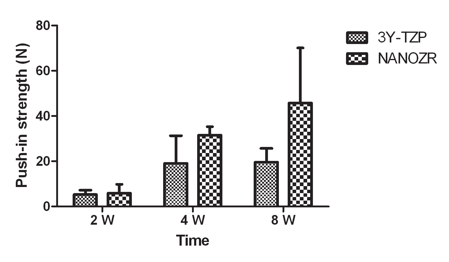
Results: The average surface roughness of 3Y-TZP (Sa, 0.788 µm) is significant higher than the NANOZR (Sa, 0.559 µm). There are similar shear strength between 3Y-TZP and NANOZR at 2 weeks, but at weeks 4, 8, the shear strength of NANOZR is higher than 3Y-TZP.
(
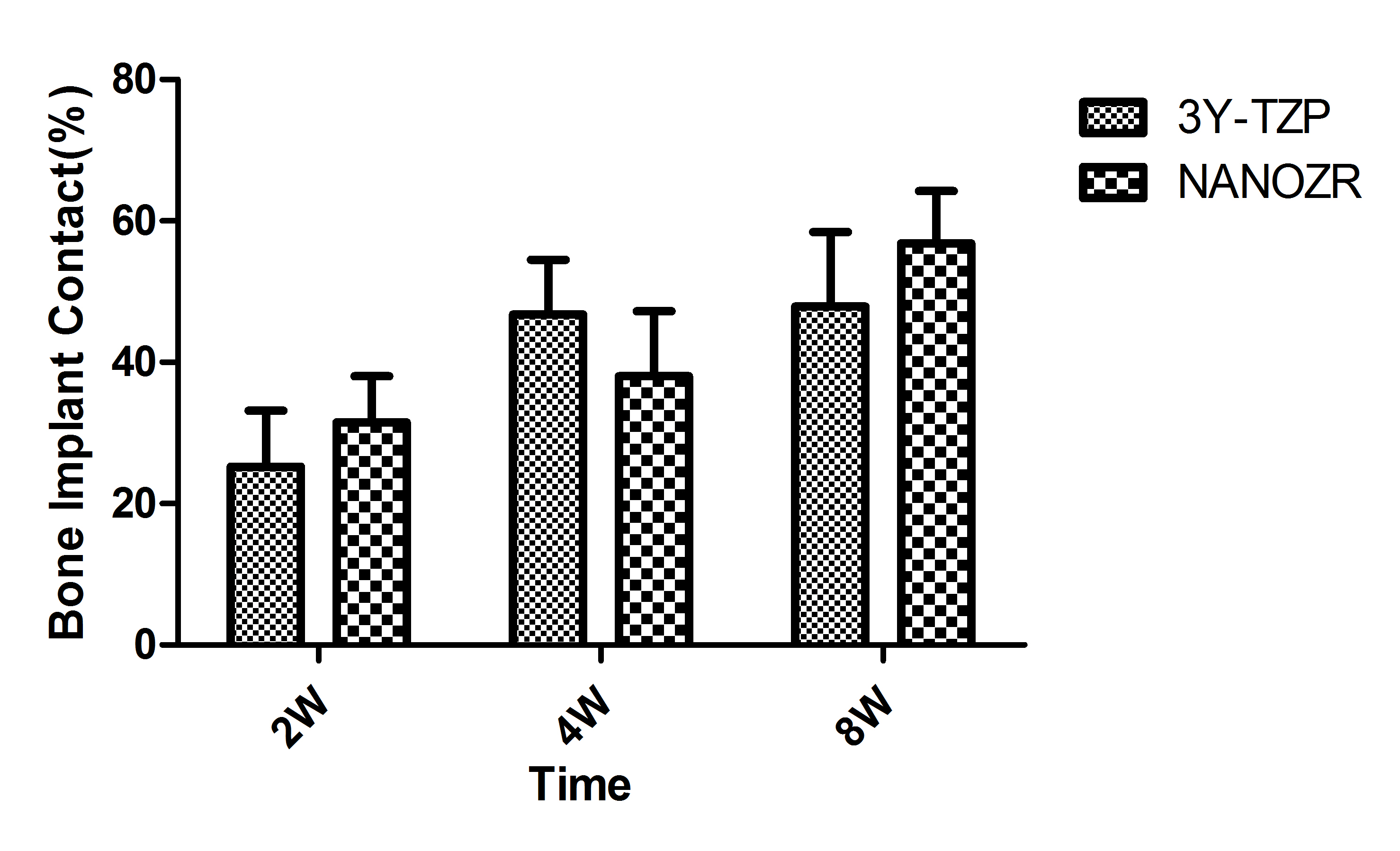
The X-ray show there is new bone formation around the both types zirconia implants after 4 weeks. The bone-implant contact is increasing during the observation periods, there is no significant difference for the bone implant contact for the two materials.
The osteoclast can be observed at 2 weeks, however, there is no osteoclast can be found at 4 weeks and 8 weeks. Both of two types zirconia had successfully osseointegration.
Conclusion: NANOZR has preferable bioactivity compare with 3Y-TZP. Further studies are necessary for potential biologic mechanisms involved.