Introduction: We developed biodegradable Mg staple used in stapler for gastrointestinal anastomosis[1] to replace current Ti staple which will be long-term left in the body and results in some adverse effects on patients[2],[3]. However, there are still two major challenges for the Mg staple: (i) there is a wide range in pH value of alimentary system (∼2-8 from gastric to intestinal fluid). (ii) non-uniform corrosion may occur resulting from the “B” shape of staple after anastomosis. Therefore, it is essential to establish standardized evaluation system on the degradation performance of Mg staple in vitro and vivo.
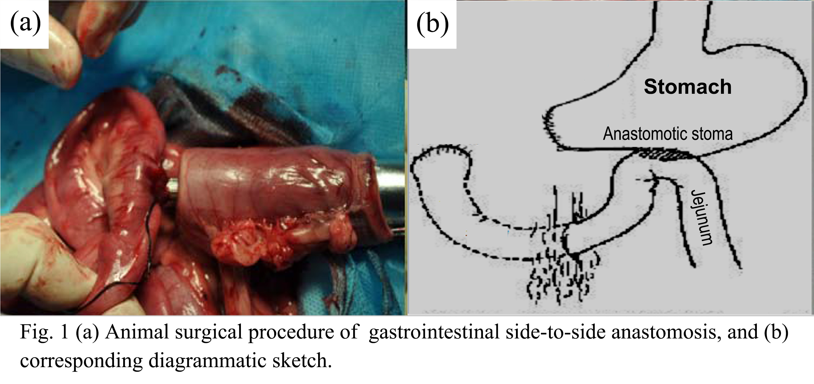
Materials and Methods: The Φ 0.3mm wires with surface coating were cut and processed into “B”-shape staples by stapler. To evaluate their in vitro degradation performance, the degradation behavior of staples in simulated gastric/intestinal/body fluid (SG/I/BF) with different pH values from 2-8 were investigated. To evaluate the in vivo degradation performance of staples, a gastrointestinal anastomosis (Fig. 1) were performed in 24 beagle with equally divided into Mg staple and Ti staple group. Some standardized indicators after surgical were compared between two groups at two time points, short-term (7 days) and long-term (90 days), respectively.
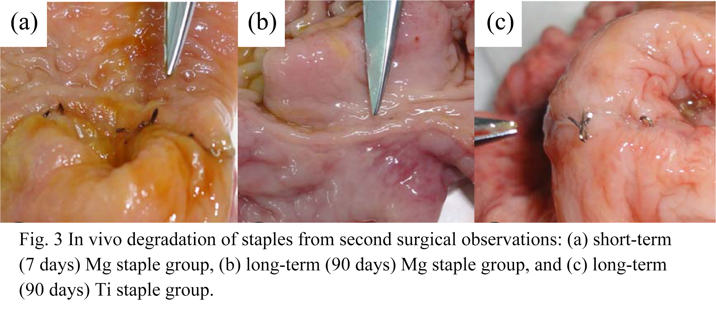
Results and Discussions: In vitro degradation evaluation results show that the pH value and ions of the corrosion medium play critical role in impacting the degradation performance of staple. The decrease of pH value leads to a rise in degradation rate, while more OH- ions also result in more serious corrosion of inner Mg substrate due to the corrosion action of OH- ions on surface poly-L-lactic acid. When pH value is above 4, the staples can maintain the structure integrity after 7 days immersion (Fig. 2). The wires also show a good mechanical stability with a ∼150MPa ultimate strength after 7 days immersion in SGF with pH=4. With prolonging time, corrosion first occurs at the ends and the bending section of staples (Fig. 2). This is attributed to the lack of protective coating at the ends of staple, and the stress corrosion associated with inner stress in bending section arising from the plastic deformation of “B”-shape staple. In vivo degradation evaluation results show staples are kept as a whole from CT and surgical observations for a short-term (7 days) degradation in animals, and disappear after 90 days (Fig. 3). The experimental group and the control group had no significant difference in anastomotic time, body weight, postoperative complications, abdominal cavity adhesion, diameter and blasting pressure of anastomotic stoma (p>0.05).

Conclusions: Mg staples can maintain the structure integrity for more than 7 days in vitro and vivo, and are degraded completely in animals in 90 days, showing a good application prospect. Standardized evaluation system of in vitro and vivo degradation performance of biodegradable Mg staples was initially established in this work.
Ph.D. Programs Foundation of Ministry of Education of China (No. 20120092120048); The National Natural Science Foundation of China (No. 31570961)
References:
[1] Robicsek F and Konstantinov I. Humer Hultl: the father of the surgical stapler. Journal of medical biography, 2001, 9(1):16-19
[2] Boffi F, Zbar A P, Pescatori M, et al. Retained staples causing rectal bleeding and severe proctalgia after the STARR procedure. Techniques in Coloproctology, 2008, 12(2):135-136
[3] Dziki A, Duncan M, Harmon J, et al. Advantages of handsewn over stapled bowel anastomosis. Diseases of the Colon & Rectum, 1991, 34(6):442-448