Introduction: Fracture fixation of small bones remains challenging for pathologies relating to tenuous retrograde blood supply. For example, acute scaphoid fractures are among the most common traumatic injuries and account for 50-80% of carpal injuries[1]. Solid screws without bone grafting are often ineffective in cases of osteonecrosis. The objectives of this study were to evaluate bone healing and strength of fixation of a new porous titanium (Ti) compression screw compared to a conventional titanium compression screw in cortical bone and in challenging models of gap-healing and osteonecrosis.
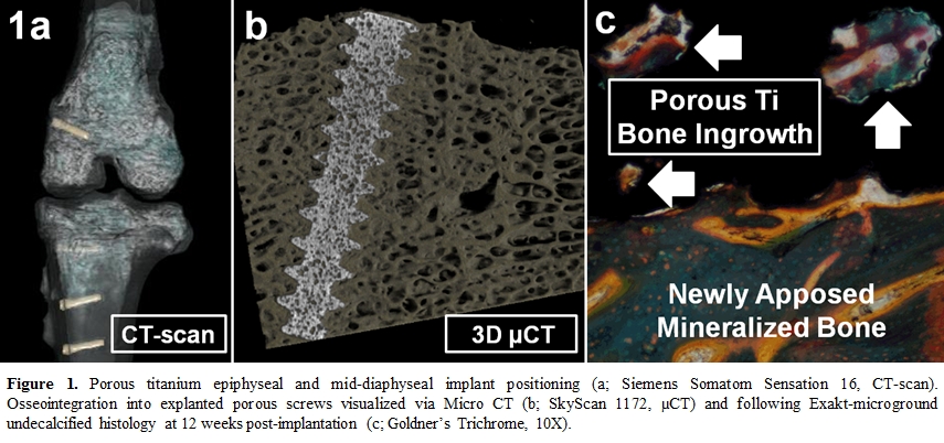
Materials and Methods: Six (6) female Dorset sheep (>18mo.; 75-90kg) were prepared for bilateral hind limb surgeries using either porous Ti (NRC) or conventional solid Ti (Acutrak) headless cannulated compression screws (Ø=3.3-3.8mm; L=15mm). Porous titanium screws presented a 40% porosity with a mean pore size of 344µm, and compressive, shear, and tensile strengths of 157 MPa, 113 MPa and 77 MPa, respectively. The 1st model evaluated non-union following removal of a bone core (Ø=8mm; L=8mm) from the proximal tibia and screw placement in its middle, resulting in a 2-mm peripheral gap. The 2nd model simulated osteonecrosis with a 2nd core removal (Ø=14mm; L=8mm) from the distal femur, submersion in liquid N2, and screw fixation of the devitalized core. The 3rd model quantified bone ingrowth and torsional strength at mid-diaphyseal tibial cortical locations. Sheep were then euthanized at 6 and 12 weeks post-implantation. Bone ingrowth was reconstructed by Micro-CT (SkyScan 1172) and quantified with histomorphometry. Explanted screws were sectioned longitudinally, processed for undecalcified histology, infiltrated with PMMA, microground (Exakt 400 CS), and stained with Goldner’s Trichrome. Digital images were analyzed for bone-implant contact and volumetric bone ingrowth. Torsional strength was recorded with a continuous digital torque meter at necropsy.
Results: Following 6 weeks of implantation in the non-union model, 75% of new bone formed adjacent to porous Ti as opposed to 38% with solid Ti screws. At 12 weeks, porous Ti screws exhibited 89% new bone formation vs. 79% for controls. Below the actual gap, bone filled 99% of the area within threads in porous Ti implants vs. 83% for controls at 12 weeks. Following bone devitalization, porous Ti showed up to 51% of new bone formation within its cannulated center as opposed to ≤3% for conventional screws. For the bone ingrowth model (Figure 1), linear bone apposition reached 55% for porous Ti vs. 36% (controls). Limited by failure of the Allen driver, the maximum torque for porous screws was 0.78Nm vs. 0.42Nm for controls. Histopathology revealed excellent safety and biocompatibility as evidenced by a mild tissue reaction.
Discussion: Superior bone apposition, volumetric bone ingrowth and torsional strength were observed using the porous titanium screw, likely attributed to its interconnected porosity and higher surface area. A beneficial bone conduction effect was pronounced within the screw cannula. The clinical implication is that porous Ti does not occlude bone ingrowth and vascularization is observed within the compression device itself, translating into bone fixation and healing.
Conclusion: Internal fixation with porous titanium compression screws increases the fixation strength and the potential for bone ingrowth in clinically challenging conditions of non-union and osteonecrosis.
The authors would like to sincerely thank Dr. Paul A. Martineau and Dr. Edward J. Harvey (McGill University Health Centre), who co-developed this porous implant with the National Research Council of Canada; Special thanks to Dr. Cathy Tkaczyk and Mr. Yannick Trudel for their respective scientific and highly-skilled surgical support
References:
[1] Hackney L.A. and Dodds S.D. Curr. Rev. Musculoskelet. Med.; 4(1): 16-22, 2011.