Introduction: The cell binding motif RGD is the most widely used peptide to improve cell binding properties of various biomaterials, including recombinant spider silk. We have previously shown a positive effect on cells when RGD was genetically coupled to the recombinant spider silk protein 4RepCT[1]. In the present study we enhanced the cell binding capacity even further by introducing the integrin binding motif from fibronectin, which contains RGD on a constrained loop.
Experimental Set-up: 4RepCT was genetically functionalized with the RGD-containing cell binding motif from fibronectin. To enable loop formation, two amino acids flanking the RGD sequence was substituted for cysteines. The positions of cysteines were carefully selected to achieve a 3D mimic of the turn loop seen in the full length fibronectin molecule. The resulting silk protein was denoted FNCC. Linear variants without cysteines were used as controls.
Primary cells of human origin (keratinocytes, endothelial cells and mesenchymal stem cells) were used to evaluate cell culture matrices of FNCC-silk. Adhesion, formation of stress fibers and focal adhesion points, as well as integrin α5β1 expression were analyzed. To survey applications, proliferation and migration assays were performed.
Results: The FNCC-silk protein allowed efficient assembly into foam and fibers, and could even be transformed into free standing films.
Human primary cells cultured on FNCC-silk showed increased attachment, spreading, stress fiber formation and focal adhesions, not only compared to RGD-silk, but also to the linear FN-silk controls. Cell binding capacity of FNCC-silk was equal to bovine fibronectin coatings, was shown to involve the α5β1 integrin, and to support proliferation and migration of keratinocytes.
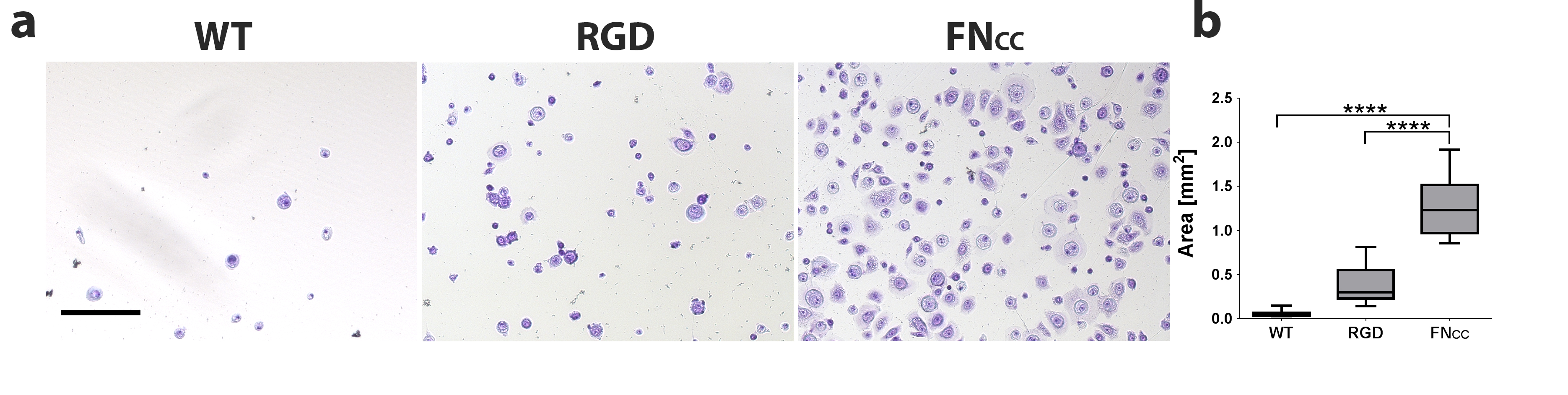
Figure 1. Keratinocytes after 60 min adhesion onto a surface coated with WT-silk, RGD-silk or FNCC-silk. (a) 10x magnification. Scalebar 50µm (b) Area measurement of attached cells.
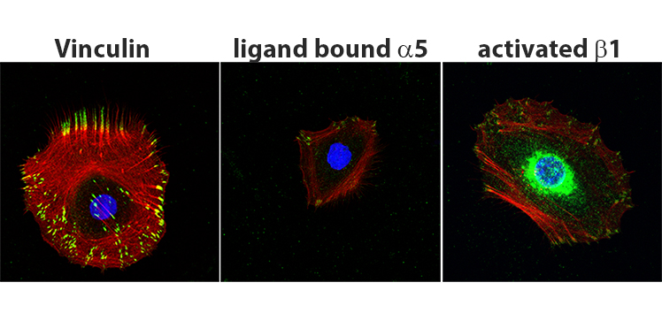
Figure 2. Confocal scans of keratinocytes after 3h adhesion onto FNCC-silk @63x magnification, f-actin red, dapi blue. Focal adhesions green: (left) vinculin (middle) ligand bound integrin α5 (right) activated integrin β1.
When culturing keratinocytes on the free standing films of FNCC-silk a monolayer could be formed. We are currently developing novel combinations of FNCC-silk and other biomaterial candidates.
Conclusion: A material based on FNCC-silk would make out a suitable candidate for coatings aiming to improve cell attachment, e.g. on stents, or in situations where cells need to be transferred as a cell sheet to e.g. a wound area. The results also suggests that FNCC-silk can efficiently attract inherent cells for migration into e.g. a wound area during healing.
The Swedish Research Council; FORMAS; Knut & Alice Wallenberg Foundation; Spiber Technologies AB (for supply of soluble proteins)
References:
[1] Widhe et al 2013, Biomaterials 34:8223-8234