Introduction: It was believed that biomaterial selection was performed based on its macroporosity, biocompatibility and degradability properties but recently its ability to withstand biomechanical stress has also been considered. Discher et al[1]. have indeed shown that material stiffness induces critical effects on cell behaviour and differentiation. Therefore, we have recently embarked on the development of mechanically-competent hydrogels for bone and cartilage tissue engineering. We prepared a cellulose based polymer capable of self cross-linkage (Si-HPMC) to avoid any toxicity issues resulting from using cross-linking chemicals or photo cross-linking. Therefore, LIOAD has been working on the development of hydrogels as ECM for bone and cartilage regeneration. The objectives of the present work are to modulate the physico-chemical and biological properties of our hydrogels by mixing two silated polysaccharides Si-HPMC and Si-Chitosan.
Experimental Methods: The self cross-linkable polymer (Si-HPMC) was prepared according to an already published method developed in LIOAD[2]. Silanol groups were linked to amine moieties of the chitosan via thio-urea bond.
Hydrogel characterizations: Hydrogels and reinforced hydrogels were characterized physico chemically to determine their elastic moduli using MARS rheometer (G’), Dynamic Mechanical Analysis (E’) and microscopy.
Biological investigations have been done with human nasal chondrocytes by determining the cell viability in 2D using MTS, 3D by confocal microscopy after Live&Dead staining.
In vivo investigations have been performed in subcutaneous sockets in nude mice and in focal defects in rabbit.
Results and Discussion: The characterizations showed that adding Si-Chitosan within Si-HPMC hydrogels increases the elastic moduli without interfering with the biocompatibility. Indeed, all the constructs that we used in this study showed no toxicity both in 2D and 3D (as shown in figure 1). Culturing cells in 3D within our hydrogel containing Si-Chitosan also induces cell adhesion onto the matrice as I can be observed on confocal micrograph in figure 2.
It is worth noted that after 5 weeks of implantation, the chondrocytes showed a good viability and still express a chondrocyte phenotype. Moreover, during the surgery, due to the presence of chitosan, our constructs showed good adhesion behaviour onto the cartilage focal defect.
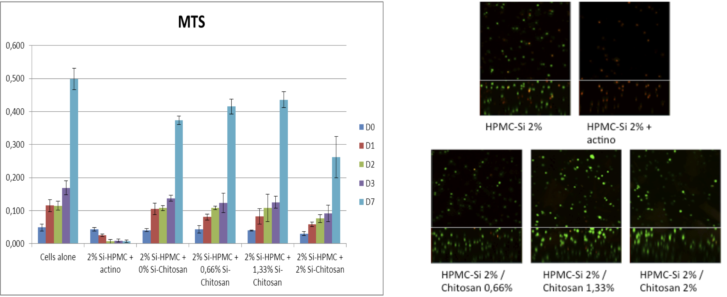
Fig 1: left : 2D hASCs viability in contact with Si-HPMC/Si-Chitosan hydrogels (Positive control : cell alone, control for cell death: actinomycin). Right : Confocal micrograph showing living cells (green) and dead cells (red) after 7 days of culture inside hydrogels.
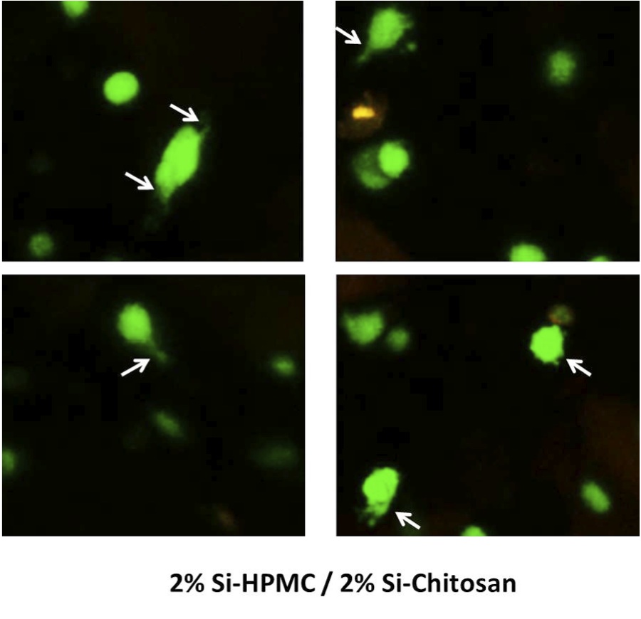
Fig 2 : Confocal micrographs showing cell adhesions after 7 days culture in Si-HPMC/Si-Chitosan hydrogel.
Conclusion: The study demonstrates the feasibility of our approach to modulate our self-setting hydrogel while keeping them biocompatible.
ANR (HYCAR); Région Pays de la Loire (BIOREGOS)
References:
[1] ENGLER AJ, SEN S, SWEENEY HL, DISCHER DE. Cell, 126, 677-689, 2006.
[2] BOURGES X, WEISS P, DACULSI G, LEGEAY G. Advances in Colloid and Interface Science, 99,3, 215-228, 2002.