Introduction: Carboxymethyl cellulose (CMC) and chitosan (CS) are natural biodegradable and biocompatible polymers. CMC is an anionic polymer obtained through chemical modification of cellulose and CS is a cationic polymer derived from chitin and hence being oppositely charged these two polymers can undergo ionotropic gelation[1]. Zirconium (Zr) is a tetravalent cationic metal extensively used in orthopedic implants to promote osseointegration in the form of zirconia and has recently been found to stimulate proliferation and differentiation of human osteoblasts by stimulating different signaling pathways[2]. This study was aimed at developing injectable, non-toxic polyelectrolyte polymer complex microparticles based on Zr as a novel cross-linker. We hypothesize the use of Zr as cross-linker will not induce any cytotoxicity and promote adhesion of osteoblasts into microparticle.
Methods: Aqueous solution of CMC (3% w/v) was prepared and added dropwise into the homogenous mixture of zirconium tetrachloride solution (5% w/v) and CS solution (1% w/v) and kept under stirring for 1 h. The volume ratio of CMC to CS was optimized at 1:2 for improved complexation between two polymers. The particles thus obtained were then dried at room temperature for 48 h before further analysis. The surface morphology of microparticles was characterized by scanning electron microscope (SEM) and energy dispersive spectroscopy (EDS) was done for elemental analysis. Fourier transform-infrared (FTIR) spectrometer in ATR mode was used to characterize the intramolecular interaction between the components in microparticle. In vitro cytotoxicity was assessed using live/dead cell assay kit after culturing the murine pre-osteoblasts (OB-6) in the presence of microparticles up to 10 days. In order to specifically determine the viability of cells attached to microparticle, the microparticles were transferred into separate well of 24 well-plate and then live/dead cell assay was performed on day 5 and 10 and imaged under fluorescence microscope. The percent area of microparticle covered with cells was determined using ImageJ taking visible portion of microparticle.
Results and Discussion: The microparticles were spherical in shape with an average diameter of 787±0.045 µm. The surface of the microparticles contained uniform grooves and ridges creating rougher surface as seen on SEM image (Fig. 1B).
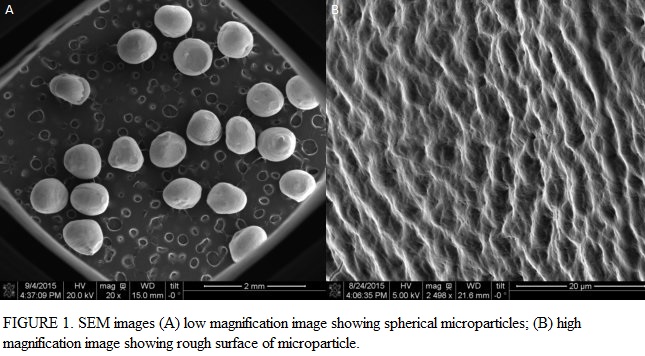
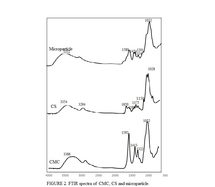
Few intact cracks were also observed on the surface of microparticles which might be due to the excessive evaporation of solvent and hence high shrinkage during drying process. EDS data obtained showed the presence of Zr along the surface of microparticles. The comparison of FTIR spectrum of CMC, CS and microparticle showed the presence of all characteristic peaks of both CMC and CS on microparticle. Moreover, absorbance band at 1580 cm-1 corresponding to the characteristic band of NH3+ and COO- suggested strong electrostatic interaction and hence complexation between two polymers (Fig. 2).
The use of Zr as a cross-linker did not induce any cytotoxicity as most of the cells were viable up to 10 days of culture with microparticles. Fluorescence images of cells seeded microparticle showed adhesion of cells to the surface of microparticle (Fig. 3).

The area percent of cells covering microparticle was significantly higher at day 10 (42%) compared to that at day 5 (15%) suggesting the proliferation of cells on microparticle.
Conclusion: In this study, an injectable CMC-CS polyelectrolyte complex microparticles with Zr as a novel crosslinker was developed. Based on the results obtained, it can be concluded that polyelectrolyte complexation occurs between CS and CMC which can be developed into injectable microparticles by crosslinking with Zr. Zr used in this study was not cytotoxic and provided a suitable surface for cell adhesion and proliferation by enhancing the surface roughness of microparticle.
National Institute of Health (NIH) (grant number R01DE023356)
References:
[1] Liuyun J, Yubao L, Chengdong X. Preparation and biological properties of a novel composite scaffold of nano-hydroxyapatite/chitosan/carboxymethyl cellulose for bone tissue engineering. Journal of Biomedical Science 2009;16:65.
[2] Chen Y, Roohani-Esfahani S-I, Lu Z, Zreiqat H, Dunstan CR. Zirconium Ions Up-Regulate the BMP/SMAD Signaling Pathway and Promote the Proliferation and Differentiation of Human Osteoblasts. PLoS ONE 2015;10:e0113426.