Introduction: An in situ fenestration technique is used to treat complex aortic aneurysms when the aneurysm involves vital arteries such as the renal arteries or left subclavian artery, and deployment of a conventional stent-graft would block blood flow to the kidneys or left arm[1]-[4]. The objective of the present study is to investigate the long term fatigue properties of this technique, by developing a fatigue test method using Bose Electroforce pulsatile fatigue tester and an accelerated cardiac motion system to reproduce the influence of cardiac and pulsatile motion to test the long-term stability of a stent graft system which is deployed and in situ fenestrated into a 3-D aortic aneurysmal model.
Materials and Methods: Four polyurethane elastomeric phantoms (Fig1) of a specific patient’s aortic arch aneurysm were fabricated by using a 3D printing technique. This patient has previously undergone stent-graft deployment and retrograde in situ fenestration via the left subclavian artery. Then four Medtronic Valiant® stent grafts were deployed inside the phantoms followed by in situ fenestration. Atrium® covered stent extensions were then deployed at the fenestration site.
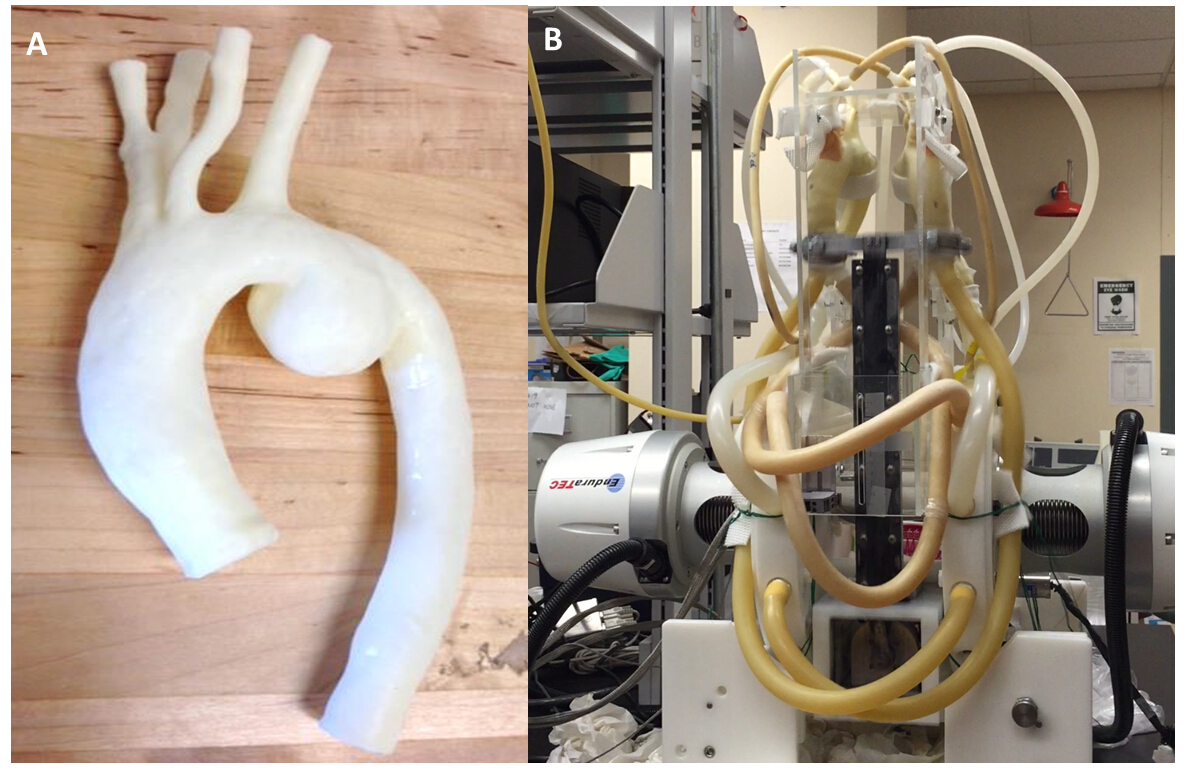
An accelerated fatigue test method (Fig1) was established by modifying the Bose Electroforce fatigue tester and fabricating a cardiac motion system. The cardiac motion system was fabricated by utilizing a high speed 3000 rpm motor driven cam, which was selected to achieve 50 Hz frequency. This test method combined two types of fatigue, a pulsatile test and a tensile test, to mimic the real fatigue situation for the stent grafts.
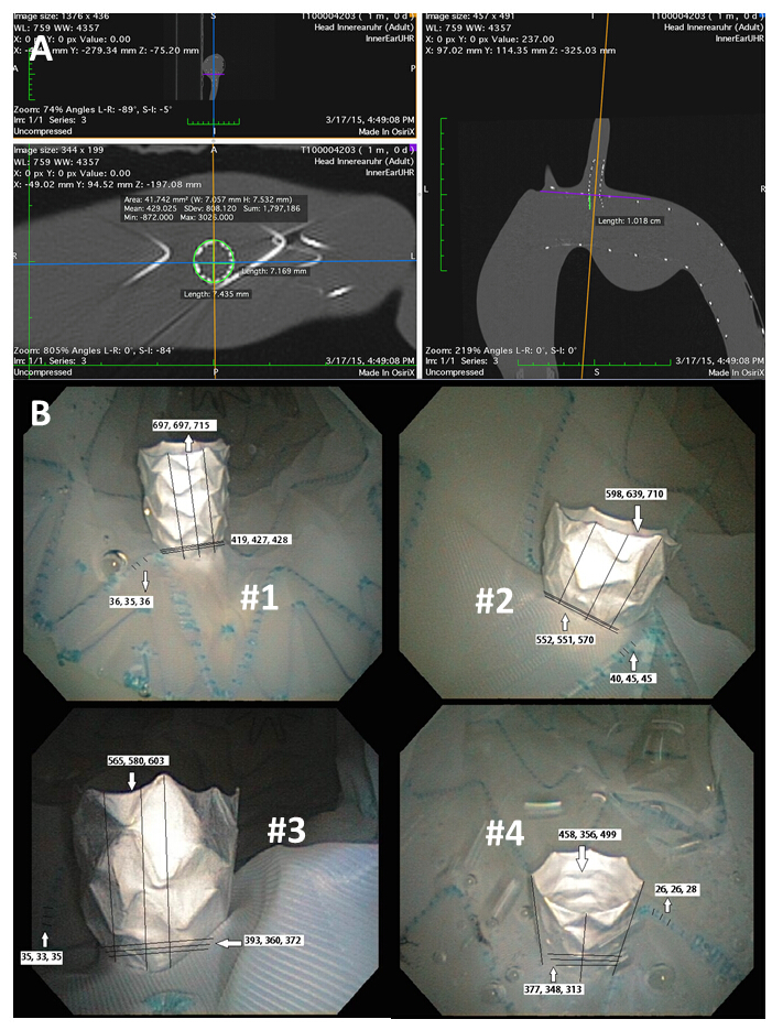
The phantoms with stent-grafts deployed inside were then attached to the fatigue tester and cycled at 50 Hz for 400 million cycles which is equivalent to 10 years’ of cardiac motion. CT scanning were used to capture change in the dimension of the covered stent and endoscopy were used to capture the appearance of tears around the covered stent and evaluate the size of the fenestration outside the covered stent (outer lumen).
Results and Discussions: The dimensions, shape and size of the fenestrated areas were measured by CT scanning and endoscopy at 0 fatigue cycles under 120 mmHg (Fig2). The dimensions of the fenestrated area measured by these two imaging modalities were compared and the results are shown below (Fig3). Following an ANOVA test (confidence interval 95%), the dimensions measured by CT scanning endoscopy had no significant difference.
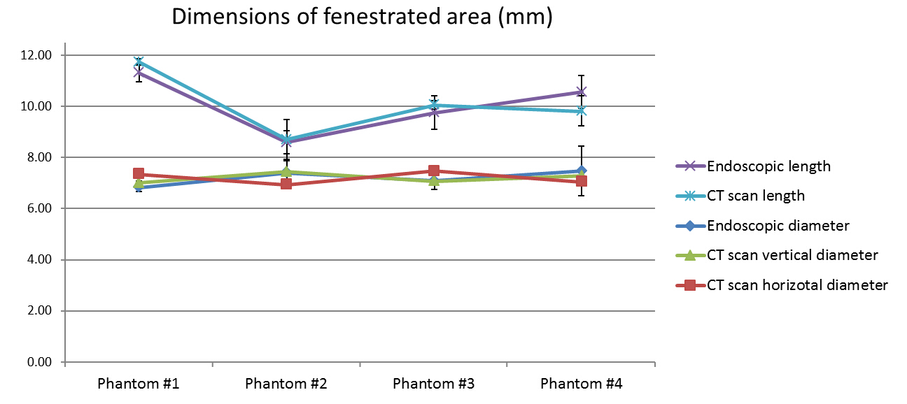
After undergoing the fatigue test for 40 million cycles (equivalent to 1-year’s time), no significant difference in fenestrated dimensions and stent graft appearance were found either by CT scanning or by endoscopy.
Conclusions: A fatigue test method has been developed using a Bose pulsatile fatigue tester and an accelerated cardiac motion at 50 Hz frequency. Dimensions measured with CT scan and endoscopy have no significant difference. Endovascular stent grafts were successfully fenestrated, and after 1 year (40 million cycles) of fatigue, there was no significant change in dimensions as measured by CT scan or endoscopy. Accelerated fatigue testing is continuing for a period equivalent to 10 years in vivo.
Boris Chayer
References:
[1] Upchurch, G. R., & Criado, E., MD. (2009). Aortic aneurysms: Pathogenesis and treatment. New York, NY: Humana Press, Inc.
[2] Bismuth, J., Duran, C., & Hassoun, H. T. (2012). In situ fenestration for branch vessel preservation during EVAR. Methodist DeBakey Cardiovascular Journal, 8(4), 33.
[3] McWilliams, R., Murphy, M., Hartley, D., Lawrence-Brown, M., & Harris, P. (2004). In situ stent-graft fenestration to preserve the left subdavian artery. Journal of Endovascular Therapy, 11(2), 170-174.
[4] Ruthrauff, A.A., King, M.W., Soulez, G., Tan, K.T., Crawford, S.A., Roche-Nagle, G., Cloutier, G., Tse, L.W. Effects of pulsatile fatigue on in situ antegrade fenestrated polyester stent grafts deployed in a patient-specific phantom model of juxtarenal aortic aneurysm. (2015) Journal Vascular Interventional Radiology, In press.