Extracellular matrix and its organization are very critical for the function and viability of the tissues. Tissues are organized in a variety of complex forms and the current approach in design for biomaterials and tissue engineering takes this into account in order to understand the cell and material interactions and also to produce successful products.
The nerves are organized in the form of fiber bundles and most of the current treatments of severed peripheral and central nervous system elements aim to provide a form of guiding element[1]. This could be a simple empty tube into which severed proximal and the distal nerve ends are introduced in order to support aligning and healing (Fig 1A). Another tissue consisting of highly aligned elements is the muscle[2]. An example of repair attempt to damaged muscle tissue after a heart attack is to employ a muscle patch with stem cells aligned on electrospun polymeric fibers prepared in the form of a mesh, a mat or a bundle (Fig 1B).
When a systematic attempt is made to determine the factors that make a surface attractive or repulsive for cells, planned creation of surfaces highly decorated with topographical and chemical/biological features using micro- and nanotechnology methods. Topographical features such as micro and nano-sized posts/pillars and wells are designed for increasing attachment on the surface and also for forcing conformational changes in the cell and in their nuclei. Physical patterns might convince cells to adhere preferentially on certain regions or constrain them in voids or gaps much smaller than their normal sizes (Figs 1C , 1D)[3],[4].
There are also chemical and biological patterning approaches that are used to create adhesive and repulsive regions or islands and posts with tips modified with cell adhesive molecules such as fibronectin or cell repulsive molecules such as PEG. The results of these intense activities find application in other fields such as biosensors.
Thus the design of the microenvironment of the cells is becoming a very important area in the field of biomaterials and tissue engineering and the newly evolving techniques contribute significantly to the advances made in these fields.
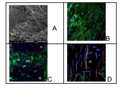
Figure 1. Various extracellular surfaces designed and used in guidance of various tissues. A) Nerve guide[1], B) Cardiac patch[2], C) Selective attachment of cells on micropillared surfaces[3], D) Micropatterned surface with cells arranging their cytoskeleton to fit the pillar dimensions for bone tissue studies[4].
BIOMATEN for the Ministry of Development funds; METU Grants
References:
[1] D. Yucel, G. T. Kose, V. Hasirci, Biomaterials, 31, 1596–1603, 2010
[2] H. Kenar, G. T. Kose, V. Hasirci, J. Mater Sci: Mater Med, 21(3), 989-997, 2010
[3] H. Ozcelik, C. Padeste, V. Hasirci, Colloids and Surfaces B:Biointerfaces, DOI:10.1016/j.colsurfb.2014.03.019
[4] M. Ermis, E. Antmen, P. Chen, U. A. Gurkan, U. Demirci, V. Hasirci, MRS Fall Mtg, Dec 2013, Boston, USA.