Introduction: Rapid ingrowth into scaffolds provides substantial advantages when regenerating large bone defects. Inverse trabeculated patterned scaffolds (Figure 1) induce more rapid bone formation compared to scaffolds with similar sized geometric pores after 6 months in a canine model. Based on these results, 3D printed biomimetic scaffolds were produced for long bone segment regeneration in a sheep critical sized defect. The goal of this study is to determine whether these scaffolds, infiltrated with calcium particles and endogenous stem cells, induce sufficiently rapid bone formation to fill a critical sized defect within 6 months.
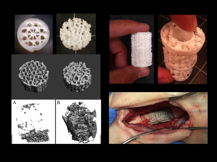
Figure 1: (Left images) 3D printed simple geometric porous scaffold design and a biomimetic porous scaffold design with µCT scans of the bone ingrowth into each at 6 months in a dog model. (Top right images) 3D printed, biomimetic, long segment regeneration scaffolds. (Bottom right) Scaffold implanted into critical sized defect.
Methods: Critical defect sized, trabecular and cortical bone samples, were collected from adult male sheep. Samples were imaged in a SCANCO µCT 20 at a 12 µm resolution. Files were combined and converted to STL, then exported to build polybutylene terephthalate scaffolds (Figure 1) in a Stratsys 1650 FDM. Structural accuracy was verified by scanning scaffolds in the µCT and comparing scans to the original bone scans.
Rosette strain gauges were attached to scaffolds that were compressed to 294 N at 49 N/s and 294 N/s in an MTS. Data was collected with LabView through a NiDaq board and exported to Excel. Stress vs. principal strain curves were drawn and strength and stiffness determined. A previously published latexing process was used to coat scaffolds with 4µm tricalcium phosphate (TCP) particles and scanning electron microscopy (SEM) was used to verify infiltration.
Male sheep underwent two IACUC approved procedures; the first was inguinal fat harvesting. Adipose derived stem cells were extracted using our published procedure. Cells were fed, passaged and perfused deep into scaffold pores. Scaffolds were placed 48 hours after perfusion. During surgery a 42 mm femoral segment was removed and a modified lockable IM rod was used to stabilize the scaffold. Sheep were held for 26 weeks. Activity was monitored with 24 hr. video. Monthly radiography and post sacrifice histology were used to determine the extent of tissue ingrowth and rate of bone formation. ANOVA’s would determine difference between groups.
Results: The final scaffold configuration was 71.6±0.2% porous. Mechanical testing indicated a linear elastic response and a stiffness of 2.8 GPa with a compressive strength of 9.44±0.94 MPa (sufficient to support physiological loads) although scaffolds were 37% as stiff as sheep bone and 10% the compressive strength. Deposition of TCP was observed deep inside the pores but cell infiltration was better near outer surfaces and pre- and post- cell counts showed 80% cell depletion following infiltration.
Scaffold placement was uneventful and sheep were load bearing within hours. Activity monitoring indicated the sheep spent 23.8±6% of their time standing or walking. After 8 weeks, radiographs showed there was 210 sq. mm. of new bone completely bridging the defect (Figure 2). Tetracycline labeling prior to sacrifice was used in conjunction with histomorphometry to accurately assess bone formation rates.
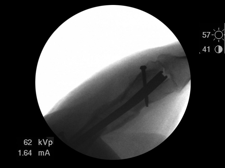
Figure 2: Radiograph showing an example of complete bridging of critical sized defect after 8 weeks.
Discussion: A long segment regeneration biomimetic scaffold infiltrated with endogenous adult sheep stem cells facilitated rapid bone growth throughout the length of a critical sized defect in a sheep femur even though sheep were very inactive. Preliminary results indicate that this design is able to support physiological loads during bone growth, which is apparent on radiographs as early as 4 weeks.