Introduction: Cornea is a transparent and avascular tissue which contributes to maintain intact vision. The cells based corneal parts such as epithelia and endothelia can be regenerated using cell therapy technologies[1]. On the other hand, M. Griffith et.al. have develop substitute for corneal stroma made by recombinant collagen and showed the efficacy in the clinical trials[2]. Even though, the material has still not enough the mechanical properties. To maintain the transparency and to keep the eyeball shape, corneal stroma has unique nano-scale anatomical features such as lattice collagen structure. Therefore, we focused on aligned nanofiber non-woven mat. We focus on silk materials and silk fibroin nanofiber has been investigated, because it is used in medical field because of its well biocompatibility. We found that electrospun aligned fibroin nanofiber non-woven mat has a potential to use for regenerative scaffold for corneal stroma[3]-[5]. In this study we evaluated the longterm tissue responses against the silk nanofibers in rabbit cornea.
Materials and Method: Aqueous silk fibroin solution (9-12 wt%) was kindly donated from National Institute of Agrobiological Sciences. For preparation of electrospinning solution, 8 wt% fibroin aqueous solution was gently mixed with a 5 wt% poly (ethylene oxide) aqueous solution in the volume ratio 4 to 1 . An electrospinning system equipped with a rotation drum collector (NANON, MECC Co., Ltd., Japan) was used to fabricate aligned non-woven mats as following setting; applied voltage was 12 kV, a feeding rate was 0.3 ml/h, a spinning distance was 185 mm, a stainless needle was 25G, a diameter of the drum collector was 100 mm, rotation speed was 3000rpm and spinning time was 3 hour. Containing poly (ethylene oxide) was eliminated by immersing into 95% ethanol and distilled water for 24 hours. Then, insolubilized fiber mat was sterilized by autoclave at 121 degree Celsius for 20minutes. Animal experiment was approved by ethical committee in National Institute for Materials Science. Four male Japanese white rabbits (body weight 2.5-3.5kg) were anesthetized by intravenous injection of sodium pentobarbital (0.6ml per 1kg body weight). After anesthesia, corneal stromal pocket was created using surgical knife and ophthalmic spatula. Sterilized fiber mat was put into the corneal stromal pocket (Fig.1). Gross observation of implanted eye was done using surgical microscope at every week after surgery till sacrifice.
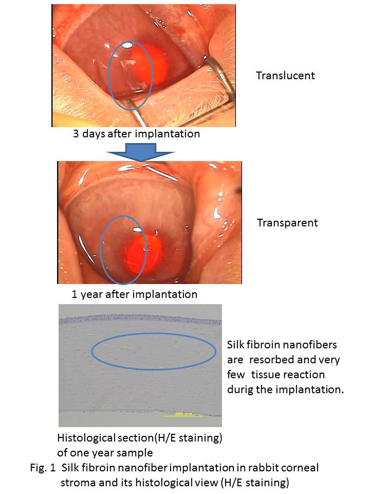
Results and Discussion: In rabbit corneal stroma, quite low inflammatory and no-immune reactions were observed till one year after surgery. And corneal epithelium was almost intact on the host cornea and a part of migration of host corneal stromal cells into the silk fibroin nanofibers was also observed by three weeks.(data is not shown) . After one year, implanted silk fibers were hardly observed by the surgical microscopic observation and the host corneas are clear and no adversed tissue reaction was observed during the period. From the histological observation, the silk fibroin nanofibers were completely degraded and resolved in the body.
Concluding Remarks: In this study, we tried to make the mimic structure of the natural corneal stroma constructing from silk fibroin nanofibrous materials. And evaluated the relatively longterm body reactions were observed. These results suggested the efficacy of materials, the nanofibrous materials was compatible to the host cornea.
References:
[1] Nishida et.al, NEW ENGLAND JOURNAL OF MEDICINE 351 (12)(2004) 1187-1196
[2] P. Fagerholm, et.al, Science Translational Medicine 2(46)(2010) 1-8
[3] D. Terada, S. Hattori, H. Kobayashi et.al, Bioinspired, Biomimetic and Nanobiomaterials, (2012) 57-61.
[4] S. Hattori, D. Terada D, H. Kobayashi et.al, Bioinspired, Biomimetic and Nanobiomaterials, (2012) 195-199.
[5] S. Hattori S, D. Terada D, H. Kobayashi H et.al, J. Silk Sci Tech Jpn, (2014) 117-123.