Introduction: The mechanism of vascular calcification is still elusive; one of the main reasons for this is the lack of effective in-vitro models that truly mimic vessel structure.[1] We fabricated 3D models using elastin (ELN), which is a main component of blood vessels and is know to be a main site for nucleation of minerals. We then studied their calcification in simulated body fluid (SBF). Such models will provide a useful platform for understanding the calcification mechanism, as well as test treatments devised to inhibit or reverse mineral deposition.
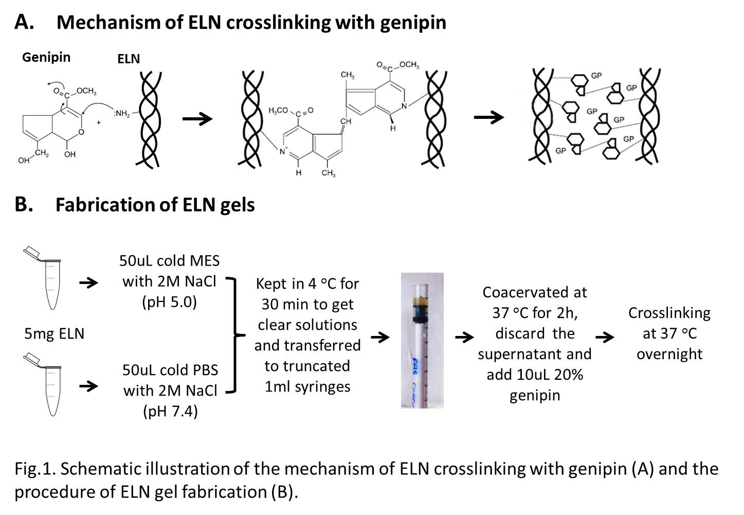
Materials and Methods: We crosslinked ELN from bovine ligament using genipin, a non-toxic natural compound. Before crosslinking, a key step is coacervation of ELN solution; the dense coacervate is considered to be a non-crosslinked precursor of the elastic fibers.[2] Since coacervation is pH dependent, we used two buffers with different pH values. We incubated the obtained gels in 1.5×SBF with stirring for 4 weeks, freeze-dried and characterized the samples by FTIR, XPS and synchrotron based X-ray absorption near edge structure (XANES) spectroscopy.
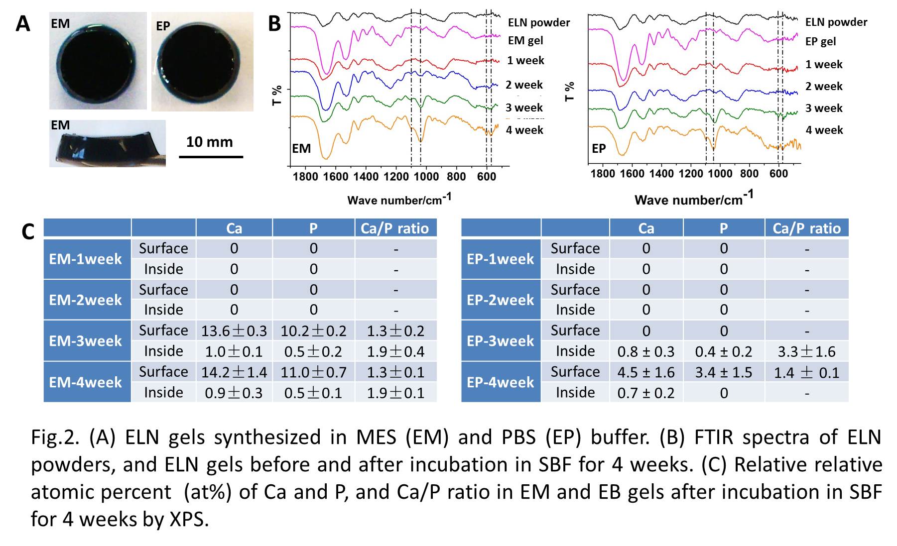
Results and Discussion: The gels synthesized in two buffers have similar sizes (Φ10mm×4mm) (Fig.1A). Fig.1B shows the FTIR spectra of the ELN gels after incubation in SBF. In the first two weeks, ELN gels synthesized in MES (EM) and PBS (EB) buffer show spectra similar to that of pure ELN powders. After 3 weeks, typical bands corresponding to hydroxyapatite (HA) at 574, 605, 1045 and 1100 cm-1 are observed. XPS results show that most of the Ca and P was distributed on the gel surfaces and the minerals were present as mixed phases since the Ca/P ratios are varied. EM gels were more calcified than EP gels.
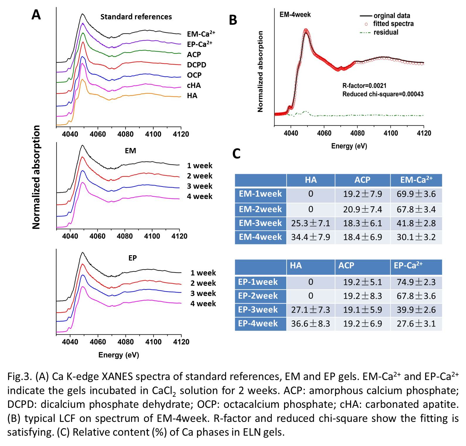
We further determined the mineral phases by XANES. We can clearly see a change in the spectra from 1 to 4 weeks (Fig.3A). Using Ca-phosphates as references, we quantified the relative contents of the various phases by linear combination fitting (LCF) (Fig.3B and C). In the first two weeks, nearly 70% of the Ca is present in a structure that resembles that of the gels incubated in CaCl2 solution (EM- or EP-Ca2+) and 20% as ACP. This indicates that most of the Ca may be adsorbed on the gels and complexed by carboxyl groups present in the ELN scaffold. HA forms and grow from ~25% at 3 weeks to ~35% at 4 weeks. At the same time, the amount of carboxyl-chelated Ca reduced significantly, going down to 30% at 4 weeks, while the relative amount of ACP remained constant. These results show that the adsorption of Ca2+ onto the gels is the main step before HA nucleation, and that ACP is probably the main precursor of HA. Given that ACP was also found in mice arteries in our lab, these results indicate that ELN gels may be a good in-vitro model of blood vessel calcification.
Conclusions:
1. 3D ELN gels can be a good in-vitro model of vascular calcification.
2. Not only HA, but also other calcium phosphate phases form on the gels after incubation in SBF. Adsorption of Ca2+ on ELN scaffold occurs prior to mineralization and ACP is the main precursor of HA.
We thank Yongfeng Hu and Qunfeng Xiao at beamline SXRMB of CLS (Canadian Light Source) for their support during the XANES experiment. This study is supported by the Canada Research Chair foundation, Les Fonds Quebecois pour la Recherche Nature et Technologie (FQRNT), and the Heart and Stroke Foundation.
References:
[1] Cloyd K. L., El-Hamamsy I., Boonrungsiman S. et al. Characterization of porcine aortic valvular interstitial cell ‘calcified’ nodules. PLoS One 2012, 7: e48154.
[2] Cox, B. A., Starcher, B. C., Urry, D. W. Coacervation of tropoelastin results in fiber formation. Journal of Biological Chemistry 1974, 249(3): 997-998.