Introduction: Interactions of prostheses, used to strengthen abdominal weaknesses, with human tissues have yet to be fully understood. Different microscopy approaches necessitate intrusive treatments of samples [1]. This project applies X-ray micro-tomography in order to acquire volumetric datasets of explanted synthetic abdominal prostheses and obtain qualitative and quantitative tri-dimensional (3D) information of the mesh and the integration of human tissue without disrupting its architecture after explantation comparing to virgin prosthesis. This imaging concept has yet to be applied for abdominal prostheses.
Materials and Methods: We imaged 5 virgin prostheses in air (Allomax, Bard mesh, Composix L/P mesh, Parietex, Surgimesh) and one in water (Bard mesh), one human flesh sample and 3 explanted prostheses samples for a total of 10 datasets. Samples were cut out of the larger sources to be approx. 5 x 5 x 5 mm3 and fitted in 5 mm diameter kapton tube, as shown in Figure 1.
Data acquisition was performed at the Center for X-ray Analytics at Empa, in Duebendorf, Switzerland. Each sample was imaged in a 360˚ manner. Source-detector distance was 1017 mm, as shown in Fig. 1. The detector was an amorphous-Silicon flat panel detector with 2048 x 2048 pixels panel. The total acquisition time varied from 30 to 60 minutes, the number of projections from 721 to 1441, the voltage from 50 to 80 kV, the current from 100 to 200 μA and the voxel size from 2.8 to 3.7 μm, providing a final resolution better than 10 μm. Data sets were reconstructed using the software Octopus and isolation using segmentation. 3D visualisation of the protheses were obtained using the software Avizo.

Results and Discussion: Imaging of the different prostheses in air resulted in clear 3D reconstructions, except for allomax, the human dermis sample. The bard mesh in water showed less clear borders due to similar densities thus low contrast between the prosthesis and water. The three explanted prostheses showed very good 3D reconstructions, probably due to the fibrosis around the fibres of the mesh.
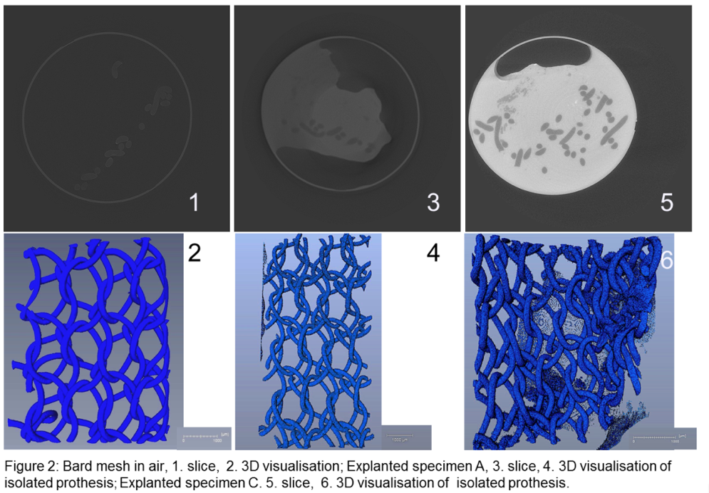
Segmentation, virtual removal of the flesh to display the mesh by itself, was successful, although challenging when the prosthesis was in contact with lipid tissues. Surfaces of the isolated prostheses lack sharpness, due to similar densities of flesh and polymer. The overall geometry of these prostheses are remarkably conserved, as shown in Fig. 2.3. Qualitative comparison of two explanted polypropylene monofilament meshes (Bard) vs. virgin indicated a directional stretching of similar magnitude in both cases. Cause of deformation could be tissue ingrowth, mechanical shearing, tensile strain at fixing. Deformation will be investigated quantitatively using image registration.
Conclusion: X-ray microtomography captures the configuration of protheses embedded in human tissues with a resolution of a few micrometer. Proper segmentation of the mesh from the tissues is achieved. This advance opens the door for full understanding of the deformation of explanted meshes.
References:
[1] Guldberg RE, Duvall CL, Peister A, Oest ME, Lin AS, Palmer AW, Levenston ME. 3D imaging of tissue integration with porous biomaterials. Biomaterials. 2008 Oct;29(28):3757-61. doi: 10.1016/j.biomaterials.2008.06.018. Epub 2008 Jul 16.