Tracing of individual hematopoietic stem cell specification events using Raman Spectroscopy and photonic crystal enhanced microscopy
-
1
University of Illinois at Urbana-Champaign, Chemical and Biomolecular Engineering, United States
-
2
University of Illinois at Urbana-Champaign, Carl R. Woese Institute for Genomic Biology, United States
-
3
University of Illinois at Urbana-Champaign, Electrical and Computer Engineering, United States
-
4
University of Illinois at Urbana-Champaign, Micro and Nanotechnology Laboratory, United States
Introduction: Hematopoietic stem cells (HSC) are rare adult stem cells residing in the bone marrow that are responsible for life-long hematopoiesis. To enhance our understanding of the underlying mechanisms of HSC regulation and facilitate the clinical use of HSCs, it is desirable to engineer their specific fate decision events (self-renewal vs. differentiation) in vitro. Such efforts are limited by the lack of functional markers that enable non-invasive, dynamic analysis of individual HSCs in situ. Raman Spectroscopy is a promising chemical imaging tool that provides unique molecular fingerprints of individual, live cells in situ in a label-free manner. Similarly, photonic crystal enhanced microscopy (PCEM) is a label-free imaging platform that enables dynamic quantification of single HSC adhesion profiles. Here, we demonstrate the feasibility of applying Raman Spectroscopy and PCEM for screening HSC phenotype via the identification of primary hematopoietic cell populations and the segmentation of primitive hematopoietic progenitor cell populations during their differentiation to granulocytes.
Materials and Methods: We isolated primary hematopoietic cell populations (long-term HSCs: LT-HSC, short-term HSCs: ST-HSC, B cells, granulocytes) from C57BL6 mouse bone marrow and analyzed individual cells with Horiba Raman confocal imaging microscope. Additionally, we analyzed individual 32D cells (a myeloid progenitor cell line) during differentiation towards granulocytes. All cells were seeded onto gold substrates decorated with or without a protein-immobilized hydrogel layer. For PCEM, cells were seeded on fibronectin-coated photonic crystals and wavelength shifts in the transmitted light from accumulation of cellular materials on cell-surface interface were quantified to visualize their adhesion profiles.
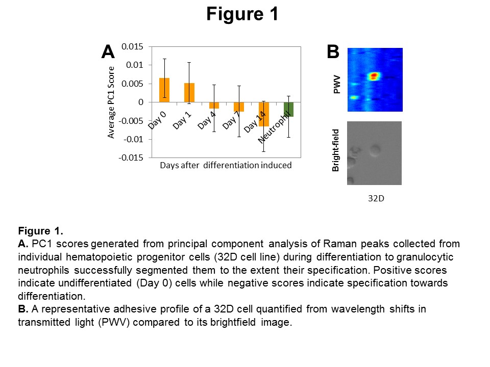
Results and Discussion: Multivariate analysis of Raman peaks from individual hematopoietic cells revealed that Raman spectra-generated molecular signatures could be used for their identification with less than 4% false identification rates. Notably, subsets of primitive HSCs (LT-HSCs, ST-HSCs) whose nuanced functional differences are difficult to segment could be easily distinguished from each other as well as from mature downstream cells (B cells, granulocytes). Additionally, cells seeded on soft vs. stiff hydrogels could be analyzed in a similar manner, indicating that Raman Spectroscopy is a promising approach for in situ screening of HSCs. Moreover, principle component analysis of Raman peaks from individual hematopoietic progenitor cells (32D cell line) was able to segment discrete stages of granulocyte specification (Fig.1A). Ongoing work with PCEM suggest that adhesive phenotype (adhesion strength, migration speed, and total displacement) of individual 32D cells during differentiation may provide unique adhesive signatures of individual cells indicative of their specification state (Fig.1B).
Conclusions: Our results show that Raman imaging and PCEM can generate unique molecular and adhesive fingerprints of individual HSCs that could be used as novel markers reflecting their functional phenotype. We therefore envision these methods may provide a means to efficiently screen HSC fate specification events in situ in real time for future bioengineering and biomanufacturing applications.
Keywords:
Cell Differentiation,
stem cell,
biosensing,
matrix-cell interaction
Conference:
10th World Biomaterials Congress, Montréal, Canada, 17 May - 22 May, 2016.
Presentation Type:
Poster
Topic:
Biomaterials in mesenchymal and hematopoietic stem cell biology
Citation:
Choi
J,
Ilin
Y,
Zhuo
Y,
Cunningham
BT,
Kraft
ML and
Harley
BA
(2016). Tracing of individual hematopoietic stem cell specification events using Raman Spectroscopy and photonic crystal enhanced microscopy.
Front. Bioeng. Biotechnol.
Conference Abstract:
10th World Biomaterials Congress.
doi: 10.3389/conf.FBIOE.2016.01.00701
Copyright:
The abstracts in this collection have not been subject to any Frontiers peer review or checks, and are not endorsed by Frontiers.
They are made available through the Frontiers publishing platform as a service to conference organizers and presenters.
The copyright in the individual abstracts is owned by the author of each abstract or his/her employer unless otherwise stated.
Each abstract, as well as the collection of abstracts, are published under a Creative Commons CC-BY 4.0 (attribution) licence (https://creativecommons.org/licenses/by/4.0/) and may thus be reproduced, translated, adapted and be the subject of derivative works provided the authors and Frontiers are attributed.
For Frontiers’ terms and conditions please see https://www.frontiersin.org/legal/terms-and-conditions.
Received:
27 Mar 2016;
Published Online:
30 Mar 2016.