Introduction: Due to its ability of non-destructive 3D imaging at high resolution, the use of micro-computed tomography (or micro-CT) in bone microstructure analysis has increased over the last decays. This technique can also be applied to implants or materials for bone augmentation e.g., calcium phosphate cements (CPCs). Despite many advantages[1], CPCs are brittle materials and are commonly not used alone, nor approved for use alone, in load-bearing applications. Nevertheless, it would be of importance to understand their fracture behaviour under different loading conditions. The aim of this study was to investigate the possibility to use micro-CT imaging to study the fracture behaviour of CPCs during compression and diametral tensile tests and to study possible correlations between mechanical and structural properties.
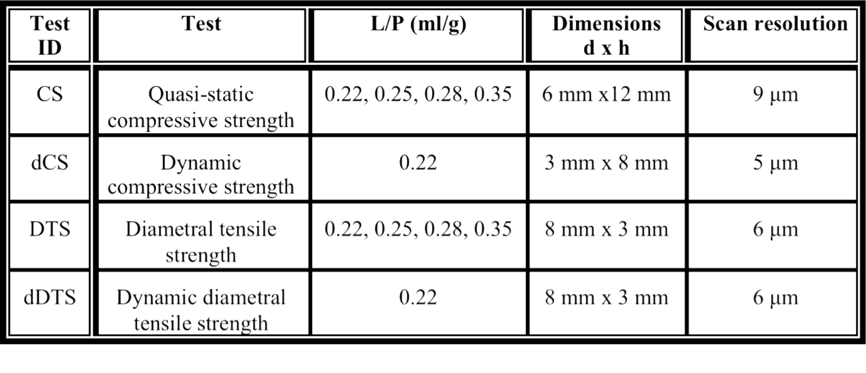
Materials and Methods: Brushite cement were prepared by mixing monocalcium phosphate monohydrate (sieved to < 75 μm) and β-tricalcium phosphate powders at a molar ratio of 45:55 (each containing 1 wt % of disodium dihydrogen pyrophosphate) with 0.5 M citric acid at different liquid to powder ratios (L/P). Samples were let to set in PBS for 24 hours. Porosity was measured with a solvent exchange method[2] and samples were scanned with a micro-CT (Skyscann 1172, Bruker), using a source voltage of 100 kV, a current of 100 μA and an aluminium and cupper filter. Wet compressive strength (CS) and diametral tensile strength (DTS) were measured. Thereafter the samples were rescanned to visualise the fracture in 3D. dCS and dDTS samples were tested and scanned using an in situ loading stage in displacement control at increasing steps of 0.01 mm. Reconstruction, 3D visualisation and analysis of the micro-CT images were performed using respectively the softwares NRecon, CTvox and CTAn (Skyscan, Bruker). Details about samples and tests are reported in Table 1. SEM was used to investigate the microstructure of CPCs.
Results and Discussion: Porosity increased linearly with L/P. Average CS decreased exponentially with L/P and the dimensions of the biggest defect (the latter calculated with CTAn). Average CS decreased with porosity but no model fitted the data. In fact, it was observed that the microstructure and the entanglement of the crystals played an important role in the CS. Materials with similar porosity but smaller crystals showed a significantly higher CS. Micro-CT imaging exposed several cracks in the samples after compression, which can be clearly visualized with this technique (Fig 1a). Similarly, DTS decreased with increased L/P and porosity. The post-scans also showed several cracks. Dynamic tests revealed that in both dCS and dDTS samples the failure did not occur at the first crack (Fig 1). Analogous results have been obtained in Portland cements[3].
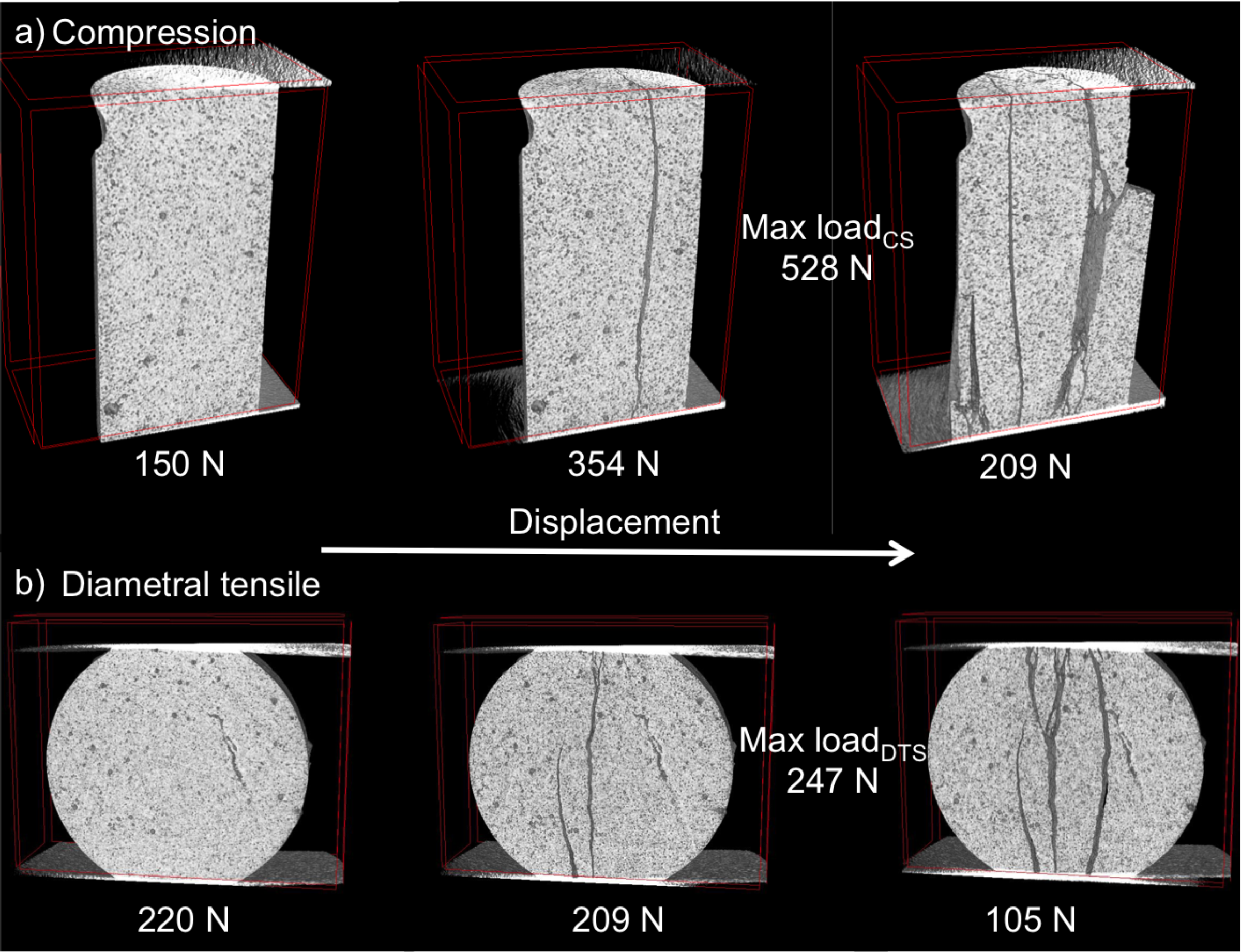
Figure 1 - Micro CT reconstructions of: a) a section of the CPC before compression (left), the first crack (mid) and the sample after failure (right); b) a section of the DTS sample before testing (left), the first cracks (mid) and the sample after failure (right). The load at each step is displayed at the bottom of each image.
Conclusions: Mechanical properties were found to be correlated to different structural parameters. Micro-CT imaging seems to be a promising method to study crack propagation and fracture behaviour of CPCs in different loading conditions.
Carl Tryggers Stiftelse (CTS 13:533); Stiftelsen Lars Hiertas Minne (FO2014-0504)
References:
[1] Ginebra MP, Calcium phosphate cements, 2006, Cambridge Ed.
[2] Ajaxon I et al., J Biomater Appl, 2015, DOI: 10.1177/0885328215594293
[3] Landis EW and Nagy EW, Eng Fract Mech, 2000, 65(2): 223-234.