Introduction: Induced pluripotent stem (iPS) cells have high potential to use as cell source for regenerative medicine. iPS cells must be cultured on/in a substrate for proliferation under maintaining undifferentiated and pluripotent state in order to utilize iPS cells for clinical applications. Further animal products, such as mouse embryonic fibroblasts as a feeder layer, bovine serum, and animal growth factors, should be avoided to use in the culture system for diminishing a contamination of antigen and virus from animals. Therefore many studies to develop a substrate and a serum-free culture medium system have been reported.
Silk have been used as a textile fiber for more than 6000 years, and is recently studied as a biomaterial because of the good biocompatibility. We reported the characteristic properties of silk fibroin film surface, that is the cell mobility on fibroin surface is significant higher than collagen, fibronectin glass, and culture dish, and the gene expression of extracellular matrix is up-regulated. We infer silk will be unique substrate for iPS cells. However the study to examine the silk fibroin as the substrate for iPS cell culture is not reported. In this paper we will present the mouse iPS cell growth behavior on silk fibroin film.
Materials and Methods: Mouse iPS cells (iPS-MEF-Ng-20D-17) were purchased from RIKEN BioResource Center. This iPS cell line express GFP by Nanog promoter as the marker for pluripotency. iPS cells were cultured on the feeder layer of SNL cells according to the typical procedure.
Silk fibroin coated dish was prepared as follows. Degummed silk was dissolved in 9M LiBr solution and the solution was dialysis against water. The fibroin aqueous solution (1w/v%) was put in 60mm culture dish and then dried. The fibroin coated dish was treated by 80% methanol for insolubilizing.
Results: Figure 1 show the iPS cell growth curve on various surfaces. iPS cells can proliferate on fibroin surface as comparable rate as on collagen, gelatin and SNL feeder layer surface. This result indicates fibroin can use as the culture substrate for iPS cells growth.

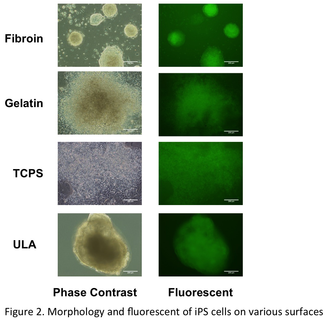
Figure 2 shows the morphology and fluorescent image of iPS cells cultured on various surfaces after 7 days incubation. iPS cells can make clear colonies on fibroin and ultra low adherent dish (Corning), while widely spreading cell layer were observed on gelatin and tissue culture dish. The fluorescent images indicate the in vitro maintenance of pluripotency of the iPS cells. The figure 2 shows clearly that iPS cell colonies on fibroin expressed strong fluorescent. This result means that the iPS cells proliferated on fibroin surface to be maintained the pluripotency. We confirmed the maintenance of pluripotency by immunostaining of Oct4 and alkaline phosphatase staining.
Conclusions: Silk fibroin substrate supports mouse iPS cells growth in the maintenance of pluripotency.