Introduction: Outright harmonization amongst the events such as infiltration of inflammatory cells, granulation tissues constitution along with angiogenesis, proliferation and migration of fibroblasts and keratinocytes maintains dynamicity of ideal and rapid wound repair. Treatment with honey has regained popularity in recent times as because a number of small clinical trials have been completed which corroborate clinical improvement with regenerative potentiality on topical application of honey on wound. Honey is thought to be providing regenerative ambience in wound bed leading to the development of an embryonic environment with its own system intelligence. In this context present study performs exhaustive estimation of honey’s regenerative potential on topical application in wound model with a special emphasis on elucidation based on histopathological study.
Materials and Methods: 10 male swiss albino mice (Mus musculus) were housed individually in a pathogen free environment and the study was performed under the Guidelines of the Institutional Animal Care and Use Committee. Two full thickness wounds were created on each animal after anaesthetizing with xylazine and ketamine hydrochloride and subsequent shaving of the dorsal part. One wound was regarded as control (povidone-iodine treated) and another was treated with physico-chemically characterized honey. The wounds were examined clinically at specific time points along with the collection of biopsy samples followed by Hematoxylin-Eosin (H and E), Periodic Acid–Schiff (PAS) and Van Gieson’s (VG) staining.
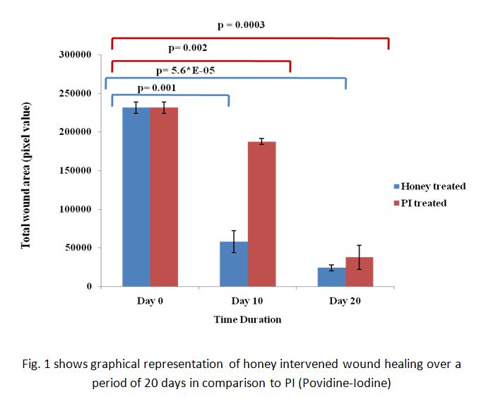
Results and Discussion: As shown in figure 1, the total wound area (pixel value) for honey treated wound as calculated by ImageJ software was found to be significantly reduced when compared to control. Higher level of significance ( p= 5.6*E-05) is noted in case of honey treated wound as an indicator of wound contraction. The long lasting effect of honey on the release of TNF-α and IL-1β and IL-6 production may explain its therapeutic interface in the harmonized process of wound healing.
On clinical evaluation, the honey treated wounds were found to show accelertaed healing comparitively with respect to control.
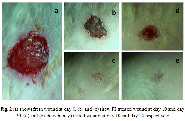
The healing progression of skin biopsies were observed by histopathological techniques On 10th and 20th days after treatment, the microscopic observations under H and E staining explored remarkably improved histological features with progressive maturation of epithelial and connective tissues. The VG staining illustrated upgraded collagen density and formation of normal skin orientation.
Conclusion: Topical application of honey escalates wound healing with exquisite dynamicity via apposite wound contraction and re-epithilialization in wound model. Results of this study may serve as a crucial step for exploration of honey in varied regenerative applications.
This work is carried out under the project "Noninvasive Characterization Of Healing And Nonhealing Wounds And Development Of Honey-Biomaterial And Stem Cell Based Wound Therapy" (NIC), sponsored by MHRD, Department of higher Education, New Delhi, Government of India. IIT Kharagpur has been acknowledged for providing infrastructural facility
References:
[1] Enoch S, Leaper DJ. Basic science of wound healing. Surgery (Oxford) 2008;26:31-7
[2] Trent JT, Kirsner RS. Wounds and malignancy. Advances in skin and wound care 2003;16:31-4
[3] Krafts KP. Tissue repair: The hidden drama. Organogenesis 2010;6:225-33
[4] Tandara AA, Mustoe TA. Oxygen in wound healing—more than a nutrient. World journal of surgery 2004;28:294-300
[5] Yang C, Zhu P, Yan L, Chen L, Meng R, Lao G. Dynamic changes in matrix metalloproteinase 9 and tissue inhibitor of metalloproteinase 1 levels during wound healing in diabetic rats. Journal of the American Podiatric Medical Association 2009;99:489-96
[6] Maharlooei MK, Bagheri M, Solhjou Z, Jahromi BM, Akrami M, Rohani L, et al. Adipose tissue derived mesenchymal stem cell (AD-MSC) promotes skin wound healing in diabetic rats. Diabetes research and clinical practice 2011;93:228-34
[7] Ito M, Yang Z, Andl T, Cui C, Kim N, Millar SE, et al. Wnt-dependent de novo hair follicle regeneration in adult mouse skin after wounding. Nature 2007;447:316-20
[8] Tonks A, Cooper R, Jones K, Blair S, Parton J, Tonks A. Honey stimulates inflammatory cytokine production from monocytes. Cytokine 2003;21:242-7