Introduction: Chitosan hydrogels can form interesting embolizing agents for vascular applications thanks to their thermosensitive properties. However they lack visibility under fluoroscopy, CT scan and magnetic resonance imaging (MRI) for follow-up. The aim of this study was to study the potential of these hydrogels as embolizing agents, and their optimization for good visualization with CT and MRI.
Materials and Methods: Different chitosan (CH; DDA 94%) thermogels were prepared by mixing CH with different combination of sodium hydrogen carbonate (SHC), beta-glycerophosphate (BGP) and phosphate buffer (PB). The results were compared to controls made with BGP and sodium tetradecylsulfate (STS) known as embolizing agents with sclerosing properties. A non-ionic isotonic iodine contrast agent (Visipaque 320, at 20-50%v/v) and/or gadolinium (Gd-BOPTA, Multihance, at 0.01- 10%v/v) were added at various concentrations. Rheological properties were determined on a Physica MCR301 (Antoon Paar) and radiodensity was measured in Hounsfield unit, before and after saline immersion, after acquisition on a cone-beam CT. Using 3.0T MRI, relaxation times of chitosan gels were measured using inversion-recovery method, conventional spin echo and gradient-recalled echo sequences with different echo times to measure T1, T2 and T2*, respectively. Preliminary in vivo embolization tests were performed in pig arteries.
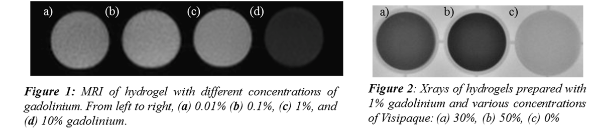
Results and Discussion: Rheology showed rapid increase of the storage modulus with time at 37°C, final modulus being significantly higher with STS as previously observed [1]. Good radiodensity was observed for VIS concentration above 30%. Nice contrast on MRI was obtained for gadolinium concentration of 0.1 and 1% (Fig. 1). T1, T2 and T2* relaxation times of chitosan 3.33% mixed with 1% gadolinium were respectively 38 msec, 18 msec, and 7 msec. T1 relaxation time is shorter than that of abdominal organs and soft tissues, making it visible after embolization. Both VIS and Gadolinium were rapidly released when immersed in saline, thus avoiding artefacts during imaging follow-up. Gadolinium had no impact on the gelation kinetic. VIS tend to slow down gelation, but increasing VIS concentration up to 50% in BGP-containing hydrogels led to good rheological properties. In vivo injection of CH-STS-VIS-GAD led to good occlusion of blood vessels and the gel was visible under fluoroscopy.
Conclusion: These chitosan hydrogels present interesting properties for endovascular applications such as the occlusion of endoleaks. They can be made radiodense and MRI visible by addition of iodine contrast agent and/or gadolinium, which are then rapidly released.
References:
[1] Fatimi, A., et al., A new injectable radiopaque chitosan-based sclerosing embolizing hydrogel for endovascular therapies. Acta Biomater, 2012. 8(7): p. 2712-21.