Case Report: A rare case of bilateral molar natal teeth in a term newborn
 B. M. Varriano
 L. Ades
  S. R. Vaughan
- Division of Neonatology and Newborn Medicine, Department of Pediatrics, Massachusetts General Hospital and Harvard Medical School, Boston, MA, United States
Reports of natal and neonatal teeth have been well documented in the literature, though the presence of natal primary molars remains rare, and a clear management strategy does not exist. This is a case study of a female newborn delivered at a gestational age of 41 weeks 1 day to a 31-year-old G1P1001 mother. Apgar scores were 8 and 9 at 1 and 5 min, respectively. The infant was delivered by cesarean section and was admitted to the Mass General Hospital Newborn Nursery, where she received routine care. The patient had two posterior molars, which warranted consultation for oral maxillofacial surgery. Due to the gross mobility of the natal teeth and the risk of aspiration in a small breastfeeding newborn, the decision was made to extract the natal teeth immediately. Understanding the management of natal teeth is important for the pediatrician.
Background
Reports of natal and neonatal teeth have been well documented in the literature in the last 70 years, although the presence of natal primary molars remains rare. Natal teeth are those that are already present at birth and neonatal teeth are those that erupt during the first 30 days of birth and are biologically normal. Natal teeth are reported to be more abundant than neonatal teeth (1, 2). Natal teeth, when present, present as mandibular primary incisors 85% of the time, maxillary incisors 11% of the time, and posterior teeth 4% of the time (3).
The etiology of natal and neonatal teeth continues to remain unknown despite suggestive causative factors, including maternal exposure to environmental toxins, hypovitaminosis, infection, endocrinal disturbances, and hereditary transmission of an autosomal dominant gene (4–8). In some studies, natal or neonatal molars identified may be associated with systemic conditions or syndromes (i.e., histiocytosis X, cleft lip and palate, Pfeiffer syndrome); however, there are no current studies that directly link a causative relationship between natal teeth and syndromes (9–11). Common presentations of natal teeth that warrant further evaluation include tooth mobility, difficulty with breast latch or suckle, and concern for aspiration (12–14). Ulceration of the ventral surface of the tongue, also known as Riga–Fede disease, is a known complication that can occur in the presence of natal or neonatal teeth, and thus it is important that these are evaluated upon initial observation (14–16). There is controversy over the prevalence of occurrence of natal and neonatal teeth in male and female infants. Some systematic reviews have reported no significant difference between male and female infants in tooth morphology (1, 2, 17); however, other authors report higher occurrence of natal teeth in female newborns compared to male newborns (2, 18).
We present a noteworthy case of a 2-day-old infant who was born with two maxillary posterior molar natal teeth. According to the American Academy of Pediatric Dentistry, the average age for primary maxillary first molars to erupt is between 11 and 18 months (19, 20). The two teeth in this case may represent the premature eruption of maxillary primary molars, which is a rare finding in newborns.
Case
Birth summary
A female newborn was delivered at a gestational age of 41 weeks 1 day to a 31-year-old G1P1001 mother. Apgar scores were 8 and 9 at 1 and 5 min, respectively. The infant was delivered by cesarean section due to concerns of category 2 tracing and fetal intolerance to labor. The physical exam at the time of birth was unremarkable. The newborn patient received vitamin K IM and erythromycin ointment at the time of birth. The patient was admitted to the Mass General Hospital Newborn Nursery, where she received routine care. Routine newborn screenings (metabolic, hearing, bilirubin, cardiac) were performed for 36 h after birth. Both parents consented to Hepatitis B vaccination. The newborn weighed 3,070 g (12.61 percentile) and was 50.8 cm in length (38.87 percentile). Her head circumference was not measured at the time of birth. The only imaging before birth was performed during the first trimester.
Family history
Family history was negative for hyperbilirubinemia in newborns, congenital heart disease, childhood kidney disease, and developmental dysplasia of the hip. The mother has a history of acquired left-sided partial hearing loss. There is no family history of dental or bone abnormalities.
Oral maxillofacial surgery management
Oral and maxillofacial surgery was consulted. A discussion of the risks and benefits of not removing the teeth was had before the parents chose to remove the teeth. Upon extraoral examination, the newborn appeared otherwise healthy and non-syndromic. An intraoral examination revealed the white crowns of two erupted primary maxillary first molars. Natal teeth #B and #I presented with sharp cusp tips, thin fragile enamel, and grade III mobility (see Figure 1). Due to the mobility of the teeth, the mother had difficulties with feeding and latching. Ultimately, given the difficulty feeding and future risk of aspiration, the parents opted to have the teeth removed. The teeth were removed using oral glucose and benzocaine topical anesthesia paste with a curved snap. Of note, the crown was removed without evidence of root formation. Hemostasis was easily achieved after extraction and the newborn patient tolerated the procedure well.
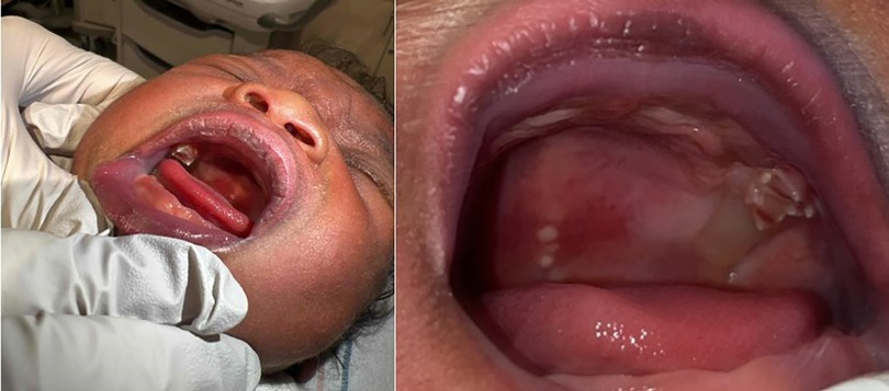
Figure 1. Intraoral examination revealed the white crowns of two erupted primary maxillary first molars. Natal tooth B (Left image) was fully erupted with class III mobility. Natal tooth I (Right image) was partially erupted with class III mobility. Of note, the newborn has Ebstein Pearls, on the midline of the soft palate.
Discussion
The presence of erupted teeth at birth or the eruption of teeth within 30 days after birth is considered a rare occurrence, with an incidence of 0.1% (20). In the case of a 2-day-old female infant delivered via cesarean section at 41 weeks of gestation, with no abnormalities detected at routine pregnancy screenings, the presence of two fully formed maxillary molar teeth were noted at birth.
According to the American Academy of Pediatric Dentistry, the average age for primary maxillary first molars to erupt is between 11 and 18 months (19, 20). In this study, maxillary molars were present from birth. The case of this 2-day-old infant is noteworthy in that the location of the two natal teeth were maxillary primary molars, as the majority of cases of natal teeth described in the literature are incisors (21). The management of incisors and molars varies. Incisors are often removed since there is little risk of removing them. The management of molars, however, varies depending on tooth maturity. Immature molars should be removed, as they are less likely to develop normally. However, mature molars should not be removed. Maturity is typically identified via radiographs or histology. However, the mobility of the teeth can also give a good estimation of the level of maturity of the teeth. The teeth in our study are most likely immature, given the high degree of mobility. Mature teeth, on the other hand, do not typically present with mobility. Lastly, this newborn was born with two molars. This is consistent with the other literature, where the average number of natal molars is one or two (21). One of the highest numbers of natal molars was documented in a study, where the baby was born with eight natal molars, four in the maxillary site and four in the mandibular site (22). In that study of eight natal molars, the patient required resuscitation, which was not an issue for our patient, likely given the number of teeth.
In some studies, natal or neonatal molars have been suggested to be associated with systemic conditions or syndromes (i.e., histiocytosis X); however, there are no current studies that directly link a causative relationship between natal teeth and syndromes (9). There are some disorders associated with natal teeth, but given the rarity, there are no conditions that are highly linked with molars specifically. In addition to systemic conditions and developmental disorders, natal teeth have been associated with the congenital defect known as cleft lip and palate, which is inherited in a multifactorial manner (23, 24). Currently, there is no known etiology of natal teeth. Some theories suggest that the development of natal teeth is a response to febrile states, endocrine disturbances, nutritional deficiencies, or a response to congenital syphilis (23). None of the above scenarios were present in this case, given our extent of knowledge of maternal health during gestation.
Finally, it is important to note that the management of natal teeth must be approached on a case-by-case basis. Erupted natal teeth may represent premature primary dentition. For this reason, they should be preserved with close monitoring by a pediatric dentist and pediatrician to ensure that they do not become an aspiration risk or impair feeding (20). However, in the present case, grade III mobility of the erupted teeth suggested that the natal teeth were immature and that not removing the teeth posed an increased risk for aspiration, especially given the small diameter of the infant airway. Ultimately, there was a decision by the parents of the newborn to remove the teeth. In our case, the teeth were causing issues with latching during breastfeeding, ultimately compromising fetal nutrition. Though this newborn did not present with ulcerations or trauma to the tongue, if the teeth remained in the mouth, there could be trauma to the adjacent soft tissue. Other complications of natal teeth can include pain and lacerations of the mother's breast, and the potential risk for hemorrhage, particularly in the setting of vitamin K deficiency (20).
Hemorrhage often occurs at the time of extraction and is a rarer occasion nowadays given current practices. Now, before teeth extraction, a consultation with a pediatrician is recommended to make sure that the newborn has received vitamin K prophylaxis after birth (20). Previously, a physician would wait until the 10th day for tooth extraction to avoid excessive hemorrhage. However, today, newborns are routinely given 1.0 mg of vitamin K after birth, which removes the need to wait until the 10th day (21). In addition, the removal of a natal tooth may impact the future development of primary and permanent dentition. Monitoring by a pediatric dentist and oral surgeon is important to ensure the normal development of primary and permanent dentition, and to exclude any other dental abnormalities.
Conclusion
Understanding the management of natal teeth is important for the pediatrician. While the cause of natal teeth is not fully understood, the positioning of the teeth can determine the need for removal, especially if the teeth are immature. In a case of a maxillary molar with grade III mobility, known complications such as problems with breastfeeding are common; therefore, the decision was made to remove the teeth.
Data availability statement
The original contributions presented in the study are included in the article/Supplementary Material, further inquiries can be directed to the corresponding author.
Ethics statement
Written informed consent was obtained from the individual(s) for the publication of any potentially identifiable images or data included in this article.
Author contributions
BV: Conceptualization, Writing – original draft, Writing – review & editing. LA: Writing – original draft, Writing – review & editing. SV: Writing – original draft, Writing – review & editing.
Funding
The authors declare that no financial support was received for the research, authorship, and/or publication of this article.
Acknowledgments
We would like to thank Dr. Romano-Clarke for her mentorship and oversight of this project. We would also like to thank the Oral Maxillofacial Surgery team for their assistance in managing this patient. Finally, we would like to express our gratitude toward the patient and their family, without whom this paper would not be possible.
Conflict of interest
The authors declare that the research was conducted in the absence of any commercial or financial relationships that could be construed as a potential conflict of interest.
Publisher's note
All claims expressed in this article are solely those of the authors and do not necessarily represent those of their affiliated organizations, or those of the publisher, the editors and the reviewers. Any product that may be evaluated in this article, or claim that may be made by its manufacturer, is not guaranteed or endorsed by the publisher.
References
1. Wang C-H, Lin Y-T, Lin Y-TJ. A survey of natal and neonatal teeth in newborn infants. J Formos Med Assoc. (2017) 116(3):193–6. doi: 10.1016/j.jfma.2016.03.009
2. Mhaske S, Yuwanati MB, Mhaske A, Ragavendra R, Kamath K, Saawarn S. Natal and neonatal teeth: an overview of the literature. Int Sch Res Notices. (2013) 2013. doi: 10.1155/2013/956269
3. Neville BW, Damm DD, Allen CM, Chi AC. Oral & Maxillofacial Pathology. 4th ed. St. Louis, MO: Elsevier (2016).
4. Bjuggren G. Premature eruption in the primary dentition–a clinical and radiological study. Sven Tandlak Tidskr. (1973) 66(4):343–55.4520015
5. Hals E. Natal and neonatal teeth: histologic investigations in two brothers. Oral Surg Oral Med Oral Pathol. (1957) 10(5):509–21. doi: 10.1016/0030-4220(57)90011-7
6. Bigeard L, Hemmerle J, Sommermater J. Clinical and ultrastructural study of the natal tooth: enamel and dentin assessments. ASDC J Dent Child. (1996) 63(1):23–31.8655747
7. Anderson R. Natal and neonatal teeth: histologic investigation of two black females. ASDC J Dent Child. (1982) 49:300–3.6956592
8. Alaluusua S, Kiviranta H, Leppäniemi A, Hölttä P, Lukinmaa P-L, Lope L, et al. Natal and neonatal teeth in relation to environmental toxicants. Pediatr Res. (2002) 52(5):652–5. doi: 10.1203/00006450-200211000-00008
9. Galassi MS, Santos-Pinto L, Ramalho L. Natal maxillary primary molars: case report. J Clin Pediatr Dent. (2004) 29(1):41–4. doi: 10.17796/jcpd.29.1.12156uq4m6u1t7r7
10. Kadam M, Kadam D, Bhandary S, Hukkeri RY. Natal and neonatal teeth among cleft lip and palate infants. Natl J Maxillofac Surg. (2013) 4(1):73. doi: 10.4103/0975-5950.117883
11. Alvarez M, Crespi P, Shanske A. Natal molars in Pfeiffer syndrome type 3: a case report. J Clin Pediatr Dent. (1993) 18(1):21–4.8110608
12. Basavanthappa NN, Kagathur U, Basavanthappa RN, Suryaprakash ST. Natal and neonatal teeth: a retrospective study of 15 cases. Eur J Dent. (2011) 5(02):168–72. doi: 10.1055/s-0039-1698875
13. DeSeta M, Holden E, Siddik D, Bhujel N. Natal and neonatal teeth: a review and case series. Br Dent J. (2022) 232(7):449–53. doi: 10.1038/s41415-022-4091-3
14. Goho C. Neonatal sublingual traumatic ulceration (Riga-Fede disease): reports of cases. ASDC J Dent Child. (1996) 63(5):362–4.8958351
15. Hegde R. Sublingual traumatic ulceration due to neonatal teeth (Riga-Fede disease). J Indian Soc Pedod Prev Dent. (2005) 23(1):51–2. doi: 10.4103/0970-4388.16031
16. Padmanabhan MY, Pandey RK, Aparna R, Radhakrishnan V. Neonatal sublingual traumatic ulceration—case report & review of the literature. Dent Traumatol. (2010) 26(6):490–5. doi: 10.1111/j.1600-9657.2010.00926.x
17. Adekoya-Sofowora C. Natal and neonatal teeth: a review. Niger Postgrad Med J. (2008) 15(1):38–41. doi: 10.4103/1117-1936.180932
18. Markou I, Kana A, Arhakis A. Natal and neonatal teeth: a review of the literature. Balkan J Stomatol. (2012) 16(3):132–40.
19. Logan WH, Kronfeld R. Development of the human jaws and surrounding structures from birth to the age of fifteen years. J Am Dent Assoc. (1933) 20(3):379–428. doi: 10.14219/jada.archive.1933.0080
20. American Academy of Pediatric Dentistry. “Management considerations for pediatric oral surgery and oral pathology”. The Reference Manual of Pediatric Dentistry. Chicago, IL: American Academy of Pediatric Dentistry (2022). p. 485–94. Available online at: https://www.aapd.org/globalassets/media/policies_guidelines/bp_oralsurgery.pdf (Accessed January 29, 2024).
21. Brandt SK, Shapiro SD, Kittle PE. Immature primary molar in the newborn. Pediatr Dent. (1983) 5(3):210–3.6579501
23. Massler M, Savara BS. Natal and neonatal teeth: a review of twenty-four cases reported in the literature. J Pediatr. (1950) 36(3):349–59. doi: 10.1016/S0022-3476(50)80105-1
Keywords: decision-making, cranio-maxillofacial surgery, prevention, risk factor(s), tooth development, natal teeth
Citation: Varriano B. M., Ades L. and Vaughan S. R. (2024) Case Report: A rare case of bilateral molar natal teeth in a term newborn. Front. Dent. Med 5:1336865. doi: 10.3389/fdmed.2024.1336865
Received: 11 November 2023; Accepted: 28 March 2024;
Published: 25 April 2024.
Edited by:
Martha J. Somerman, Frontiers in Dental Medicine, United StatesReviewed by:
Sivakumar Nuvvula, Narayana Dental College and Hospital, IndiaAshish Shrestha, B.P. Koirala Institute of Health Sciences, Nepal
© 2024 Varriano, Ades and Vaughan. This is an open-access article distributed under the terms of the Creative Commons Attribution License (CC BY). The use, distribution or reproduction in other forums is permitted, provided the original author(s) and the copyright owner(s) are credited and that the original publication in this journal is cited, in accordance with accepted academic practice. No use, distribution or reproduction is permitted which does not comply with these terms.
*Correspondence: B. M. Varriano bvarriano@mgb.org