 Mylenna de Cássia Neves Guimarães1
 Maria Nazaré Oliveira Freitas1Alana Watanabe de Sousa1Marcos Antônio Correia Rodrigues da Cunha2Gilton Luiz Almada2Alessandro Pecego Martins Romano3Maria Guadalupe Dias Pestana Santos4Gilsa Aparecida Pimenta Rodrigues2Lívia Caricio Martins1Jannifer Oliveira Chiang1
 Livia Medeiros Neves Casseb1*
- 1Department of Arbovirology and Hemorrhagic Fevers, Evandro Chagas Institute, Ananindeua, Brazil
- 2Center for Strategic Information and Responses in Health Surveillance, Espírito Santo State Department of Health, Vitória, Brazil
- 3Department of Health Surveillance, Ministry of Health, Brasília, Brazil
- 4Department of Health of the Municipality of Venécia, Health Surveillance, Venécia, Brazil
Many human arboviruses are also pathogenic for horses, and some of these have emerged recently. A descriptive cross-sectional observational study was conducted to assess the prevalence of West Nile virus (WNV) and other arboviruses among 77 horses on the rural properties of the Espirito Santo state, Brazil. Serum samples were screened for arbovirus-reactive antibodies using the hemagglutination inhibition technique and subsequently a plaque reduction neutralization test for the confirmation of exposure from sera was used to detect heterotypic immune reactions. Overall, the total antibodies against at least one arbovirus of Alphavirus, Flavivirus, and Orthobunyavirus genera were detected in 39 (50.6%) animals. The antibodies to Phlebovirus were not detected in any sample. When the 24 WNV hemagglutination inhibition (HI)-positive samples were tested by the plaque-reduction neutralization test 90%, 9 (32.1%) were positive for WNV antibodies and 14 (50%) for Saint Louis encephalitis virus. Our findings indicate that the region provides ideal conditions for the emergence of arboviruses, reinforcing the need for further surveillance of mosquito-transmitted diseases in domestic animals.
Introduction
Over the past four decades, the rate of infectious disease emergence has increased—in both humans and animals (1). Between a quarter and two-thirds of human infectious pathogens are zoonotic, and there is evidence to suggest that increased agricultural intensification is linked to the emergence of zoonotic diseases while it has expanded the geographic range of livestock diseases with major economic repercussions (2, 3).
In the Americas, several endemic and emerging arboviruses cause clinically indistinguishable systemic and neurologic diseases in equids and humans (4). As horses can become infected and seroconvert to the arboviruses of public health importance, they also serve as sentinels for these viruses (5).
The Flaviviridae family contains the largest number of viral species that can cause encephalitis in horses. The most significant are the West Nile virus (WNV) and Japanese encephalitis virus (JEV) (6). The serological evidence of flavivirus activity has been vastly reported in many countries, including South Korea (7), Canada (8), Spain (9), France (10), Argentina (11), and China (12).
The epidemic potential of this viral genus reflects many factors related to the unique characteristics of their insect vectors, the geographical expansion of vectors, changing environmental conditions, extensive global travel, and the consequences of poorly planned urbanization that creates ideal arthropod-breeding habitats (13).
Espírito Santo is a state located on the southeastern coast of Brazil, the second smallest region in the country but the most populous one, inserted in the Atlantic Forest biome (14). Due to a high degree of destruction and fragmentation, the Atlantic Forest is considered one of the most threatened biomes in the world and has been classified as one of the five largest hotspots (15).
On April 25, 2018, the first isolation of WNV in Brazil occurred from a horse with neurological disease from the region of Pedra Grande in the municipality of São Mateus, Espírito Santo (16). Due to the case, an extensive scientific expedition was carried out in the state, including different actors in the transmission chain, such as domestic and wild birds and equines. Here, we conducted a serosurvey to investigate the presence of arboviruses in horses from the State of Espírito Santo.
Materials and Methods
Ethics Statement
The procedures were in accordance with fundamental ethical and scientific requirements contained in the Research Regulatory Guidelines and Norms of the Ethics Committee on the Use of Animals of Evandro Chagas Institute, Ananindeua, Pará, Brazil.
Regardless, no animal ethics approvals were required for animal samples, considering that they were obtained from a continuous public health surveillance of a mandatory reporting disease.
Study Site and Sample Collection
From July 4–13, 2018, serological surveys were conducted in Espírito Santo State, Brazil, and 6 municipalities were visited in areas belonging to 22 different rural properties distributed across different parts of the region (Figure 1).
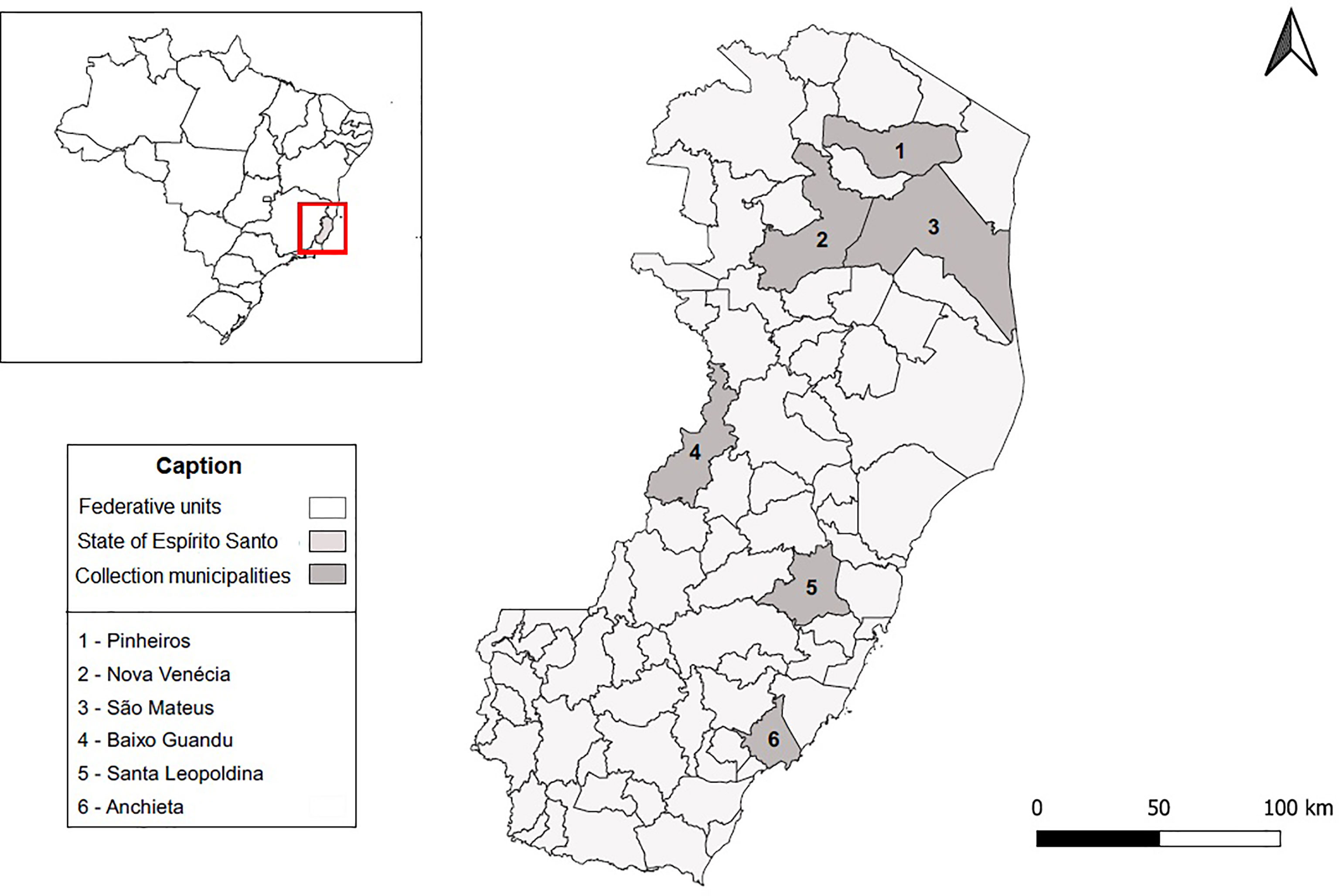
Figure 1 Schematic representation of the Espírito Santo State. Espírito Santo located in southeastern Brazil is shown in light gray. Municipalities where sampling occurred are shown in dark gray.
Blood was collected from 77 horses by jugular venipuncture in a microtube without an anticoagulant. After collection, the clot was retracted at room temperature and subsequently, each sample was subjected to centrifugation at 3,000 rpm for 5–10 min. The separated serum was transferred to another microtube, immediately stored in liquid nitrogen, then at -70°C freezer, and later transported on dry ice.
All of the animals enrolled in the serologic survey were apparently healthy and without a history of traveling outside of the state. The data collected for each animal included the sex, age, vaccination history, and utility (work purposes).
Hemagglutination Inhibition Test
Arbovirus infections were evaluated by screening for total antibodies with the hemagglutination inhibition (HI) test (17) adapted to microplates (18), using sucrose-acetone (19).
The test was performed with a standardized panel of 19 arbovirus antigens, including four genera: Flavivirus: Yellow fever virus (YFV), West Nile virus (WNV), Saint Louis encephalitis virus (SLEV), Cacipacore virus (CPCV), Bussuquara virus (BSGV), Rocio virus (ROCV), Ilheus virus (ILHV); Alphavirus: Eastern equine encephalitis (EEEV), Western equine encephalomyelitis virus (WEEV), Mayaro virus (MAYV), Mucambo virus (MUCV); Orthobunyavirus: Maguari virus (MAGV), Utinga virus (UTIV), Belem virus (BELV), Oropouche virus (OROV), Catu virus (CATUV), Caraparu virus (CARV), Tacaiuma virus (TCMV); and Phlebovirus: Icoaraci virus (ICOV).
Samples were initially treated with 100% acetone, aiming to remove possible non-specific inhibitors of hemagglutinant activity, and subsequently, the test was performed in two steps: screening and titration. Serum samples were placed to react in equal parts of serum and antigen, with the introduction of a revealing system [composed by white goose red cells (Anser cinereus) diluted in dextrose, gelatin, and barbital solution] to test for the formation of an antigen–antibody complex. The positive samples at the screening step were diluted with bovine serum albumin (0.4%) and titrated up to a dilution of 1:40 to 1:1,280.
Positive reactions were classified as either monotypic reactions, referring to the presence of antibodies with titers ≥1:20 for one arbovirus; heterotypic or cross reactions were the samples with the presence of antibodies with titers ≥1:20 for more than one arbovirus of one same viral genus.
Plaque Reduction Neutralization Test (PRNT90)
For samples with cross-reactive results in the HI test to SLEV (ChimeriVax - SLEV) and WNV (BeAn854747 strain), we performed the plaque-reduction neutralization test 90% (PRNT90)test, since it is more specific than HI, and due the purpose of the surveillance action in the study area. It was executed using Vero cells at a concentration of 1.6 × 10⁵/cm2 in 6-well plates, as previously described (20).
Horse sera were inactivated at 56°C for 30 min and diluted to 1:5 in Medium 199 with Earle′s salts. Samples were tested at twofold serial dilutions (1:10 to 1:80), mixed with viruses and allowed to incubate for 1 h at 37°C, at which point the samples were added onto Vero cells, overlaid, and incubated for 5– 7days (21, 22).
Serum samples were considered positive when a serum dilution of 1:10 or greater reduced the viral formation of plaques by at least 90%. In heterotypic patterns, specific virus exposures were assigned if a fourfold or greater titer to one virus than to all other flaviviruses tested for simultaneously was demonstrated.
Statistical Analysis
Results were tabulated by simple percentage and descriptive statistics implemented by GraphPadPrism 6.0 for Windows (GraphPad software, San Diego, CA, USA). They were also analyzed using the BioEstat 5.0 software (23), assuming statistical significance for p ≤ 0.05.
Results
The seroepidemiological survey was carried out in 77 horses, 34 (44.1%) of which were males and 34 (44.1%) females, 9 did not have detailed sex data. Their ages ranged from 5 months to 19 years. Moreover, those animals were used in many different ways, such as for riding, leisure, sports, reproduction, and general work. Most horses were used for work (48.0%) (Table 1).
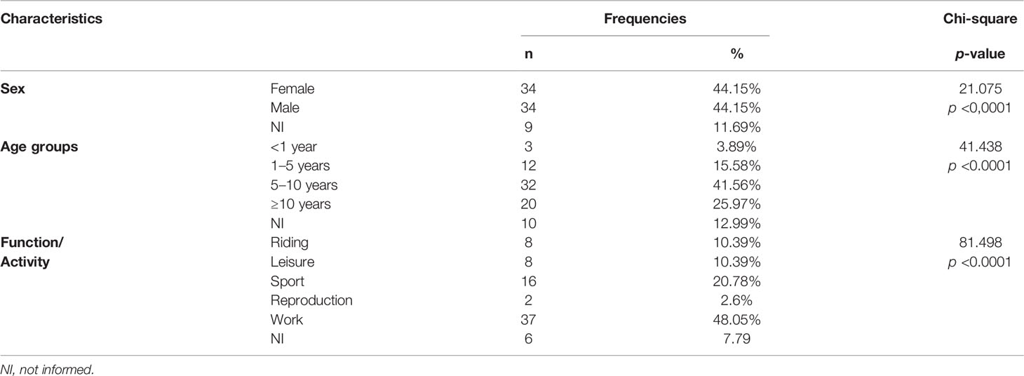
Table 1 Result of statistical analysis of horses attributes tested for arbovirus in Espírito Santo, Brazil.
Of the serum samples analyzed by the HI test, 39/77 (50.6%) had total antibodies to at least one arbovirus tested in the study, considering both monotypic and heterotypic reactions, and 38 (49.3%) were seronegative. Overall, 8 (20.5%) samples had antibodies to the arboviruses of the Alphavirus genera, 28 (71.8%) to Flavivirus, and 21 (53.8%) to the Orthobunyavirus genus.
Monotypic HI reactions occurred in 19/39 (48.7%) of the samples, 5 (12.8%) for EEEV (with titers ranging from 1:40 to 1:160), 8 (20.5%) for MAGV (with titers ranging from 1:20 to 1:80), 3 (7.6%) for TCMV (with titers ranging from 1:20 to 1:640), 3 (7.6%) for SLEV (with titers ranging from 1:20 to 1:80) and 1 (2.5%) for WNV (with a titer of 1:20).
On data collection about equine vaccination, it was reported that 43 (55.8%) animals had been immunized against equine encephalitis. From those, we could observe that 04 (9.3%) and 02 (4.6%) were seropositive for EEEV and WEEV on HI, respectively.
For neutralizing antibody detection, PRNT90 tests were performed for all samples of animals that showed antibodies to WNV (24/39) or SLEV (27/39) in the HI, and 28 samples were selected (Table 2).

Table 2 Results of PRNT90 for WNV and SLEV in horses of Espírito Santo, Brazil (n = 28).
Regarding the reactivity by HI and the sex of the individuals, when informed, 19 (48.7%) were females and 16 (41%) were males. Considering the detection of total antibodies and the age group, it was possible to observe that 1 (2.5%) animal with <1 year was reactive, while in the age group of 1–5 years, there were 4 (10.2%), in the 5–10 years, there were 17 (43.5%), and in those aged ≥10 years, there were 10 (25.6%), which suggests greater reactivity to arbovirus in animals from 5 years of age.
When comparing the use of animals in samples reactive by HI, a higher percentage of reactivity was observed in those used for work practice (46.1%). However, this utility also represents the majority of those investigated.
Discussion
Serological monitoring has been carried out in recent years in domestic animals to assess whether these species can act as sentinels for specific arboviruses (8). Horses are large domestic animals that live outside the protection of homes and are therefore common victims of mosquito bites. Consequently, they are a suitable source for studies on the viruses transmitted by these arthropods (24).
In horses, the age pattern for morbidity reflects a high risk of disease in the very young and middle-aged, that is, during the ages of use or more intense activity. Indeed, the prevalence of diseases is high in the population of animals aged ≥15 years, with the majority presenting multiple anomalies with increasing age (25). Herein, we could observe that the seropositivity in the HI was higher in animals from 5 years of age and in spite of those used for the work practice. Furthermore, an increase of flavivirus seroprevalence with age in horses has been justified by low-level enzootic transmission, where the risk of infection increases over time (26).
The HI test has often been used in serological research because it can detect antibodies for a long time after natural infection. It is considered a test of high sensitivity and low specificity when compared to other serological tests (27). The heterotypic reactions found can be explained by the greater sensitivity than specificity present in the HI test (28).
The Flavivirus genus was responsible for the majority of seropositivity in the investigated equine population. Similar results of seropositivity were previously observed in Brazil (24, 29). In addition to the presence of the hemagglutination inhibitors of the HI test, monotypic reactions were observed for WNV and SLEV, which suggests that these viruses circulate among this population, corroborating with what was observed in the northern (30), northeast (31), and central-west regions (32). These results provide an evidence of the circulation of these closely related flaviviruses in different regions of the country.
With the recent increase in suspected cases of West Nile fever and the first isolation of WNV from an infected horse, there were concerns with the possibility that this virus would establish itself (33). However, it was determined that WNV has been circulating silently in Brazil for many years, probably between 2001 and 2005 (34).
In April 2018, epizootics in horses with meningoencephalitis were notified to the Ministry of Health by the Espírito Santo State Health Department–which culminated in the first isolation of WNV in the country, from an equine with neurological disease (16). Between March 2018 and June 2019, there was a confirmation of the occurrence of infection in equines in the municipalities of São Mateus, Nova Venécia, and Baixo Guandu (35). This report, together with our description of PRNT90 titers against WNV in the same region, suggests that measures should be taken to monitor WNV activity in Brazil, especially with a permanent surveillance program of domestic birds and horses.
Furthermore, the possibility of the circulation of other flaviviruses cannot be excluded since it was observed cross-reactions for this genus. In addition, the HI test is characterized by a high cross-reactivity that generally allows only a qualitative conclusion about the presence of antibodies against flaviviruses (36).
It was possible to observe that the percentage of animals investigated with antibodies to the genus Alphavirus showed differences when comparing previous studies with 66.6% (37) and 14.6% (38).The divergence between the percentages observed between these studies may have occurred due to the different inclusion criteria adopted in the sampling, such as the non-vaccination of those investigated against arboviruses or, finally, access to the forest.
As for the Orthobunyavirus genus, monotypic reactions were observed for MAGV and TCMV. Previously neutralizing antibodies to MAGV (28.2%) and TCMV (15.7%) were detected in horses under 2 years of age in the Pantanal, indicating recent circulation (39). The serological evidence for both viruses was also found in the state of Pará in buffaloes, with a higher prevalence for MAGV with 7.33%, while the antibodies to TCMV in these animals corresponded to 1.37% of the total reactions in the HI (40). Most of the monotypic reactions detected in the study were for MAGV and TCMV.
There was no positivity for the Phlebovirus genus, represented by ICOV, nor for MAYV, CPCV, BSGV, BELV, or CARV. Therefore, we assume that these viruses did not circulate in the population tested. However, among the viruses that were associated with seronegativity, some have already had serological evidence in Brazilian equids, such as MAYV (30, 41–43) and ICOV (44), and CPCV (26, 29).
Study limitations included the small number of animals per species; thus, a more extensive evaluation of the animals that developed antibody titers is needed. Moreover, we included the viruses that are part of a routine serological test and PRNT90 was performed only for WNV and SLEV, considering the epidemiological importance of the viruses in the period evaluated in the study region.
For a PRNT titer to be considered specific for a given flavivirus, a fourfold or greater titer to that virus than to all other flaviviruses tested for simultaneously must be demonstrated (45). However, the interpretation of heterotypic patterns is complex and this conservative criterion could be limited to provide the distinction of the most recent infection (46).
One of the fundamental aspects in controlling the transmission of arboviruses is the early detection of the virus or the identification of the increase in its activity. For WNV, monitoring is carried out mainly in bird and equine epizootics (47). Moreover, many horses live in the region; according to federal data, the effective herd of horses in the state is of 47,503 (48). Additional surveillance methods and collaboration among veterinary and human health services are essential in providing early warning and adequate protection of human and animal health against arboviruses.
Mammals can be better sentinels than birds when the objective is the early detection of diseases that threaten humans (49). As an example, the Venezuelan equine encephalitis virus has an epizootic cycle during which the amplification of the virus in the horse is sufficient to result in mosquito infection and is believed to significantly increase the risk of human infection (50). Contrarily, for WNV, horses and humans are considered to be dead-end hosts, so the virus is not directly contagious from horse to horse or horse to human (51). Therefore, the results from laboratory-based experiments should not be viewed in isolation (52).
Because horses are more prone to WNV infection than humans, the signs of illness are often observed and reported quickly, demonstrating the importance of the horse as a sentinel of epizootic WNV activity (53).
Accordingly, long-term studies are needed to increase the understanding of the role of horses and other vertebrate hosts in arbovirus circulation.
Data Availability Statement
The original contributions presented in the study are included in the article/supplementary material. Further inquiries can be directed to the corresponding author.
Ethics Statement
No animal ethics approvals were required for stored animal samples, considering that they were obtained from a continuous public health surveillance of a mandatory reporting disease. Written informed consent was obtained from the owners for the participation of their animals in this study.
Author Contributions
LC contributed in sample collection and carried out statistical analysis. MF and AS performed serological tests. AR, MC, GA, MS, and GR conducted the investigation process. MG wrote the article and did data curation. LM and JC took part in critically reviewing the study. All authors contributed to the article and approved the submitted version.
Funding
Sources of funding included Instituto Evandro Chagas (IEC), linked to the Secretaria de Vigilância em Saúde (SVS) of the Ministério da Saúde (MS); Coordenação Geral de Vigilância de Arboviroses (CGARB) and Secretaria de Estado da Saúde do Espírito Santo (SES/ES).
Conflict of Interest
The authors declare that the research was conducted in the absence of any commercial or financial relationships that could be construed as a potential conflict of interest.
Publisher’s Note
All claims expressed in this article are solely those of the authors and do not necessarily represent those of their affiliated organizations, or those of the publisher, the editors and the reviewers. Any product that may be evaluated in this article, or claim that may be made by its manufacturer, is not guaranteed or endorsed by the publisher.
Acknowledgments
The authors would like to thank the Conselho Nacional de Desenvolvimento Científico e Tecnológico (CNPq) for financial support and to Evandro Chagas Institute for structural support to accomplish serological tests.
References
1. Atlas R, Rubin C, Maloy S, Daszak P, Colwell R, Hyde B. One Health-Attaining Optimal Health for People, Animals, and the Environment. Microbe (2010) 5:383–9. doi: 10.1128/MICROBE.5.383.1
2. Rushton J, Bruce M. Using a One Health Approach to Assess the Impact of Parasitic Disease in Livestock: How Does it Add Value? Parasitology (2017) 144:15–25. doi: 10.1017/S0031182016000196
3. Guthid S, Hanley KA, Althouse BM, Boots M. Ecological Processes Underlying the Emergence of Novel Enzootic Cycles: Arboviruses in the Neotropics as a Case Study. PloS Negl Trop Dis (2020) 14:e0008338. doi: 10.1371/JOURNAL.PNTD.0008338
4. León B, Käsbohrer A, Hutter SE, Baldi M, Firth CL, Romero-Zúñiga JJ, et al. National Seroprevalence and Risk Factors for Eastern Equine Encephalitis and Venezuelan Equine Encephalitis in Costa Rica. J Equine Vet Sci (2020) 92:103140. doi: 10.1016/J.JEVS.2020.103140
5. Bayeux JJM, Silva ASG, de Queiroz GA, da S Santos BSÁ, Rocha MN, Rehfeld IS, et al. Epidemiological Surveillance of West Nile Virus in the World and Brazil. Braz J Vet Res Anim Sci (2019) 56:e164335–e164335. doi: 10.11606/ISSN.1678-4456.BJVRAS.2019.164335
6. Barba M, Fairbanks EL, Daly J. Equine Viral Encephalitis: Prevalence, Impact, and Management Strategies. Vet Med (Auckland NZ) (2019) 10:99–110. doi: 10.2147/VMRR.S168227
7. Jeoung HY, Yang SJ, Choi Y k, Lee JH, Seo HJ, Kim SH, et al. Surveillance of Encephalitis-Causing Arboviruses in Horses in South Korea. J Equine Vet Sci (2016) 37:11–6. doi: 10.1016/J.JEVS.2015.11.004
8. Rocheleau JP, Michel P, Lindsay LR, Drebot M, Dibernardo A, Ogden NH, et al. Emerging Arboviruses in Quebec, Canada: Assessing Public Health Risk by Serology in Humans, Horses and Pet Dogs. Epidemiol Infect (2017) 145:2940–8. doi: 10.1017/S0950268817002205
9. Vanhomwegen J, Beck C, Desprès P, Figuerola A, García R, Lecollinet S, et al. Circulation of Zoonotic Arboviruses in Equine Populations of Mallorca Island (Spain). Vector-Borne Zoonotic Dis (2017) 17:340–6. doi: 10.1089/vbz.2016.2042
10. Beck C, Leparc-Goffart I, Desoutter D, Debergé E, Bichet H, Lowenski S, et al. Serological Evidence of Infection With Dengue and Zika Viruses in Horses on French Pacific Islands. PloS Negl Trop Dis (2019) 13:e20190089. doi: 10.1371/JOURNAL.PNTD.0007162
11. Tauro L, Marino B, Diaz LA, Lucca E, Gallozo D, Spinsanti L, et al. Serological Detection of St. Louis Encephalitis Virus and West Nile Virus in Equines From Santa Fe, Argentina. Mem Inst Oswaldo Cruz (2012) 107:553–6. doi: 10.1590/S0074-02762012000400019
12. Lan DL, Wang CS, Deng B, Zhou JP, Cui L, Tang C, et al. Serological Investigations on West Nile Virus in Birds and Horses in Shanghai, China. Epidemiol Infect (2013) 141:596–600. doi: 10.1017/S0950268812001094
13. Pierson TC, Diamond MS. The Continued Threat of Emerging Flaviviruses. Nat Microbiol (2020) 5:796–812. doi: 10.1038/S41564-020-0714-0
14. Instituto Brasileiro de Geografia e Estatística. IBGE. Cidades@ | Espírito Santo | Panorama. Available at: https://cidades.ibge.gov.br/brasil/es/panorama (Accessed February 20, 2022).
15. Moreira DDO, Coutinho BR, Mendes SL. O Status do Conhecimento Sobre a Fauna De Mamíferos do Espírito Santo Baseado Em Registros De Museus E Literatura Científica. Biota Neotrop (2008) 8:163–73. doi: 10.1590/S1676-06032008000200017
16. Martins LC, da Silva EVP, Casseb LMN, da Silva SP, Cruz ACR, de Sousa Pantoja JA, et al. First Isolation of West Nile Virus in Brazil. Mem Inst Oswaldo Cruz (2019) 114:e180332. doi: 10.1590/0074-02760180332
17. Clarke DH, Casals J. Techniques for Hemagglutination and Hemagglutination-Inhibition With Arthropod-Borne Viruses. Am J Trop Med Hyg (1958) 7:561–73. doi: 10.4269/AJTMH.1958.7.561
18. Shope RE. The Use of a Micro Hemagglutinationinhibition Test to Follow Antibody Response After Arthropod-Borne Virus Infection in a Community of Forest Animals. Microbiol (1963) 11:167–9.
19. Lennette DA. General Principles of Laboratory Diagnostic Methods for Viral, Rickettsial and Chlamydial Infections. In: Lennette EH, Schmidt NJ, editors. Diagnostic Procedures for Viral, Rickettsial and Chlamydial Infections. Washington: American Public Health Association (1995). p. 3–25.
20. Earley E, Peralta PH, Johnson KM. A Plaque Neutralization Method for Arboviruses. Exp Biol Med (1967) 125:741–7. doi: 10.3181/00379727-125-32194
21. OIE - World Organisation for Animal Health. Manual of Diagnostic Tests and Vaccines for Terrestrial Animals. Available at: https://www.oie.int/en/what-we-do/standards/codes-and-manuals/terrestrial-manual-online-access/ (Accessed March 21, 2022).
22. Stefano I, Sato HK, Pannuti CS, Omoto TM, Mann G, Freire MS, et al. Recent Immunization Against Measles Does Not Interfere With the Sero-Response to Yellow Fever Vaccine. Vaccine (1999) 17:1042–6. doi: 10.1016/S0264-410X(98)00320-X
23. Ayres MJ. BioEstat 2.0: Aplicações Estatísticas Nas Áreas Das Ciências Biológicas E Biomédicas. In: Ayres M, editor. BioEstat 2.0: Aplicações Estatísticas Nas Áreas Das Ciências Ebiológicas E Médicas. Belém: Sociedade Civil Mamirauá (2000). p. 12–259.
24. Silva JR, Romeiro MF, de Souza WM, Munhoz TD, Borges GP, Soares OAB, et al. A Saint Louis Encephalitis and Rocio Virus Serosurvey in Brazilian Horses. Rev Soc Bras Med Trop (2014) 47:414–7. doi: 10.1590/0037-8682-0117-2014
25. Ireland JL, Clegg PD, McGowan CM, McKane SA, Chandler KJ, Pinchbeck GL. Disease Prevalence in Geriatric Horses in the United Kingdom: Veterinary Clinical Assessment of 200 Cases. Equine Vet J (2012) 44:101–6. doi: 10.1111/j.2042-3306.2010.00361.x
26. Pauvolid-Corrêa A, Campos Z, Juliano R, Velez J, Nogueira RMR, Komar N. Serological Evidence of Widespread Circulation of West Nile Virus and Other Flaviviruses in Equines of the Pantanal, Brazil. PloS Negl Trop Dis (2014) 8:e2706. doi: 10.1371/journal.pntd.0002706
27. Batista PM, Andreotti R, de Almeida PS, Marques AC, Rodrigues SG, Chiang JO, et al. Detection of Arboviruses of Public Health Interest in Free-Living New World Primates (Sapajus Spp.; Alouatta Caraya) Captured in Mato Grosso do Sul, Brazil. Rev Soc Bras Med Trop (2013) 46:684–90. doi: 10.1590/0037-8682-0181-2013
28. Catenacci LS, Ferreira M, Martins LC, de Vleeschouwer KM, Cassano CR, Oliveira LC, et al. Surveillance of Arboviruses in Primates and Sloths in the Atlantic Forest, Bahia, Brazil. EcoHealth (2018) 15:777–91. doi: 10.1007/s10393-018-1361-2
29. Pauvolid-Corrêa A, Morales MA, Levis S, Figueiredo LTM, Couto-Lima D, Campos Z, et al. Neutralising Antibodies for West Nile Virus in Horses From Brazilian Pantanal. Mem Inst Oswaldo Cruz (2011) 106:467–74. doi: 10.1590/S0074-02762011000400014
30. Gomes FA, Jansen AM, Machado RZ, Jesus Pena HF, Fumagalli MJ, Silva A, et al. Serological Evidence of Arboviruses and Coccidia Infecting Horses in the Amazonian Region of Brazil. PloS One (2019) 14:e0225895. doi: 10.1371/journal.pone.0225895
31. Löwen Levy Chalhoub F, Maia de Queiroz-Júnior E, Holanda Duarte B, Eielson Pinheiro de Sá M, Cerqueira Lima P, Carneiro de Oliveira A, et al. West Nile Virus in the State of Ceará, Northeast Brazil. Microorganisms (2021) 9:1699. doi: 10.3390/microorganisms9081699
32. Melandri V, Guimarães AÉ, Komar N, Nogueira ML, Mondini A, Fernandez-Sesma A, et al. Serological Detection of West Nile Virus in Horses and Chicken From Pantanal, Brazil. Mem Inst Oswaldo Cruz (2012) 107:1073–5. doi: 10.1590/S0074-02762012000800020
33. de Castro-Jorge LA, Siconelli MJL, dos Ribeiro BS, de Moraes FM, de Moraes JB, Agostinho MR, et al. West Nile Virus Infections Are Here! Are We Prepared to Face Another Flavivirus Epidemic? Rev Soc Bras Med Trop (2019) 52:e20190089. doi: 10.1590/0037-8682-0089-2018
34. Siconelli MJL, de Jorge DMM, de Castro-Jorge LA, Fonseca-Júnior AA, Nascimento ML, Floriano VG, et al. Evidence for Current Circulation of an Ancient West Nile Virus Strain (NY99) in Brazil. Rev Soc Bras Med Trop (2021) 54:e0687–2020. doi: 10.1590/0037-8682-0687-2020
35. BRASIL, Ministério da Saúde, Secretaria de Vigilância em Saúde, Departamento de Imunização e Doenças Transmissíveis, Coordenação Geral de Vigilância de Arboviroses. Monitoramento Da Febre do Nilo Ocidental No Brasil, 2014 a 2019. Informe No 1, Julho, 2019 (2019). Available at: https://antigo.saude.gov.br/images/pdf/2019/julho/08/Nota-Informativa-Febre-do-Nilo.pdf (Accessed February 21, 2022).
36. Auerswald H, Ruget A-S, Ladreyt H, In S, Mao S, Sorn S, et al. Serological Evidence for Japanese Encephalitis and West Nile Virus Infections in Domestic Birds in Cambodia. Front Vet Sci (2020) 7:15. doi: 10.3389/fvets.2020.00015
37. Casseb A do R, Chiang JO, Martins LC, da Silva SP, Henriques DF, Casseb LMN, et al. Alphavirus Serosurvey in Domestic Herbivores in Pará State, Brazilian Amazon. Rev Pan-Amazônica Saúde (2012) 3:43–8. doi: 10.5123/S2176-62232012000400005
38. do S da SS Nunes J, Casseb LMN, de PS Guimarães RJ, Reis WDM, de CV de Barros B, Ferreira MS, et al. Serological Evidence of Eastern Equine Encephalitis Circulation in Equids in Pará State, Brazil. Braz J Vet Med (2021) 43:e001720. doi: 10.29374/2527-2179.bjvm001720
39. Iversson LB, Silva RAMS, da Rosa APAT, Barros VLRS. Circulation of Eastern Equine Encephalitis, Western Equine Encephalitis, Ilhéus, Maguari and Tacaiuma Viruses in Equines of the Brazilian Pantanal, South America. Rev Inst Med Trop Sao Paulo (1993) 35:355–9. doi: 10.1590/S0036-46651993000400009
40. Casseb AR, Silva SP, Casseb LMN, Chiang JO, Martins LC, Vasconcelos PFC. PREVALÊNCIA DE ANTICORPOS CONTRA ARBOVÍRUS DA FAMÍLIA Bunyaviridae EM BÚFALOS DE ÁGUA. Ciec Anim Bras (2015) 16:428–36. doi: 10.1590/1089-6891v16i327208
41. Araújo FAA, Andrade MA, Jayme VS, Santos AL, Romano APM, Ramos DG, et al. Anticorpos Antialfavírus Detectados Em Equinos Durante Diferentes Epizootias De Encefalite Equina, Paraíba, 2009. Rev Bras Ciec Veterinária (2012) 19:80–5. doi: 10.4322/rbcv.2014.086
42. do R Casseb A, Brito TC, da Silva MRM, Chiang JO, Martins LC, da Silva SP, et al. Prevalence of Antibodies to Equine Alphaviruses in the State of Pará, Brazil. Arq Inst Biol (Sao Paulo) (2016) 83:e0202014. doi: 10.1590/1808-1657000202014
43. Pauvolid-Corrêa A, Juliano RS, Campos Z, Velez J, Nogueira RMR, Komar N. Neutralising Antibodies for Mayaro Virus in Pantanal, Brazil. Mem Inst Oswaldo Cruz (2015) 110:125–33. doi: 10.1590/0074-02760140383
44. do Casseb A, Nunes MR, Rodrigues S, Travassos da Rosa E, Casseb LM, Casseb SM, et al. Diagnosis of Arboviruses Using Indirect Sandwich IgG ELISA in Horses From the Brazilian Amazon. J Venom Anim Toxins Incl Trop Dis (2014) 20:29. doi: 10.1186/1678-9199-20-29
45. Johnson BW, Kosoy O, Martin DA, Noga AJ, Russell BJ, Johnson AA, et al. West Nile Virus Infection and Serologic Response Among Persons Previously Vaccinated Against Yellow Fever and Japanese Encephalitis Viruses. Vector-Borne Zoonotic Dis (2005) 5:137–45. doi: 10.1089/vbz.2005.5.137
46. Morales MA, Fabbri CM, Zunino GE, Kowalewski MM, Luppo VC, Enría DA, et al. Detection of the Mosquito-Borne Flaviviruses, West Nile, Dengue, Saint Louis Encephalitis, Ilheus, Bussuquara, and Yellow Fever in Free-Ranging Black Howlers (Alouatta Caraya) of Northeastern Argentina. PloS Negl Trop Dis (2017) 11:e0005351. doi: 10.1371/journal.pntd.0005351
47. Silva ASG, Rehfeld IS, da S Santos BSÁ, de S Franklin LF, Teixeira RBC, Lobato ZIP, et al. Febre do Nilo Ocidental No Brasil: O Novo Desafio Aos Médicos-Veterinários. Rev Educ Contin em Med Veterinária e Zootec do CRMV-SP (2021) 19:e38082. doi: 10.36440/recmvz.v19i1.38082
48. Instituto Brasileiro de Geografia e Estatística. IBGE. Censo Agro 2017 | Espírito Santo | Equinos. Available at: https://censos.ibge.gov.br/agro/2017/templates/censo_agro/resultadosagro/pecuaria.html?localidade=32&tema=75665 (Accessed February 21, 2022).
49. Dórea FC, Elbers ARW, Hendrikx P, Enoe C, Kirkeby C, Hoinville L, et al. Vector-Borne Disease Surveillance in Livestock Populations: A Critical Review of Literature Recommendations and Implemented Surveillance (BTV-8) in Five European Countries. Prev Vet Med (2016) 125:1–9. doi: 10.1016/j.prevetmed.2016.01.005
50. Chapman GE, Sherlock K, Hesson JC, Blagrove MSC, Lycett GJ, Archer D, et al. Laboratory Transmission Potential of British Mosquitoes for Equine Arboviruses. Parasit Vectors (2020) 13:413. doi: 10.1186/s13071-020-04285-x
51. Benjelloun A, el Harrak M, Belkadi B. West Nile Disease Epidemiology in North-West Africa: Bibliographical Review. Transbound Emerg Dis (2016) 63:e153–9. doi: 10.1111/tbed.12341
52. van den Hurk AF, Hall-Mendelin S, Webb CE, Tan CSE, Frentiu FD, Prow NA, et al. Role of Enhanced Vector Transmission of a New West Nile Virus Strain in an Outbreak of Equine Disease in Australia in 2011. Parasit Vectors (2014) 7:586. doi: 10.1186/s13071-014-0586-3
Keywords: equids, serology, flavivirus, arboviral disease, zoonoses
Citation: Guimarães MCN, Freitas MNO, Sousa AW, Cunha MACR, Almada GL, Romano APM, Santos MGDP, Rodrigues GAP, Martins LC, Chiang JO and Casseb LMN (2022) Serological Evidence of Arboviruses in Horses During West Nile Fever Monitoring Surveillance in Southeastern Brazil. Front. Trop. Dis 3:881710. doi: 10.3389/fitd.2022.881710
Received: 23 February 2022; Accepted: 04 April 2022;
Published: 17 May 2022.
Edited by:
Flavia Barreto Dos Santos, Oswaldo Cruz Foundation (Fiocruz), BrazilReviewed by:
María Alejandra Morales, Instituto Nacional de Enfermedades Virales “Dr. Julio I. Maiztegui" (INEVH)- ANLIS, ArgentinaErik Hofmeister, Erik Hofmeister, United States
Copyright © 2022 Guimarães, Freitas, Sousa, Cunha, Almada, Romano, Santos, Rodrigues, Martins, Chiang and Casseb. This is an open-access article distributed under the terms of the Creative Commons Attribution License (CC BY). The use, distribution or reproduction in other forums is permitted, provided the original author(s) and the copyright owner(s) are credited and that the original publication in this journal is cited, in accordance with accepted academic practice. No use, distribution or reproduction is permitted which does not comply with these terms.
*Correspondence: Livia Medeiros Neves Casseb, liviacasseb@iec.gov.br