Analysis of the main indicators and risk factors of ultrasonic shear wave elastography for the diagnosis of osteoarthritis among adults
 Jiong Zhang1
  Xiaozuo Zheng
 Ying Zhao
- 1Department of Ultrasound, The Third Hospital of Hebei Medical University, Shijiazhuang, China
- 2Department of Osteoarthritis, The Third Hospital of Hebei Medical University, Shijiazhuang, China
Objective: This study was conducted to explore the main indicators of ultrasonic shear wave elastography (SWE) for the diagnosis of osteoarthritis (OA) and its influencing factors.
Methods: We collected 910 patients between January 2018 and November 2023 from the department of ultrasound, Third Hospital of Hebei Medical University. Logistic regression was used to analyze the effects of age, gender, body mass index (BMI), smoking, alcohol, hypertension and diabetes on the diagnosis of OA by SWE.
Results: The results showed that medial meniscal projection distance (MMPD) and OA had a positively correlated dose–response relationship (OR = 2.12, 95%CI (1.53, 3.95), trend p < 0.05). Also, medial meniscus elastometry (MME) had a positive dose–response correlation with OA (OR = 8.98, 95%CI (3.89, 11.52), trend p < 0.05). In addition, regarding the analysis of factors influencing the diagnosis of OA, the risk of OA was significantly higher in the older age group [OR = 1.11, 95%CI (1.01, 1.25)], and the risk of diagnosis in OA was high in the high BMI group [OR = 1.8, 95%CI (1.23, 3.01)].
Conclusion: In diagnosing OA, MMPD and MME can be used as reliable indicators, while people of advanced age and high BMI have a high possibility diagnosed with OA.
Introduction
Osteoarthritis (OA) is a chronic joint disease that is highly prevalent worldwide and a whole joint disease involving all joint tissues such as menisci, subchondral bone, infrapatellar fat pad and synovial membrane (1, 2). The 2015 World Health Organization Global Report on Aging and Health highlights OA as the leading cause of disability in adults aged more than 60 (3–5). Globally, the proportion of joint-related disability due to OA increased by 114.5% from 1990 to 2019. Given the aggravating trend of global population aging and the continued increase in the prevalence of OA, global healthcare expenditure is also expected to be huge in the future (6, 7).
The most common positions affected by OA are the hip, knee and hand. The knee is the most frequently affected joint, and knee OA is associated with excessive joint loading and injury (8, 9). X-ray radiography is frequently used for the diagnosis of OA, but x-ray is an indirect and insensitive way of detecting osteoarticular cartilage (10). Arthroscopy, although a reliable and sensitive method, is an invasive technique (11). Magnetic resonance imaging (MRI) is a non-invasive method that provides multiplanar imaging, and excellent soft tissue contrast, however, it is expensive and time consuming (12). Ultrasound (US) is a reliable tool for assessing the integrity and thickness of articular cartilage. Its advantages include being inexpensive, non-invasive, rapid and easily accessible (13).
Ultrasonic shear wave elastography (SWE) is a recently developed technique that is based on the measurement of shear wave velocity generated by ultrasound pulses (13). The technique can be used to assess tissue elasticity. SWE was first applied in breast lesions and thyroid gland lesions, and was subsequently used to assess liver fibrosis (14). More recently, SWE has also been used to assess the mechanical properties of musculoskeletal tissues (12). The SWE technique identifies differences in stiffness between tissues, which is a good indicator of damage to articular cartilage.
Although elastic ultrasound has been previously reported to be useful in the diagnosis of OA (11, 13), there is limited research on the effective indicators of OA diagnosis in SWE and their risk factors, so we conducted this study.
Methods
Patients selection
The data in this study were obtained from the Department of Ultrasound, Third Affiliated Hospital of Hebei Medical University. We obtained raw data from January 2018 to November 2023 for 910 patients, including 210 patients with OA and 700 samples without OA. The data of this study were approved by the Ethics Committee from the Third Affiliated Hospital of Hebei Medical University (No. 1354064). All patients/subjects signed the informed consent and that the study was performed in accordance with the ethical standards of the 1964 Declaration of Helsinki as revised in 2013. The patient inclusion and exclusion flow chart was shown in Supplementary Figure S1. The main inclusion criteria were as follow: (a) Adults patients ≥20 years old receiving ultrasonic shear wave elastography; (b) Patients had eligible health and diagnosis criteria; (c) Patients did not receive any surgery; (d) Patients agree to participate in the study. The main exclusion criteria were as follow: (a) Patients with meniscal injuries (based on MRI evaluation); (b) Patients lack information of diagnosis or other covariates; (c) Patients do not want to join in the study.
Definition of OA
The typical diagnosis of OA include (1): (A) Joint pain: It is mostly a slight dull pain at rest, while a little bit of activity can reduce the pain. (B) Joint swelling: Most of the patients may have joint swelling, bleeding or fluid. (C) Joint stiffness: Joints become inflexible and stiff when they are at rest, and stiffness occurs when they wake up or sit upright. (D) Restriction of activities: With the development of the disease, patients gradually develop muscle atrophy and joint deformity around the joints, resulting in dysfunction. (E) Bone friction sensation: When the articular cartilage is destroyed and the joint surface is incomplete, the joint activities will have the feeling of joint friction or creaking.
Ultrasonic shear wave elastography
SWE were performed using an Aixplorer ultrasound machine equipped with a 12 MHz shallow linear transducer (SuperSonic Imagine, France). The first set of two ultrasound images was obtained in the flat lying position when the subject model was in the flat lying position with the ultrasound probe located in the position (Figure 1A), and then another set of two ultrasound images was obtained in the bipedal natural standing position. When the ultrasound probe was located in the position from anterior angle (Figure 1B). The medial meniscus of the knee was shown in the lying and standing positions, at which point the images were frozen and the line connecting the medial femoral cortex to the tibial cortex was immediately drawn using the electronic caliper function on the ultrasound machine. The distance of the MME over the tibiofemoral line was measured using the electronic caliper function on the ultrasound machine, while the SWE function on the ultrasound machine was applied to measure the elasticity values associated with the medial meniscus.
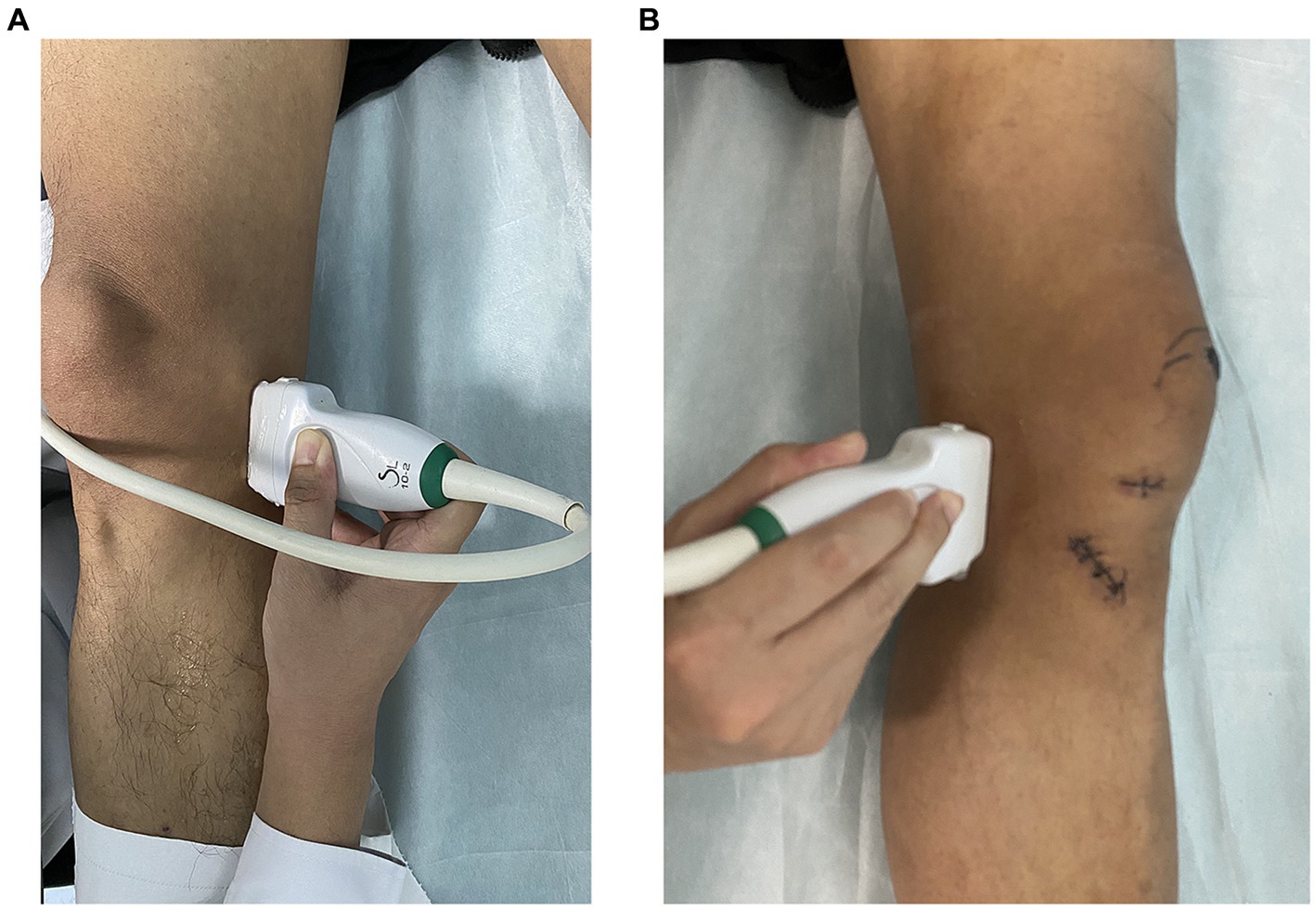
Figure 1. Ultrasonic shear wave elastography operation. (A) Ultrasound measurement of meniscus in a supine position with knee joint in an unloaded state. (B) Measurement of meniscus position in supine position using ultrasound in patients after knee joint surgery.
During the scanning process, the angle of the probe was adjusted so that the incident sound wave was perpendicular to the surface of the articular cartilage as much as possible. Finally, we collected the medial meniscal projection distance (MMPD) and the medial meniscus elasticity (MME). The operation of SWE is shown in Figure 1. In addition, the images for MMPD and MME in OA diagnosis and a healthy control is shown in Figure 2.
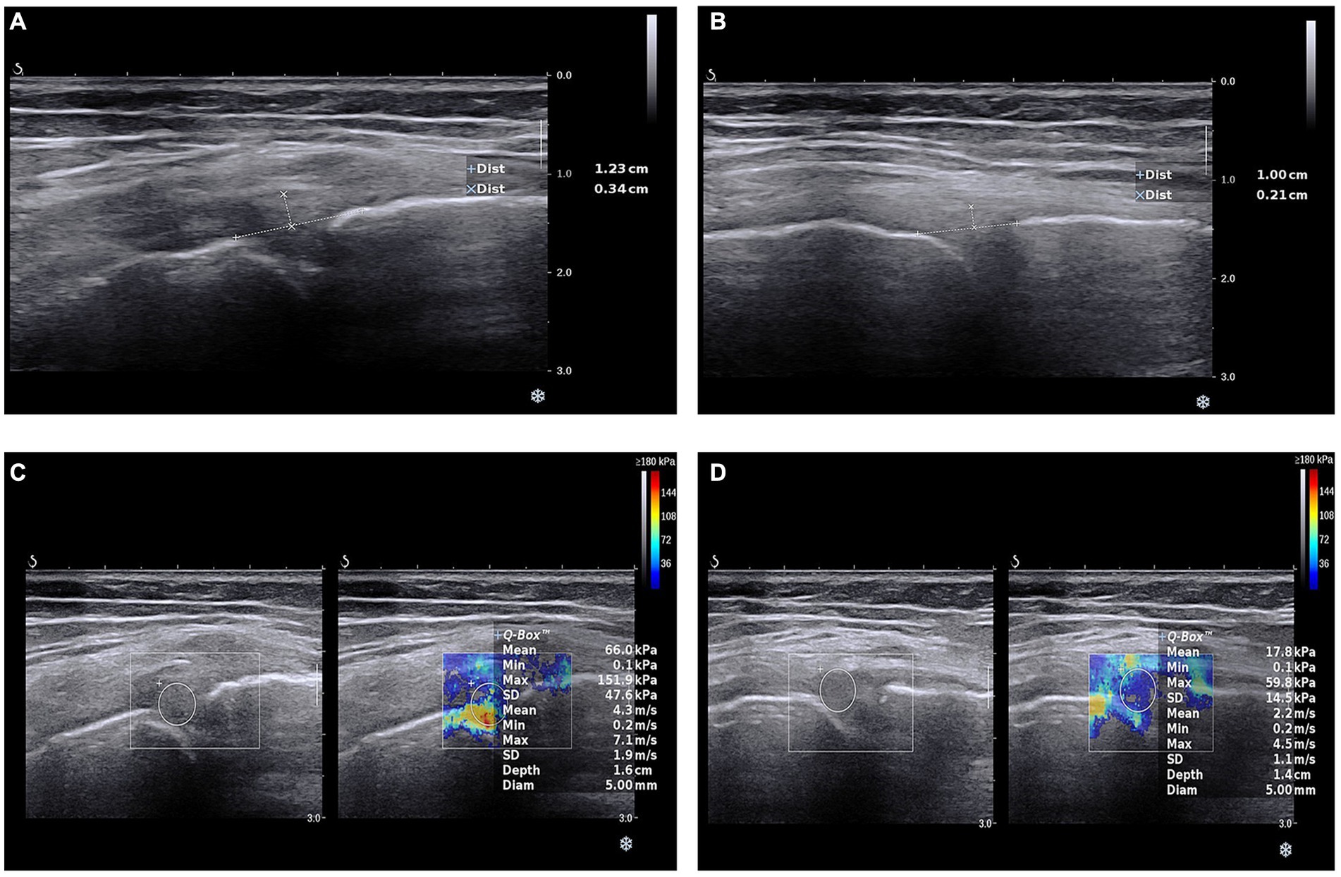
Figure 2. Ultrasonic shear wave elastography images. (A) Medial meniscal projection distance in osteoarthritis patients. (B) Medial meniscal projection distance in health people. (C) Medial meniscus elasticity in osteoarthritis patients. (D) Medial meniscus elasticity in health people.
Covariates
To resilient the SWE main indicator for OA, we also retrospectively collected data on participants’ age, gender, BMI, smoking, alcohol consumption, hypertension, and diabetes from the hospital’s electronic medical record system.
Logistic regression
To analyze whether there was a dose–response association between MMPD, MME and OA with different covariates, we used stepwise logistic regression (15). There were 4 models including, model 1 = no adjustment, model 2 = model 1 plus sex, age (years, continuous), and BMI, model 3 = model 2 plus smoking and alcohol, and model 4 = model 3 plus hypertension and diabetes. The effects of each covariate on OA were further analyzed by multifactor logistic regression and forest plots were drawn. The MMPD and MME were orderly cut into 4 quartiles and 2–4 quartiles were compared with quartile 1 to assess the dose response relationship between MMPD, MME and OA. Odds ratio (OR) was the main indicator to assess the associations, and OR < 1 represented negative relationship with OR > 1 represented positive relationship, meanwhile OR = 1 standed for meaningless.
The R software 4.1.2 (The R Foundation for Statistical Computing, United States) was used to perform all of the aforementioned analysis. p < 0.05 on both sides was regarded as statistically significant.
Results
Based on the presence or absence of OA, we analyzed the baseline data. As shown in Table 1, there were differences between the two groups in age, gender, BMI, and smoking and alcohol, MMPD and MME. While there was no difference in hypertension and diabetes (Table 1).
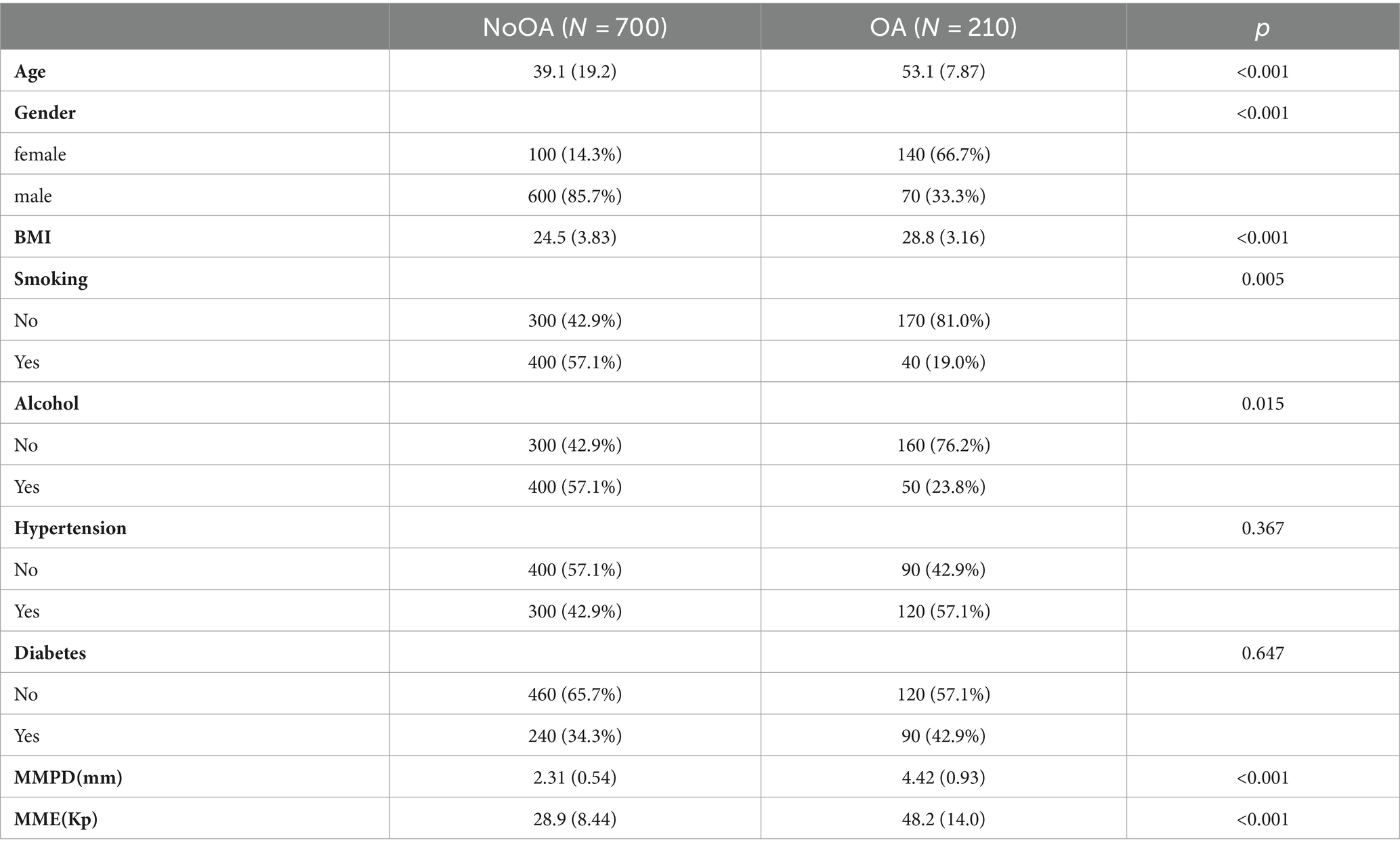
Table 1. Characteristics of included participants.
Using the Q1 group as a reference, we performed stepwise logistic regression (Table 2) to analyze whether the risk of OA changed with increasing SWE indicators in the Q2–Q4 groups. It can be seen in model 1, when only MMPD was used as the independent variable, there was a positive correlation dose–response relationship between this value and OA (trend p < 0.05) and the OR for Q4 was much greater than 1, [OR = 5.97, 95% CI (2.81, 4.98)], implying that the higher the level of MMPD, the higher the probability of having OA.
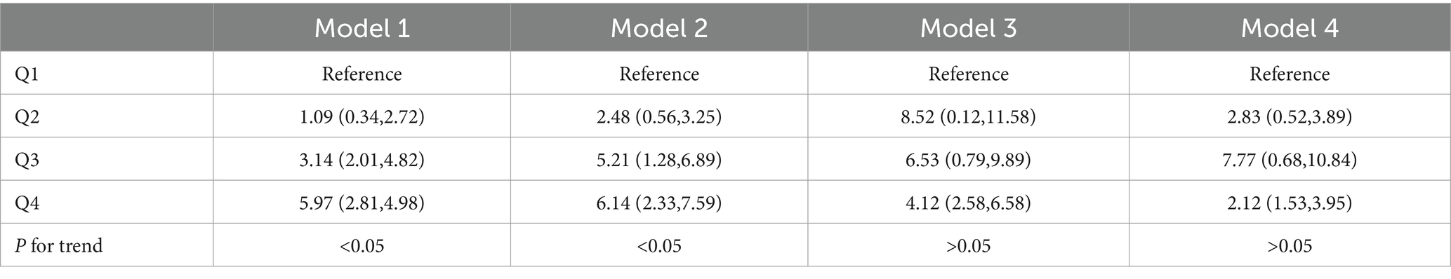
Table 2. Association between MMPD and OA.
Model 2 was constructed after demographic data were included as covariates and a positive correlation dose–response relationship remained. Model 3 had a positive correlation between high levels of MMPD and OA after lifestyle habits were put into the model [OR = 4.12, 95% CI (2.58, 6.58)], but there was no longer a dose–response relationship (Table 2). Model 4 also showed a positive association between high levels of MMPD and OA [OR = 2.12, 95% CI (1.53, 3.95)] after incorporating history of illness.
We also performed an analysis of MME and OA, which showed a dose–response relationship for all models 1 to 4 (trend p < 0.05). Moreover, with the gradual inclusion of the variables, the ORs increased minutely, reaching an OR of 8.98 [OR = 8.98, 95% CI (3.89, 11.52)] for Q4 in model 4 (Table 3).
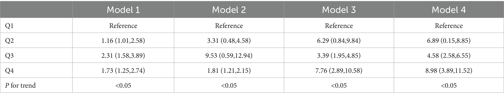
Table 3. Association between MME and OA.
In addition to analysing the relationship between elastic ultrasound measurements and OA, we also analyzed the relationship between the included covariates and OA. As shown in Figure 1, the risk of OA was significantly higher in the older age group [1.11, 95% CI (1.01, 1.25)] and the risk of diagnosing OA was high in the high BMI group [1.8, 95% CI (1.23, 3.01)] (Figure 3).

Figure 3. Forest plot of association between covariates and OA.
Discussion
In this study, we assessed knee OA lesions by using two metrics including MMPD and MME. We found that the correlation between the two measurements for assessing knee OA was high. Subsequently, exploring the factors influencing the SWE measurement of OA, people with advanced age and high BMI had a higher probability of being diagnosed with OA.
MME is strongly associated with knee OA and meniscal tears, which have also been described as radial displacement of the medial meniscus or medial radial displacement of the medial meniscus (16). Longitudinal studies have shown that the value of MME increases with increasing Kellgren/Lawrence radiological degrees in both supine and standing assessments of the knee (17). Similarly, studies have confirmed a significant correlation between an MME greater than or equal to 3 mm on MRI and an increase in the severity of cartilage damage, and a close association between MME and joint space narrowing (18). MME has been shown to be an independent predictor of disease progression in OA and is strongly associated with tibial and femoral cartilage loss (19).
However, there are limited studies on MMPD for the diagnosis of OA, and our study also supports the validity of MMPD for the diagnosis and prediction of OA, which needs to be confirmed by more relevant studies.
Previous reports have stated that there is a significant correlation between age and tendon, muscle and joint stiffness (20–22). This study also found a significant correlation between age and OA, while the correlation between degeneration and stiffness was equally significant. In moderate to severe OA, the meniscal matrix showed severe fibrocartilage detachment and extensive wear and tear, with reduced cellularity and cellular hypertrophy. Degeneration of the meniscus in OA is also found in people with high BMI, as evidenced by the accumulation of meniscal proteoglycans (23–25). These micro and macro changes in the degenerative meniscus are reflected by increased tissue elastic stiffness values measured by SWE (26, 27). Several previous researches supported that shear wave elastography can serve as an adjunctive tool to aid in the assessment of knee meniscal elasticity, especially can detect meniscus pathologies that may develop due to obesity and aging (28–30). These findings were totally consistent with ours. Ultrasound evaluation with SWE is an easily accessible, inexpensive imaging modality that also provides real-time dynamic evaluation and guides percutaneous interventional procedures. However, evaluation of the meniscus by ultrasound and SWE may be technically difficult due to adjacent bone and ligamentous structures and narrowing of the joint space due to degenerative disease (31).
In conclusion, these preliminary findings suggest that MME and MMPD correlate with the degree of meniscal degeneration, which is seen to reflect knee OA lesions. So, it is reasonable to assume that SWE could be used as a potential tool for assessing OA, especially in aging and high BMI people.
There are several limitations of this study. Firstly, the sample size was small, which reduced the reliability and validity. Secondly, there was a lack of low-age samples. Thirdly, this study only involved measurements of the medial meniscus of the knee and lacked values for the lateral meniscus and other joint measurements. Further studies are needed to improve the reliability of our findings.
Data availability statement
The raw data supporting the conclusions of this article will be made available by the authors, without undue reservation.
Ethics statement
The studies involving humans were approved by this study were approved by the Ethics Committee from the Third Affiliated Hospital of Hebei Medical University (No. 1354064). The studies were conducted in accordance with the local legislation and institutional requirements. The participants provided their written informed consent to participate in this study.
Author contributions
JZ: Data curation, Formal analysis, Investigation, Writing – original draft. XZ: Methodology, Software, Writing – original draft. YZ: Conceptualization, Formal analysis, Methodology, Validation, Writing – review & editing.
Funding
The author(s) declare that no financial support was received for the research, authorship, and/or publication of this article.
Conflict of interest
The authors declare that the research was conducted in the absence of any commercial or financial relationships that could be construed as a potential conflict of interest.
Publisher’s note
All claims expressed in this article are solely those of the authors and do not necessarily represent those of their affiliated organizations, or those of the publisher, the editors and the reviewers. Any product that may be evaluated in this article, or claim that may be made by its manufacturer, is not guaranteed or endorsed by the publisher.
Supplementary material
The Supplementary material for this article can be found online at: https://www.frontiersin.org/articles/10.3389/fmed.2024.1366793/full#supplementary-material
References
1. Yokuş, A, Toprak, M, Arslan, H, Toprak, N, Akdeniz, H, and Gündüz, AM. Evaluation of distal femoral cartilage by b-mode ultrasonography and shear wave elastography in patients with knee osteoarthritis: a preliminary study. Acta Radiol. (2021) 62:510–4. doi: 10.1177/0284185120930642
2. Hayashi, D, Roemer, FW, Link, T, Li, X, Kogan, F, Segal, NA, et al. Latest advancements in imaging techniques in oa. Ther Adv Musculoskelet Dis. (2022) 14:1759720X–2211466X. doi: 10.1177/1759720X221146621
3. Steinmetz, JD, Culbreth, GT, Haile, LM, Rafferty, Q, Lo, J, Fukutaki, KG, et al. Global, regional, and national burden of osteoarthritis, 1990–2020 and projections to 2050: a systematic analysis for the global burden of disease study 2021. Lancet Rheumatol. (2023) 5:e508–22. doi: 10.1016/S2665-99103(23)00163-7
4. Zhang, Y, Lin, C, Liu, M, Zhang, W, Xun, X, Wu, J, et al. Burden and trend of cardiovascular diseases among people under 20 years in China, western pacific region, and the world: an analysis of the global burden of disease study in 2019. Front Cardiovasc Med. (2023) 10:1067072. doi: 10.3389/fcvm.2023.1067072
5. Zhang, Y, Luo, Z, Yi, J, Zhu, J, Qiu, Y, Xu, X, et al. Burden and trends of stroke attributable to dietary risk factors from 1990 to 2019 in the belt and road initiative countries: an analysis from the global burden of disease study 2019. Front Nutr. (2023) 10:1235271. doi: 10.3389/fnut.2023.1235271
6. Long, H, Liu, Q, Yin, H, Wang, K, Diao, N, Zhang, Y, et al. Prevalence trends of site-specific osteoarthritis from 1990 to 2019: findings from the global burden of disease study 2019. Arthritis Rheumatol. (2022) 74:1172–83. doi: 10.1002/art.42089
7. Wang, D, Liang, R, Yang, M, Ma, J, Li, W, Mu, M, et al. Incidence and disease burden of coal workers' pneumoconiosis worldwide, 1990–2019: evidence from the global burden of disease study 2019. Eur Respir J. (2021) 58:58. doi: 10.1183/13993003.01669-2021
8. Satake, Y, Izumi, M, Aso, K, and Ikeuchi, M. Association between infrapatellar fat pad ultrasound elasticity and anterior knee pain in patients with knee osteoarthritis. Sci Rep. (2023) 13:20103. doi: 10.1038/s41598-023-47459-0
9. Park, JY, Kim, JK, Cheon, JE, Lee, MC, and Han, HS. Meniscus stiffness measured with shear wave elastography is correlated with meniscus degeneration. Ultrasound Med Biol. (2020) 46:297–304. doi: 10.1016/j.ultrasmedbio.2019.10.014
10. Laubach, M, Hellmann, JT, Dirrichs, T, Gatz, M, Quack, V, Tingart, M, et al. Anterior knee pain after total knee arthroplasty: a multifactorial analysis. J Orthop Surg (Hong Kong). (2020) 28:616579235. doi: 10.1177/23094990209108947
11. Chen, W, Li, C, Wang, Y, Shen, X, Wu, Z, Li, J, et al. Comparison of the asymmetries in muscle mass, biomechanical property and muscle activation asymmetry of quadriceps femoris between patients with unilateral and bilateral knee osteoarthritis. Front Physiol. (2023) 14:1126116. doi: 10.3389/fphys.2023.1126116
12. Ciloğlu, O, Karaali, E, Görgülü, FF, and Toğrul, E. Evaluation of the relationship between stiffness and thickness of the sciatic nerve and clinical outcomes after total hip arthroplasty: a prospective casecontrolled study. Acta Orthop Traumatol Turc. (2021) 55:500–7. doi: 10.5152/j.aott.2021.20324
13. Martinez, JA, Taljanovic, MS, Nuncio, ZA, Wertheim, BC, Roe, DJ, Ehsani, S, et al. Feasibility trial to evaluate tendon stiffness obtained from shear wave elastography imaging as a biomarker of aromatase inhibitor-induced arthralgias. J Clin Med. (2022) 11:11. doi: 10.3390/jcm11041067
14. Li, F, Wang, Z, Zhang, Z, Shen, S, Guo, J, Guo, Y, et al. In hamstring muscles of patients with knee osteoarthritis an increased ultrasound shear modulus indicates a permanently elevated muscle tonus. Front Physiol. (2022) 12:12. doi: 10.3389/fphys.2021.752455
15. Wang, K, Xia, F, Li, Q, Luo, X, and Wu, J. The associations of weekend warrior activity patterns with the visceral adiposity index in us adults: repeated cross-sectional study. JMIR Public Health Surveill. (2023) 9:e41973. doi: 10.2196/41973
16. Deng, W, Lin, M, Yu, S, Liang, H, Zhang, Z, and Liu, C. Quantifying region-specific elastic properties of distal femoral articular cartilage: a shear-wave elastography study. Appl Bionics Biomech. (2022) 2022:9406863. doi: 10.1155/2022/9406863
17. Zhang, X, Lin, D, Jiang, J, and Guo, Z. Preliminary study on grading diagnosis of early knee osteoarthritis by shear wave elastography. Contrast Media Mol Imaging. (2022) 2022:42291081. doi: 10.1155/2022/42291081
18. Åkesson, KS, Sundén, A, Stigmar, K, Eek, F, Pawlikowska, T, and Hansson, EE. Empowerment and enablement and their associations with change in health-related quality of life after a supported osteoarthritis self-management programme – a prospective observational study. Arch Physiother. (2023) 13:18. doi: 10.1186/s40945-023-00172-7
19. Wu, J, Wang, K, Tao, F, Li, Q, Luo, X, and Xia, F. The association of blood metals with latent tuberculosis infection among adults and adolescents. Front Nutr. (2023) 10:1259902. doi: 10.3389/fnut.2023.1259902
20. Xia, F, Li, Q, Luo, X, and Wu, J. Association between urinary metals and leukocyte telomere length involving an artificial neural network prediction: findings based on nhanes 1999–2002. Front Public Health. (2022) 10:963138. doi: 10.3389/fpubh.2022.963138
21. Li, W, Chen, D, Ruan, W, Peng, Y, Lu, Z, and Wang, D. Associations of depression, sleep disorder with total and cause-specific mortality: a prospective cohort study. J Affect Disord. (2022) 298:134–41. doi: 10.1016/j.jad.2021.10.131
22. Li, W, Ruan, W, Peng, Y, Lu, Z, and Wang, D. Associations of socioeconomic status and sleep disorder with depression among us adults. J Affect Disord. (2021) 295:21–7. doi: 10.1016/j.jad.2021.08.009
23. Zhang, Y, Wang, K, Zhu, J, and Wu, J. A network suspected infectious disease model for the development of syphilis transmission from 2015 to 2021 in Hubei province, China. J Appl Microbiol. (2023) 134:lxad311. doi: 10.1093/jambio/lxad311
24. Xia, F, Li, Q, Luo, X, and Wu, J. Machine learning model for depression based on heavy metals among aging people: a study with national health and nutrition examination survey 2017–2018. Front Public Health. (2022) 10:939758. doi: 10.3389/fpubh.2022.939758
25. Jinyi, W, Zhang, Y, Wang, K, and Peng, P. Global, regional, and national mortality of tuberculosis attributable to alcohol and tobacco from 1990 to 2019: a modelling study based on the global burden of disease study 2019. J Glob Health. (2024) 14:04023. doi: 10.7189/jogh.14.04023
26. Wang, D, Cao, L, Zhou, M, Ma, J, Wang, B, Yuan, J, et al. Alcohol intake, beverage type, and lung function: a multicohort study of chinese adults. Ann N Y Acad Sci. (2022) 1511:164–72. doi: 10.1111/nyas.14744
27. Zheng, X, Shi, J, and Wu, J. Analysis of factors and corresponding interactions influencing clinical management assistant ability using competency model in China. Medicine (Baltimore). (2020) 99:e23516. doi: 10.1097/MD.0000000000023516
28. Gurun, E, Akdulum, I, Akyuz, M, Tokgoz, N, and Ozhan, OS. Shear wave Elastography evaluation of Meniscus degeneration with magnetic resonance imaging correlation. Acad Radiol. (2021) 28:1383–8. doi: 10.1016/j.acra.2020.12.013
29. Ye, R, Xiong, H, Liu, X, Yang, J, Guo, J, and Qiu, J. Assessment of knee menisci in healthy adults using shear wave Elastography. J Ultrasound Med. (2023) 42:2859–66. doi: 10.1002/jum.16326
30. Dag, N, and Sinanoglu, MS. Evaluation of meniscal elasticity using shear wave elastography in obese children and adolescents: a preliminary cross-sectional study. Pediatr Radiol. (2024) 54:293–8. doi: 10.1007/s00247-023-05836-x
31. Altman, R, Asch, E, Bloch, D, Bole, G, Borenstein, D, Brandt, K, et al. Development of criteria for the classification and reporting of osteoarthritis. Classification of osteoarthritis of the knee. Diagnostic and therapeutic criteria Committee of the American Rheumatism Association. Arthritis Rheum. (1986) 29:1039–49. doi: 10.1002/art.1780290816
Keywords: ultrasonic shear wave elastography, ultrasound, osteoarthritis, adults, diagnosis
Citation: Zhang J, Zheng X and Zhao Y (2024) Analysis of the main indicators and risk factors of ultrasonic shear wave elastography for the diagnosis of osteoarthritis among adults. Front. Med. 11:1366793. doi: 10.3389/fmed.2024.1366793
Edited by:
Elisa Belluzzi, University of Padua, ItalyReviewed by:
Adem Yokuş, Yuzuncu Yil University, TürkiyeEnes Gürün, Samsun University, Türkiye
Yong Lu, Shanghai Jiao Tong University, China
Copyright © 2024 Zhang, Zheng and Zhao. This is an open-access article distributed under the terms of the Creative Commons Attribution License (CC BY). The use, distribution or reproduction in other forums is permitted, provided the original author(s) and the copyright owner(s) are credited and that the original publication in this journal is cited, in accordance with accepted academic practice. No use, distribution or reproduction is permitted which does not comply with these terms.
*Correspondence: Ying Zhao, zhaoying0227@hebmu.edu.cn