 Diana Espadinha1,2
 Rita G. Sobral3
 Catarina Inês Mendes4
 Guillaume Méric5
 Samuel K. Sheppard5,6
 João A. Carriço4
 Hermínia de Lencastre2,7
 Maria Miragaia1*
- 1Laboratory of Bacterial Evolution and Molecular Epidemiology, Instituto de Tecnologia Química e Biológica António Xavier, Universidade Nova de Lisboa, Oeiras, Portugal
- 2Laboratory of Molecular Genetics, Instituto de Tecnologia Química e Biológica António Xavier, Universidade Nova de Lisboa, Oeiras, Portugal
- 3Laboratory of Molecular Microbiology of Bacterial Pathogens, UCIBIO/REQUIMTE, Faculdade de Ciências e Tecnologia, Universidade Nova de Lisboa, Costa de Caparica, Portugal
- 4Molecular Microbiology and Infection Unit, Instituto de Medicina Molecular, Faculdade de Medicina de Lisboa, Universidade de Lisboa, Lisbon, Portugal
- 5The Milner Centre for Evolution, University of Bath, Bath, United Kingdom
- 6MRC CLIMB Consortium, Bath, United Kingdom
- 7Laboratory of Microbiology and Infectious Diseases, The Rockefeller University, New York, NY, United States
Background: Staphylococcus epidermidis is a common skin commensal that has emerged as a pathogen in hospitals, mainly related to medical devices-associated infections. Noteworthy, infection rates by S. epidermidis have the tendency to rise steeply in next decades together with medical devices use and immunocompromized population growth. Staphylococcus epidermidis population structure includes two major clonal lineages (A/C and B) that present contrasting pathogenic potentials. To address this distinction and explore the basis of increased pathogenicity of A/C lineage, we performed a detailed comparative analysis using phylogenetic and integrated pangenome-wide-association study (panGWAS) approaches and compared the lineages’s phenotypes in in vitro conditions mimicking carriage and infection.
Results: Each S. epidermidis lineage had distinct phenotypic signatures in skin and infection conditions and differed in genomic content. Combination of phenotypic and genotypic data revealed that both lineages were well adapted to skin environmental cues. However, they appear to occupy different skin niches, perform distinct biological functions in the skin and use different mechanisms to complete the same function: lineage B strains showed evidence of specialization to survival in microaerobic and lipid rich environment, characteristic of hair follicle and sebaceous glands; lineage A/C strains showed evidence for adaption to diverse osmotic and pH conditions, potentially allowing them to occupy a broader and more superficial skin niche. In infection conditions, A/C strains had an advantage, having the potential to bind blood-associated host matrix proteins, form biofilms at blood pH, resist antibiotics and macrophage acidity and to produce proteases. These features were observed to be rare in the lineage B strains. PanGWAS analysis produced a catalog of putative S. epidermidis virulence factors and identified an epidemiological molecular marker for the more pathogenic lineage.
Conclusion: The prevalence of A/C lineage in infection is probably related to a higher metabolic and genomic versatility that allows rapid adaptation during transition from a commensal to a pathogenic lifestyle. The putative virulence and phenotypic factors associated to A/C lineage constitute a reliable framework for future studies on S. epidermidis pathogenesis and the finding of an epidemiological marker for the more pathogenic lineage is an asset for the management of S. epidermidis infections.
Background
Staphylococcus epidermidis is one of the most abundant commensal bacteria of healthy human skin and mucosa. This organism has emerged in recent decades as an important opportunistic pathogen, being the main cause of nosocomial infections associated to indwelling medical devices such as peripheral or central intravenous catheters (CVCs) (Otto, 2009). Infections by S. epidermidis usually occur due to a breach in the skin barrier resulting from the insertion of the medical devices, allowing S. epidermidis to penetrate the host tissues.
The progression to infection after skin penetration depends on the ability of S. epidermidis to rapidly change from a commensal to a pathogenic state. As skin commensals, S. epidermidis survive and grow under nutrient limitation, at a low temperature (<37°C) and pH (∼4.5–6.4) (Schmid-Wendtner and Korting, 2006) and at diverse osmotic pressures resulting from the production/evaporation of sweat and fluctuations in environmental humidity (Wilson, 2005). Furthermore, they have to cope with cell desquamation, and oxidative stress resulting from UV exposure (Kammeyer and Luiten, 2015). Once in the bloodstream, upon skin barrier breach, the environmental landscape changes dramatically and S. epidermidis suddenly face a nutrient-rich and alkaline environment with a higher temperature, pro-inflammatory molecules and reactive oxygen species (ROS) generated by immune cells (Akira et al., 2006; Weiss and Schaible, 2015) and eventual antibiotic pressure (Ciofu et al., 2017). However, the factors of S. epidermidis that contribute for the transition from health to disease state are not completely understood.
One of the factors thought to be crucial for transition from skin to blood is the formation of biofilms on the surface of medical devices, which can be composed of a mesh of proteins, exopolysaccharides and extracellular DNA (Qin et al., 2007; Otto, 2009). These biofilms can confer protection against the host immune system (Vuong et al., 2004) and resistance to antibiotics (Farber et al., 1990; Khardori et al., 1995; Singh et al., 2010), making infections extremely difficult to treat (Otto, 2009). Other mechanisms that have been shown to be important for S. epidermidis pathogenicity include the ability to evade human innate immunity, namely through processes involved in resistance to antimicrobial peptides (AMP) (Cheung et al., 2010). Besides the ica operon, which is directly involved in biofilm formation (Heilmann et al., 1996) and the insertion sequence IS256, shown to modulate biofilm formation and antibiotic resistance (Kozitskaya et al., 2004), other virulence factors have been proposed. These include the primary attachment to host extracellular matrices and intercellular aggregation, toxins, proteases and lipases, all suggested to be implicated in invasive potential (Otto, 2012).
Whole genome sequence analysis (Conlan et al., 2012; Meric et al., 2015) has shown that the S. epidermidis population was composed of two main phylogenetic clusters: lineage A/C, containing most of the isolates from colonization and infection and lineage B, comprising mainly colonization isolates. In spite of their clinical relevance, the factors associated to the success of A/C strains, both as colonizers and pathogens, are not fully understood.
Here we hypothesize that the success of A/C strains as colonizers and as pathogens is associated with an increased ability to adapt to environmental constraints imposed by both colonization and infection states. To address this, we analyzed the genomes of representative isolates from A/C and B clusters and characterized their phenotypic traits in conditions that mimic the colonization and infection scenarios.
Results and Discussion
S. epidermidis Population Structure Is Composed of Two Clusters With Different Pathogenic Potential
In previous studies we have characterized a collection of 1714 S. epidermidis from different isolation dates, geographic and clinical origins (Miragaia et al., 2007; Rolo et al., 2012). Based on multilocus sequence typing (MLST) data we selected a collection of 83 isolates that represent the diversity of strains in terms of genetic backgrounds (71 sequence types, between 1996 and 2001). To understand how isolates of different origins were related, we generated a pan genome based on the annotated genomes from 82 S. epidermidis strains under study (one genome was excluded from the genomic analysis due to possible contamination, see Materials and Methods) and compared the genomic content of the strains according to their isolation source. The pangenome obtained had a total size of 5.0 Mbp with 31.3% of GC content, encoding a total of 6682 genes. From these, approximately one third (n = 1653) comprised the core genome (present in all strains in this study), with the remainder (5029) comprising the accessory genome. Gene accumulation curves that plot pan genome size as a function of the number of genomes sequenced, suggest an open pan genome that is characteristic of populations that undergo frequent horizontal gene transfer (HGT) (Meric et al., 2015) (Supplementary Figure S1).
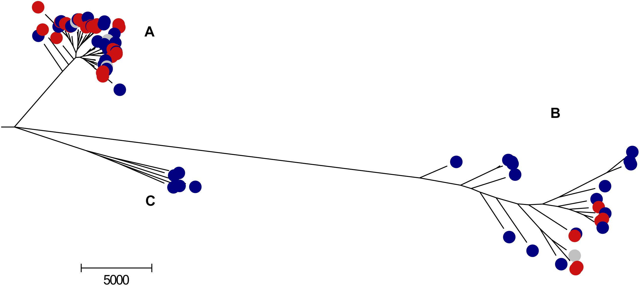
Figure 1. Maximum-likelihood tree of the 82 S. epidermidis strains. The tree was generated by RaxML (Stamatakis et al., 2012) embedded in Gubbins (Croucher et al., 2015), based on the core gene alignment with removal of the recombining regions. In blue are depicted the colonization isolates, in red the infection isolates and in gray are the isolates for which no epidemiological information was available. (A–C) Correspond to S. epidermidis clonal clusters, according to Meric et al. (2015).
Phylogenetic reconstruction, based on the Maximum-likelihood (ML) tree derived from the core genome corrected for recombining regions, confirmed that the S. epidermidis population is divided into three clusters (Figure 1). Consistent with previous studies (Meric et al., 2015), Clusters A (66%) and C (6%) were joined in a single cluster and were analyzed together in further analysis. The remainder of the isolates belonged to cluster B (28%). The A/C cluster contained mainly nosocomial isolates (n = 49/59, 83%) whereas the B cluster contained a higher proportion of isolates from healthy people in the community (n = 13/23, 56%) (p = 0.0007), confirming previous findings by Conlan et al. (2012) for a much smaller and geographically delimited isolate collection. A/C cluster isolates almost equally originated from infection (n = 28/59, 47%) and colonization (n = 31/59, 53%) sources, while B cluster isolates were mostly from colonization (17/23, 70%) (p = 0.127) even though this different distribution did not reach statistical significance. These results suggest that although both A/C and B strains are carried asymptomatically, A/C strains are, apparently, more implicated in infection events. This observation is corroborated by multiple studies wherein S. epidermidis from infection were in their majority from A/C cluster (which comprises clonal complex 2 as defined by MLST) (Miragaia et al., 2007; Widerstrom et al., 2009; Gordon et al., 2012; Hellmark et al., 2013; Cherifi et al., 2014).
Evidence for the Clonal Dissemination of A/C Cluster Isolates
An important characteristic of some opportunistic pathogens is the ability to disseminate in different hosts and environments. This can lead to broad distribution of closely related strains. The mean number of SNP differences between the core genomes of A/C and B clusters was 39889, which correspond to 1.6% of the average genome size of S. epidermidis. Within each cluster, the average number of SNPs was similar for strains of cluster B (average number of SNP differences = 16786, ranging between 31 and 36796) and for strains of cluster A/C (average number of SNP differences = 17696 nucleotides, ranging between 1 and 37617). The lower number of differences (<100 SNPs) were found between 11 pairs of A/C isolates and only one pair in cluster B, suggesting that A/C isolates are more clonal than B isolates. Furthermore, these pairs included isolates from different countries, isolated either from the hospital or the community, demonstrating the dissemination of this cluster through these settings.
In contrast with other known nosocomial pathogens, including S. aureus (Ruimy et al., 2009; Grundmann et al., 2010), we found no evidence of geographic clustering in S. epidermidis, since each branch in the ML tree, corresponding either to A/C or B clusters (considering a maximum of 50 SNPs difference), contained isolates from multiple countries (see Supplementary Table S1). Furthermore, there were eight pairs of strains from different countries that, with one exception, belonged to the A/C cluster that diverged in less than 100 SNPs and that were isolated within the same year. This was the case for strains from Denmark and Portugal (68 SNPs; DEN161, 966N), Denmark and Iceland (60 SNPs; DEN116, ICE122) and Argentina and Hungary (17 SNPs; AGT24, HUR105), among others (see Supplementary Table S1). These examples suggest extensive and rapid geographic dissemination of strains belonging to the A/C cluster, likely promoted by skin-to-skin contact between travelers. This is consistent with previous studies that hypothesized broad geographic dissemination of this species (Miragaia et al., 2002; Widerstrom et al., 2006).
Evidence for Ecological Isolation and Distinct Biological Functions of A/C and B Clusters
The success of a bacterial clone may be influenced by its capacity to adapt to different environments and this is frequently accomplished by the acquisition of exogenous genetic elements (Uhlemann et al., 2012; Walther et al., 2018), which will compose the accessory genome. To assess differences in gene content of the two S. epidermidis clusters, we analyzed and quantified accessory genome variation. The matrix for presence/absence of all the genes in the genomes is represented in Supplementary Table S2. The majority of A/C and B pan genomes was composed of accessory genes (A/C: 74%, 3723/5029; B: 67%, 3381/5029). Among this accessory genome, 1648 genes were exclusive to A/C cluster and these genes varied in frequency from 93% to 2% and 1306 genes were exclusive to cluster B and ranged from 100 to 4%, suggesting high genome plasticity and cluster-specific functions. The number of accessory genes shared between strains of the same cluster (3723 in A/C and 3381 genes in B) is 1.8–1.6 higher than the number of genes shared between the two clusters (2075 genes). This difference could be due to the existence of a barrier to genetic transfer between them. However, in contrast to S. aureus, this apparently low frequency of genetic transfer is not evidently associated with the presence of Restriction/Modification (R-M) systems (Waldron and Lindsay, 2006; Corvaglia et al., 2010) and/or the clustered regularly interspaced short palindromic repeats (CRISPR) (Marraffini and Sontheimer, 2008). There was no clear association of a hsdR/hsdM system to any of the clusters and cas and csm genes were equally distributed among strains of the two clusters [A/C: 12% and B: 17% (p > 0.05)] (Supplementary Table S2). An alternative explanation for the lower level of genetic exchange between the clusters, in this apparently recombinogenic species (Miragaia et al., 2007; Meric et al., 2015), might be the existence of tropism of each of these lineages to specific skin niches (Grice et al., 2009; Oh et al., 2014) that could have lead to some ecological isolation of the two genetic clusters. Actually, skin microbiota composition was previously shown to vary according to the environmental characteristics of the skin ecological niche sampled (Costello et al., 2009; Grice et al., 2009). To test the hypothesis of ecological isolation we have examined the seven MLST genes, which constitute a good representation of the core genes, either in terms of genome distribution and genetic diversity (Gomes et al., 2005; Miragaia et al., 2007). Data showed an apparent allelic segregation, where aroE gene presented no common alleles between the clusters and the remaining genes (arcC, gtr, mutS, pyrR, tpi, yqiL), comprising between 11 and 20 alleles, had a maximum of two alleles shared by the two clusters.
Furthermore, we looked for genes that were significantly associated to each cluster using a pan genome wide-association approach, that scores genes for associations with specific epidemiological features. In particular, 166 genes were identified to have a strong positive association (Benjamini–Hochberg p < 0.05) with cluster A/C (see Supplementary Table S3) and 244 genes with a strong association with cluster B (see Supplementary Table S4). Tables 1, 2 include the genes among these, which have functions that were either present exclusively in one of the clusters or that were present in both but in significantly different frequencies. Strains of the A/C cluster were enriched for genes involved in processes such as biofilm formation and adhesion to host matrix proteins, proteolysis, resistance to antibiotics and adaptation to low pH. Conversely, strains from cluster B were enriched for genes involved in the detoxification of formate and formaldehyde, oxidative stress response, host interaction through type VII secretion system, lipid metabolism and cell wall biosynthesis. This is consistent with S. epidermidis clusters A/C and B performing distinct biological functions and possibly occupying different skin niches.

Table 1. Genes positively associated to cluster A/C (Benjamini–Hochberg p < 0.05 and OR > 1).
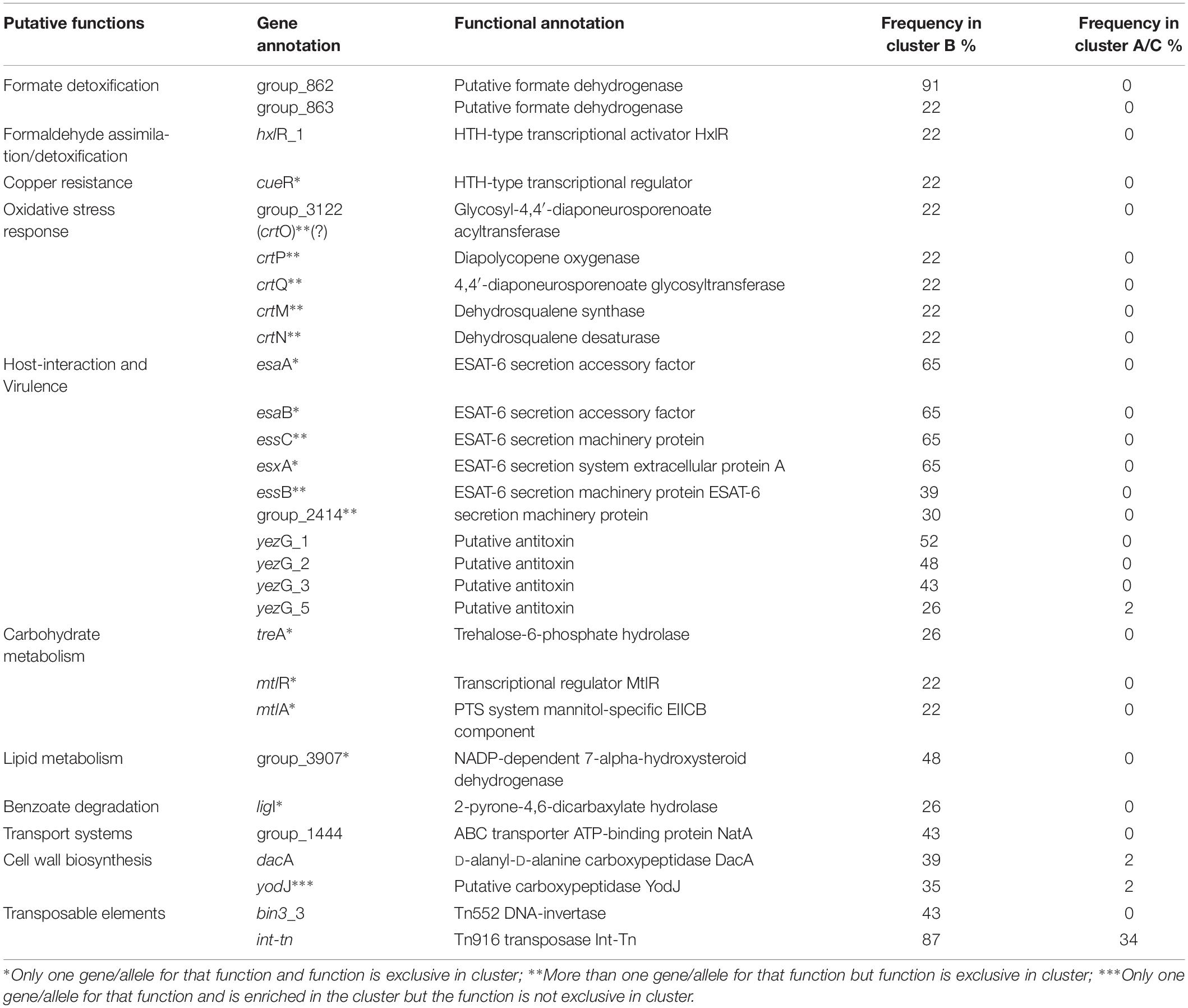
Table 2. Genes positively associated to cluster B (Benjamini–Hochberg p < 0.05 and OR > 1).
S. epidermidis of A/C and B Clusters Have Adapted to Skin Environment Conditions Using Different Strategies
Skin is the first barrier of the human body against external aggressions and contains molecules that derive from the metabolism of skin cells and our microbiota, but also from the environment (Wilson, 2005). On the skin, S. epidermidis have to deal with nutrient limitation, harsh and variable environmental conditions and mechanic stresses. Perspiration is one of the central contributors for the composition of skin milieu and a major physiological function that assists in thermoregulation, skin surface hydration and immune defense. Perspiration is composed mainly of water, but contains several other metabolites that contribute for its multiple functions, such as antimicrobial peptides (AMP), immunoglobulins, natural moisturizing factors (lactate, urea, electrolytes, amino acids), vitamins, metals and salts (Wilson, 2005).
To understand if the S. epidermidis clusters differ in their ability to adapt to skin we have compared their ability to grow and produce biofilm in conditions that mimic the skin environment (acidic pH and increased salt concentrations). Moreover, we looked in more detail for the genes that were associated by the pan genomic wide-association approach to each lineage and that could provide an advantage in the skin environmental conditions.
Growth and Biofilm Formation at Acidic pH
Staphylococcus epidermidis belonging to A/C cluster appear to have a distinctive ability to adapt to the two acidic pH values tested (pH 4.5 and pH 5.5), as strains from the A/C cluster reached, in average, higher cell densities in stationary phase (∼12% higher in both cases) and presented either a greater (pH4.5: μA/C = 0.335; μB = 0.274, p = 0.043) or similar average growth rate (pH 5.5: μA/C = 0.626; μB = 0.594, p = 0.273) when compared to strains from the B cluster (see Figure 2A and Table 3). Additionally, strains belonging to the A/C cluster were associated to a gene involved in the arginine deiminase metabolism (arcD, an arginine/ornithine antiporter) (see Table 1) usually carried by the arginine catabolic mobile element (ACME). Although a genomic copy of arcD gene with a similar function is known to be ubiquitous in the S. epidermidis genome, this extra copy together with the remaining arc operon within ACME was previously suggested to be important for pH homeostasis in S. epidermidis and to contribute to additional tolerance to low-pH in community-associated Staphylococcus aureus USA300 clone (Thurlow et al., 2013; Lindgren et al., 2014). Other functions associated to A/C cluster strains that can eventually contribute for the observed increased growth rate is the flavin-containing oxidoreductase (YwqN), the YdjH sugar kinase; and the hexose-6-phosphate:phosphate antiporter (UhpT) (Park et al., 2015), which lead either to the production of reducing power (NADH) for the biosynthesis of major cell components or to the rescue of alternative sugar sources.
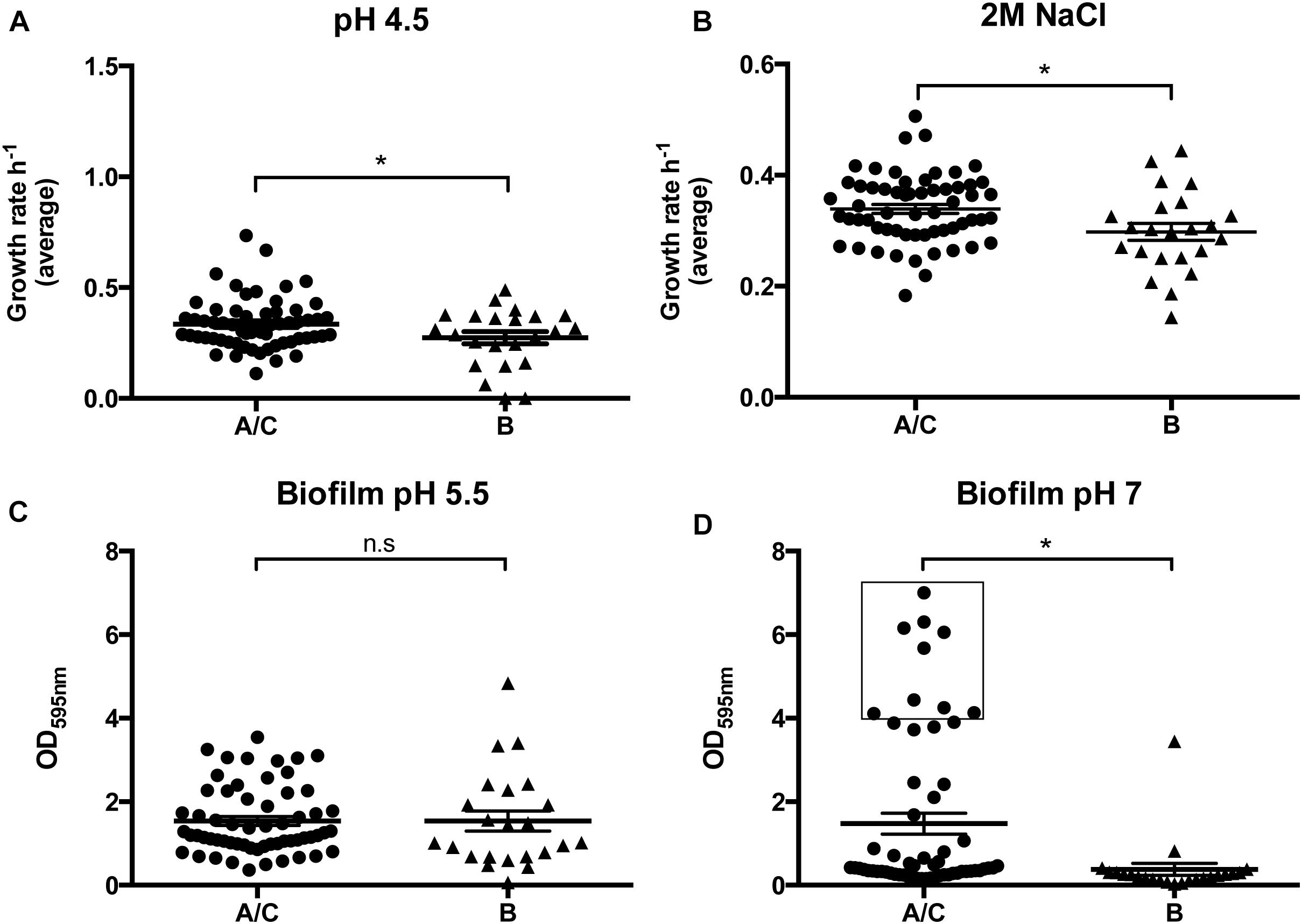
Figure 2. Performance of cluster A/C and B strains in the growth and biofilm assays. (A) Average growth rates for A/C and B strains grown in liquid medium at pH 4.5; (B) Average growth rates for A/C and B strains grown in liquid medium with 2M NaCl; (C) Average biofilm formation capacity for A/C and B strains at pH 5.5; (D) Average biofilm formation capacity for A/C and B strains at pH 7.
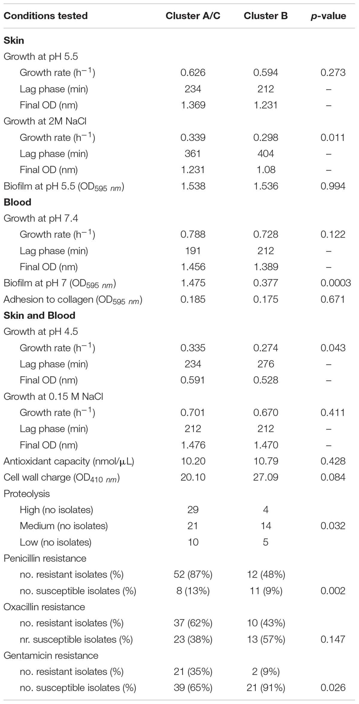
Table 3. Performance of cluster A/C and cluster B strains in skin and blood conditions.
The two groups could not be distinguished by their ability to produce biofilm at pH 5.5 (ODA/C = 1.538 vs. ODB = 1.536, p = 0.994), since almost all strains, irrespective of their genetic background, produced biofilm in this condition, suggesting that biofilm is important for the growth of this commensal at the surface of the skin (see Table 3 and Figure 2C).
Growth in Osmotic Stress Conditions
The better capacity of the strains from the A/C cluster to withstand perspiration, osmotic stress and desiccation conditions frequently imposed at the skin surface was apparent from their capacity to grow at sodium chloride concentrations characteristic of the skin (0.15M NaCL) (Schittek et al., 2001) and in high salinity stress (2M NaCl) (Price-Whelan et al., 2013). Whereas at a physiological concentration (0.15M NaCl), strains from cluster A/C and B had equivalent average growth rates (μA/C = 0.701 vs. μB = 0.670, p = 0.411) (see Table 3), at osmotic stress conditions (2M NaCl), the medium growth rate of A/C cluster strains was significantly higher (μA/C = 0.339 vs. μB = 0.298, p = 0.011; see Figure 2B). Furthermore, they showed a much shorter lag phase (A/C: 361 min vs. B: 404 min), and reached higher final ODs (14% higher) (see Table 3). The results suggest that, in contrast to S. epidermidis strains of B cluster, strain from A/C cluster are well adapted to a range of salinity concentrations suggesting improved survival in a broader range of niches.
We found that different genes indirectly involved on resistance to osmotic stress were specifically associated to either cluster A/C or B. This is the case of genes which were associated to A/C cluster that are involved in the production of polysaccharide intercellular adhesion (PIA) (icaADBCR operon), which through the production of biofilm induce protection against fast osmotic changes by decreasing the rate of water loss and rehydration (Roberson and Firestone, 1992; de Goffau et al., 2009) (see Table 1). Furthermore, genes associated to B cluster that could induce osmoprotection include those involved in: peptidoglycan cross-linking (dacA - a pbp4 homolog, yodJ) that provides extra rigidity to the bacterial cell wall (Wyke et al., 1981; Loskill et al., 2014); and the uptake of mannitol (mtlA, mtlR), a known osmolyte (Kets et al., 1996; Zahid et al., 2015). The absence of mtlD, responsible for mannitol degradation, suggests that strains of B cluster use mannitol mainly as an osmolyte rather than as a carbon or energy source (da Costa et al., 1998).
Although strains of both clusters carry genes that are associated to resistance to osmotic stress, overall, the processes used by strains of the A/C cluster appear to be more efficient than those of strains from the B cluster, as illustrated by its higher growth rate in vitro.
Genetic Traits Associated to Evasion of Skin Innate Immunity
Skin is continuously exposed to commensal microorganisms and challenged to defend the human body from invasion by pathogens. To deal with this extensive microbial exposure, skin tissues produce antimicrobial peptides (AMPs) and free fatty acids that target the bacterial cell wall or cell membrane structures (Kenny et al., 2009; Gallo and Hooper, 2012). To survive, bacteria have developed multiple strategies, including the production of carotenoid pigments that protect from free fatty acids and the inactivation of AMPs by proteolysis.
We found that strains belonging to the B cluster more commonly contained the operon involved in staphyloxanthin biosynthesis (crtOPQMN) that was described to confer resistance to free fatty acids produced by skin cells (Chamberlain et al., 1991). The production of the carotenoid was confirmed phenotypically for the group of five strains carrying this operon through the observation of production of a yellow color by colonies grown on agar growth medium. Other pigments had been found to be synthesized by S. epidermidis (Ogo, 1985), but this is the first time that staphyloxanthin operon is described in this species. We also found a serine-protease (SplF) positively associated to strains from cluster A/C (Table 1). Staphylococcal proteases have already been implicated in the mechanism of defense against the human innate immune system, as some of these proteases have the ability to cleave and inactivate human AMPs, namely dermcidin (Lai et al., 2007) and LL-37 (Sieprawska-Lupa et al., 2004) that are produced by epithelial cells and although this could be the case also for S. epidermidis SplF, this has yet to be proven. Altogether, the results suggest that both S. epidermidis of A/C and B cluster are equipped to evade the host immune system and that could be a major advantage for survival in both skin and blood.
Interaction With the Host and Pathogens
Commensal bacteria are in constant interaction with the surrounding environment and the host. Secretion systems are central elements in these interactions but have been investigated mostly from the perspective of pathogen-host interactions. Recent data suggest that these systems may play a role in mutualistic relationships between bacteria and the host (Silver et al., 2007) or in mediation of bacterial interactions (Basler et al., 2013). We found that S. epidermidis strains belonging to B cluster were associated to genes related to type VII secretion system (T7SS) (Lai et al., 2007). In particular, all the conserved T7SS structural genes (esaA, esaB, essA, essB, essC) were co-located in the chromosome. Moreover, there was considerable strain-to strain variation in the number and type of secreted toxin/antitoxin-homologs downstream this operon (see Table 2).
T7SS were previously described in S. epidermidis and were proposed to secrete an inhibitory toxin against Propionibacterium acnes (Christensen et al., 2016), another common commensal of the skin. Yet, in S. aureus, the functions of T7SS are broader, involving not only intraspecies competition (Cao et al., 2016), but also persistence and pathogenicity (Burts et al., 2008; Kneuper et al., 2014; Korea et al., 2014). The identification of a T7SS in S. epidermidis belonging to B cluster is consistent with the hypothesis that S. epidermidis of cluster B and Propionibacterium may share a similar niche in the skin.
S. epidermidis Genetic Content Reflects Adaptation to Anthropogenic Activities
Previous studies have shown that human skin composition is also defined by our daily routines (Bouslimani et al., 2015), including activities that imply frequent contact with the skin, like hygiene habits and the application of skin care products and clothing. Moreover, sweat has been considered for long as a cleanser, allowing the excretion of toxic substances that could have been absorbed due to acute or chronic exposure to contaminated environment (air, water, food) or to consumed products. We found that S. epidermidis of either A/C or B cluster were associated to six loci related to the uptake, detoxification or regulation of metabolites (gluconate, trehalose, copper, and formaldehyde) that according to the Human Metabolome Database1 (Wishart et al., 2018) are present in the skin tissues and that are known to be simultaneously present in the composition of food, pharmaceutical products, detergents/disinfectants and/or cosmetics (see Tables 1, 2).
Degradation of Sugar and Sugar Derivatives
Genes involved in the degradation of sugars or sugar derivatives, namely trehalose (treA) and gluconate (gno), were associated with B and A/C clusters respectively, suggesting that strains from the two clusters use these alternative sugars as carbon and energy sources. The use of gluconate as a carbon source and its central role in bacterial cell division and colonization was previously described for other bacteria like Streptococcus suis (Shi et al., 2014) and Escherichia. coli (Sweeney et al., 1996).
Trehalose is not commonly used by S. epidermidis as a carbon source. Actually, the absence of trehalose fermentation has been considered a signature of S. epidermidis used to distinguish it from other bacterial species (Kloos and Schleifer, 1975). A possible origin for trehalose found in the skin, besides food and cosmetics (Ohtake and Wang, 2011), are bacteria of the genus Propionibacterium (also part of skin natural flora), which are described to be capable of synthesizing this sugar (Piwowarek et al., 2018). The results suggest once more that Propionibacterium and S. epidermidis of B cluster might share the same niche, possibly hair follicles and sebaceous regions wherein Propionibacterium are enriched (Scharschmidt and Fischbach, 2013) and the trehalose produced by Propionibacterium can be used by S. epidermidis as a carbon and energy source.
Having the capacity to metabolize gluconate and trehalose, either from anthropogenic or microbial origin, can be highly advantageous for S. epidermidis in an environment of nutrient limitation like the skin.
Resistance and Modulation of Copper
Staphylococcus epidermidis strains belonging to A/C cluster and B cluster were associated to genes involved in copper regulation, although using different mechanisms. While strains from the A/C cluster were associated to multicopper oxidase (mco), an enzyme involved in the oxidation of Cu+ to Cu2+ (Sitthisak et al., 2005), B cluster strains were associated to cueR, a MerR-family metalloregulatory transcriptional activator that senses intracellular Cu+ and upregulates copA and cueO, two genes involved in copper efflux and oxidation in response to increasing copper concentrations (Chaturvedi and Henderson, 2014). Copper is utilized by bacteria for several essential metabolic processes, but is also critical for the maintenance of a healthy skin, namely for the synthesis and stabilization of extracellular matrix skin proteins and angiogenesis (Raju et al., 1982; Philips et al., 2012). Although an important enzyme cofactor and signaling molecule, the intracellular levels of free copper can be damaging for bacteria, leading to ROS-related oxidative stress derived from its reduction and oxidation cycles, reason by which it is frequently added to cosmetics as a biocide. Copper oxidation performed by systems like Mco and Cue probably help S. epidermidis to tightly regulate and maintain copper homeostasis in the skin (Festa and Thiele, 2012; Chaturvedi and Henderson, 2014).
Detoxification of Formaldehyde
Staphylococcus epidermidis belonging to B cluster were additionally associated with a gene (hxlR) involved in detoxification of formaldehyde, a metabolite that can be highly deleterious to human cells. The formaldehyde-responsive transcriptional factor hxlR is part of the formaldehyde detoxification/assimilation via a ribulose monophosphate (RuMP)-dependent pathway, through which formaldehyde is converted into fructose-6 phosphate and subsequently incorporated into the usual carbon metabolism pathway (Chen et al., 2016). Formaldehyde, is ubiquitously found in nature and is an important metabolite of both bacteria and human cells (Burgos-Barragan et al., 2017), but is also one of the most synthesized compounds as the result of anthropogenic activities. Toxicity of formaldehyde to animals, humans and bacteria (Tang et al., 2009; Chen et al., 2016) is due to the fast reaction of formaldehyde with free thiol (−SH) and amine groups (−NH2) on proteins and DNA (Feldman, 1973; Paget and Buttner, 2003), making formaldehyde one of the most potent protein and DNA cross-linking agents.
The presence of genes for the detoxification in S. epidermidis strains belonging to B cluster probably allowed them to adapt to formaldehyde resulting either from human and bacterial cells metabolism or from continuous exposure to formaldehyde containing products (dyes, synthetic textiles, disinfectants, and cosmetics). In this regard, S. epidermidis might be functioning not only for its own benefit but also in the benefit of the host by avoiding that these compounds reach toxic concentrations to human cells.
Strains of A/C Cluster Have a Higher Capacity of Survival in Hospital and Infection Environmental Conditions
During infection S. epidermidis has to adapt to a new environment, the bloodstream, wherein the pH is stabilized at 7.4, the concentration of salt is steady (0.15 M), specific host matrix proteins are produced, the immune response is exacerbated, and inhibitory concentrations of antibiotics may be present. One of the strategies to deal with such stresses is the attachment to the surface of a device, followed by the development of the biofilm, where bacterial cells are protected.
To evaluate if S. epidermidis belonging to A/C and B clusters have developed different capacities to adapt to an infection environment, we compared their ability to bind collagen I, to grow and produce biofilm in blood-similar pH and salt conditions (growth: pH 7.4; 0.15 M NaCl; biofilm: pH 7), to resist oxidative stress and to evade the immune system (by measuring the surface charge and proteolytic activity). Moreover, we searched for specific genes within these lineages that could be associated to a higher infectious capacity.
Binding to Host Matrix Proteins and Biofilm Formation at Blood pH
When a medical device is introduced into the human body, its surface becomes coated with a layer of host proteins (Vaudaux et al., 1995; von Eiff et al., 2002; Arrecubieta et al., 2007; Singh et al., 2012). Foreign-body associated infections are attributed to the attachment of bacteria to these host proteins, such as collagen I from tissues, and fibronectin and fibrinogen from blood, through binding of surface proteins (Patti et al., 1994; Foster and Hook, 1998) followed by the production of biofilm. To understand if S. epidermidis belonging to the two genetic lineages differed in their ability to cause device-associated infection, we tested their ability to bind collagen I (see Table 3), the collagen type more abundant in human tissues (Singh et al., 2012), rather than the skin. However, our results showed that binding to collagen was not a distinctive factor contributing for their different infectious capacity (ODA/C = 0.185 vs. ODB = 0.175, p = 0.067) (see Supplementary Figure S2A). Moreover, we identified two adhesins or microbial surface components recognizing adhesive matrix molecules (MSCRAMMs), namely SdrF and SdrI, that were associated to A/C strains and that have been described to be involved in binding to collagen I and fibronectin (Hartford et al., 2001; Sakinc et al., 2005; Arrecubieta et al., 2007), respectively. In spite of the identified association between SdrF and A/C cluster, this did not impact directly the phenotype of collagen binding, probably due to the existence of known redundancy of proteins other than SdrF that bind collagen I. On the other hand, the comparison of the two lineages regarding the ability to form biofilm at pH 7 (similar to blood’s pH) showed that A/C strains produced significantly more biofilm (ODA/C = 1.475; ODB = 0.377, p = 0.0003) (see Table 3 and Figure 2D) and were simultaneously associated to the presence of the ica operon, involved in the production of polysaccharide intercellular adhesion (PIA) (Benjamini–Hochberg p = 0.029). These results suggest that upon skin disruption the strains that are able to produce biofilm and that survive better in blood environment are those belonging to A/C cluster that in addition to the ability of binding collagen I have the potential to bind fibronectin (mainly found in the blood after injury) and to produce PIA-dependent biofilms at blood’s pH.
Resistance to Host Immune System During Infection
Once in the bloodstream, bacteria are immediately recognized by monocytes and neutrophils through several Pattern recognizing receptors (such as Toll-Like receptors, Nod-Like receptors and C-type lectin receptors), leading to the expression of AMPs, phagocytosis, degranulation, respiratory burst and killing and the formation of neutrophil extracellular traps that capture bacteria inducing a rapid inflammatory response (cytokines and chemokines) (Akira et al., 2006). Bacterial phagocytosis by macrophages occurs via the formation of a membrane-enclosed phagosome containing the microbe in an acidic environment (can be as low as pH 4.5), extremely rich in bactericidal enzymes and toxic compounds (reactive oxygen and nitrogen species and copper) (Weiss and Schaible, 2015).
According to our data, S. epidermidis strains have developed several mechanisms that allow them to survive within macrophages. In particular, we observed that although A/C strains had a significantly higher medium growth rate at pH 4.5 (pH of macrophages milieu) (μA/C = 0.335; μB = 0.274, p = 0.043) than B strains, they did not vary significantly in their total antioxidant capacity (see Supplementary Figure S2B). The lack of difference in antioxidant capacity might be due to the fact that both A/C and B clusters were associated to genes that provide resistance to oxidative stress (A/C cluster: mco; B cluster: crtOPQMN operon) and tolerance to copper (A/C cluster: mco; B cluster: cueR) (see Tables 1, 2). Especially, staphyloxanthin produced by crtOPQMN was described in S. aureus to participate in the scavenging of free radicals and to have impact in virulence (Clauditz et al., 2006; Mishra et al., 2011).
We have also compared the surface charge and proteolytic activity of the two clusters, as a measure of the capacity to evade the host immune system. According to our data, strains of the A/C and B clusters showed a similar average value of surface charge measured by the relative percentage of unbound cytochrome C, but the values obtained varied from 0 and 78% among strains (see Supplementary Figure S2C and Supplementary Table S5). In particular, closely related strains in the phylogenetic tree (DEN116 and ICE21, 101 SNPs) showed completely distinct relative surface charges (72 and 36%), suggesting that although S. epidermidis clusters could not be differentiated based on this feature, modulation of the surface charge appears to be a strategy frequently used by S. epidermidis during host-microbe interaction. This ability might confer bacteria with a range of different susceptibilities to human AMPs. Still, S. epidermidis clusters appear to differ in their proteolytic capacity when grown on top of skimmed milk agar containing casein (pH = 7), a test utilized to assess production of excreted proteases such as staphylococcal serine proteases (Reed et al., 2001). We found that the proportion of strains that had a high proteolytic capacity was significantly higher in the A/C cluster (48%, p = 0.0321) (see Supplementary Figure S2D and Supplementary Table S5) when compared to B cluster (17%). Moreover, we found by pangenomic analysis that a homolog of SplF, an extracellular serine protease-like protein (Reed et al., 2001), was associated to S. epidermidis of A/C cluster (77% vs. 35% in B cluster; p = 0.008). SplF has already been described to be highly immunogenic and to be involved in allergic reactions in S. aureus (Zdzalik et al., 2012; Stentzel et al., 2017) and may also interact with the immune system during S. epidermidis medical device infections. In S. aureus this gene is part of the spl operon carried by the pathogenicity island νSaβ (Paharik et al., 2016), however, analysis of the genes in the vicinity of the splF homolog in S. epidermidis strains from our study showed that this gene was not included within any operon or mobile genetic element.
Resistance to Antibiotics
Staphylococcus epidermidis colonizing healthy people in the community are usually susceptible to antibiotics (Rolo et al., 2012; Cherifi et al., 2013) but isolates that cause infections within the hospital are typically resistant to multiple antibiotic classes. In particular, around 80% are resistant to β-lactams (Santos Sanches et al., 2000; May et al., 2014). To understand if clusters A/C and B differed in their susceptibility to antibiotics we analyzed their susceptibility profiles to a panel of 14 antibiotics. S. epidermidis strains showed resistance to almost all classes of antibiotics tested, varying between 4% and 88% of resistant isolates (see Supplementary Figure S3A) with the exception of linezolid, quinupristin-dalfopristin or vancomycin for which no resistance was observed. Regarding resistance to oxacillin, the frequency and proportion of isolates with high-level resistance (>192 μg/ml) was similar between the two groups (A/C cluster: 53%; B cluster: 50%) (see Supplementary Figure S3B and Table 3). Overall, A/C cluster showed a higher frequency of resistance to penicillin and gentamicin when compared to B cluster (p-value < 0.05) (see Table 3). Although in the case of penicillin we could observe a positive association of the blaZ and blaI genes with the A/C group, no specific genes related to resistance to gentamicin were found associated to this lineage. This was probably due to the fact that diverse resistance mechanisms were detected among isolates of both clusters. In particular, we observed that gentamicin resistant strains belonging to A/C and B groups carried the genes ant(4′)-Ib, aac(6′)-Ie-aph(2′′)-Ia and aph(3′)-IIIa. Resistance of A/C strains to gentamicin has probably emerged as a means of survival since gentamicin is frequently used for the treatment of biofilm-associated infections (van de Belt et al., 2001). However, the reason for the increased resistance to penicillin is not so obvious, since this antibiotic is not frequently used to treat device-related infections. A possibility is that blaZ regulators are maintained associated to A/C strains because they are important for SCCmec acquisition by S. epidermidis as was previously described for S. aureus (Katayama et al., 2003). Actually, all strains that were resistant to methicillin contained blaZ.
Identification of Epidemiological Markers for S. epidermidis A/C and B Clusters
The results from this study highlighted the phenotypic and genotypic differences between the A/C and B clusters and provided several lines of evidence supporting the higher pathogenic potential of A/C strains. Thus, the finding of good epidemiological markers for these two clusters would assist in a better prognosis of the infection and would improve clinical decision-making (namely more appropriate treatment approach) and, ultimately, patient outcomes.
To identify good epidemiological markers for the A/C and B clusters we looked for genes that were associated to the clusters and had frequencies above 90% (see Supplementary Table S3 and Table 2). We found two genes meeting these criteria: a gene encoding a hypothetical protein with a putative DUF1641 domain with a formate dehydrogenase activity associated to cluster B; and one gene encoding for a hydrolase (yxeP) of unknown function associated to cluster A/C. In this latter, there were only three isolates belonging to B cluster that contained yxeP. These three strains cluster together in the tree, with two strains being more closely related (70 SNPs) than the other (∼2500 SNPs of difference), all collected from nasal colonization in Portugal from draftees sharing the same environment, suggesting that the acquisition of yxeP by strains of this lineage might be a rare genetic event.
Our results confirmed the presence of a putative formate dehydrogenase gene (group_862) as highly specific of strains belonging to B cluster, as previously suggested (Conlan et al., 2012). Formate dehydrogenase was described to be important for formate detoxification and NADH/H + production during anaerobic fermentative growth in E. coli (Knappe and Sawers, 1990; Sawers, 2005), but in S. aureus this enzyme is only expressed in a microaerobic environment (Leibig et al., 2011). It is thus tempting to speculate that fdh might enable cluster B strains to survive in specific microaerobic niches in the skin, as hair follicles and sebaceous glands, like Propionibacterium spp.
Conclusion
Staphylococcus epidermidis is the main colonizer of the human skin but also one the most important opportunistic pathogens associated to medical device-related infections that occur when the skin barrier is disrupted and the host is immunocompromised.
In this study we have compared the two unique clusters that compose S. epidermidis population (A/C and B) regarding their genomic content, using genome-wide association studies, and regarding their phenotypic performance in conditions that mimic colonization and infection, which allowed us to create a portrait of each cluster that sheds some light into their ecology and adaptation to commensalism or pathogenicity (see Figure 3). Herein we have shown that strains from cluster A/C and cluster B differ extensively in both their core and accessory genomes and are associated to different genes enrolled in biological functions that were frequently related to their phenotypic performance in the skin and blood conditions, suggesting that the two clusters have been ecologically isolated. Strains from cluster A/C grew faster or reached higher final cell densities in all skin acidic pH (4.5–5.5) and salt concentrations tested (0.15 M, 2 M), whereas B cluster strains were less fit under acidic and osmotic stress. Moreover, B strains were specifically associated to functions related to resistance to free fatty acids, lipid metabolism, microaerobic environment and interspecies interaction (Propionibacterium spp.). In infection conditions, however, A/C strains were superior in their ability to resist antibiotics, to grow in macrophage milieu pH conditions (pH 4.5) and to form biofilm at blood pH (∼pH 7). Moreover, they were associated to functions related to binding to host-matrix proteins. Overall, B strains seem to have adapted to survive in a microaerophilic and lipid rich environment such as hair follicles and sebaceous glands; whereas A/C strains probably occupy a more superficial and broader niche in the skin as they were better adapted to the changing osmotic and pH conditions of the skin surface.
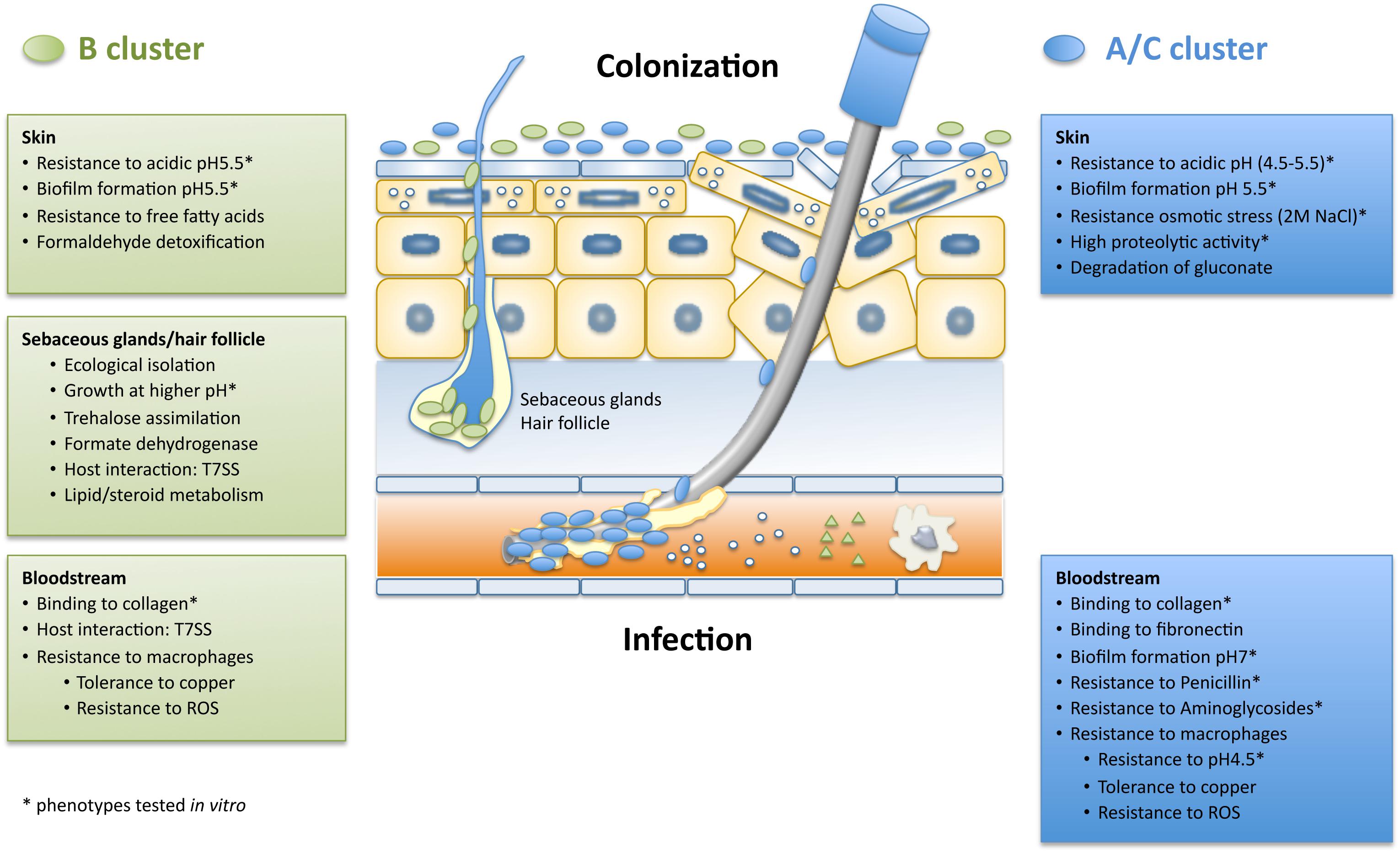
Figure 3. Ecological model of cluster A/C and B strains during a catheter-associated infection. This model was based on the pangenomic wide association studies and phenotypic results performed within this study. In each text box are listed the traits associated to each cluster that are important for survival and adaptation in a specific niche. A/C strains are represented by blue circles and B strains are represented by green circles.
Herein we have also produced a population and genome-wide based catalog of genes associated to A/C cluster and identified environmental conditions that favor the growth of A/C strains that could serve as the basis for future studies on S. epidermidis pathogenesis and eventually for the development of precision antimicrobial strategies. Additionally, the finding of a genetic marker for the more pathogenic genetic lineage can improve significantly the prognosis and management of medical-device related infections caused by this bacterium.
Materials and Methods
Bacterial Isolates
The bacterial collection comprised a total of 83 S. epidermidis isolates, collected from different countries (Argentina, n = 1; Bulgaria, n = 2; Cape Verde, n = 4; Colombia, n = 1; Denmark, n = 14; Greece, n = 3; Hungary, n = 2; Iceland, n = 13; Italy, n = 2; Japan, n = 2; Mexico, n = 5; Portugal, n = 31; Taiwan, n = 2; and Uruguay, n = 1) between 1996 and 2001 and representative of the population found in the hospital (n = 60) and community settings (n = 23). These isolates were selected to represent all the diversity in sequence types previously found among a temporal and geographically diverse collection of nosocomial and community-associated S. epidermidis isolates (Miragaia et al., 2007; Rolo et al., 2012). Isolates were classified as being from colonization (n = 45) or infection (n = 34) according to defined clinical parameters. Colonization isolates were recovered from several products including nasal swabs (n = 35), wounds (n = 6), sputum (n = 1), urine (n = 1) and blood (n = 1). Infection products were recovered from blood (n = 15), wounds (n = 8), urine (n = 3), sputum (n = 2), catheter (n = 2), and cerebrospinal fluid (n = 1). Four isolates had no information regarding the clinical significance (infection/colonization) and seven isolates had no information regarding the clinical source.
Whole-Genome Sequencing
All strains were previously sequenced (Meric et al., 2015) using a HiSeq 2500 machine (Illumina, San Diego, CA, United States) and genome sequences are available on NCBI BioProject PRJNA320931 and BIGSdb2 (Jolley and Maiden, 2010). One sample, ICE120 (assembly ID 293_SS_376) although aligning with the reference genome, had a very fragmented assembly and a cumulative length much superior to the expected for this species, suggesting a contamination of the DNA. For this reason, the sample was removed from the dataset and not included in the genomic analysis but still included in the phenotypic analysis.
Phylogeny Determination
The phylogeny of the strains was reconstructed by generating a Maximum-likelihood (ML) tree with RaxML v8.2.11 (Stamatakis et al., 2012), based on the core genome alignment without the recombining regions identified by Gubbins v2.3.4 (Croucher et al., 2015). Gubbins was run using the default parameters on the complete core genome alignment of the 82 strains. For the first iteration of the program, a Neighbor-joining tree of the same alignment was provided, being previously obtained with FastTree v2.1.10 (Price et al., 2010) using default parameters. The core genome alignment with filtered polymorphic sites was used with RaxML, using the GTRGAMMA substitution model and a bootstrap of 500 simulations. The final tree was visualized using MEGA v7.10.18 (Kumar et al., 2016). The final alignment resulted in 78512 bp polymorphic positions.
Average Nucleotide Distance
The average nucleotide distance within clusters and between clusters was calculated by averaging the number of single nucleotide polymorphisms (SNPs) of each pair of strains within a cluster or between all strains of each cluster, based on a matrix of distances obtained by Geneious vR8.1.9 based on the core genome alignment, after removal of the recombination regions generated with Gubbins.
Genomic Variation Assessment
To assess accessory genomic variation a reference pan genome approach was implemented. The 82 genomes were annotated with PROKKA (Seemann, 2014) using a database of eight published complete genomes of S. epidermidis: RP62A (Genbank: NC_002976.3); PM221 (Genbank: NZ_HG813242.1); SEI (Genbank: NZ_CP009046.1); 14.1.R1 (Genbank: CP018842.1); 1457 (Genbank: NZ_CP020463.1); ATCC 12228 (Genbank: NC_004461.1), ATCC 12228 (Genbank NZ_CP022247.1) and BPH0662 (Genbank: NZ_LT571449). The annotated genomes were then used in the pan genome creation using Roary v3.11.0 (Page et al., 2015), with locus defined by alleles with a minimum of 85% blastp identity. Scoary v.1.6.16 (Brynildsrud et al., 2016) was used to find significant associations between the genetic loci and the different phylogenetic clusters. If several genes within each cluster were attributed the same functional annotation, a unique gene was considered for cluster comparison, as most probably this correspond to different alleles of the same gene that fell below the 85% cut-off. The only exceptions were paralogs that were considered as different genes although having the same annotation. Only loci with a Benjamini–Hochberg p < 0.05 and an odds ratio (OR) > 1 were considered to be positively associated to a cluster. Additionally, they were only considered for analysis if they had no matching function in the genes positively associated to the other cluster.
Antimicrobial Susceptibility Testing
All bacterial isolates were tested for antimicrobial susceptibility trough disk diffusion method (Kirby-Bauer), according to the Clinical and Laboratory Standards Institute (CLSI) guidelines of 2014 (CLSI, 2014). Susceptibility was tested to a panel of 12 antibiotics: penicillin, erythromycin, clindamycin, linezolid, ciprofloxacin, quinupristin-dalfopristin, sulfamethoxazole-trimethoprim, tetracycline, fusidic acid, rifampicin, chloramphenicol, and gentamicin. In the case of fusidic acid, the European Committee on Antimicrobial Susceptibility Testing (EUCAST) guidelines from 2016 were used. Strains were considered as multidrug-resistant (MDR) if they were non-susceptible to at least three or more antimicrobial classes (Magiorakos et al., 2012).
The minimum inhibitory concentration (MIC) for oxacillin and vancomycin was determined by e-test (BioMérieux, Marcy-l’Étoile, France). Bacterial suspensions (0.5 McFarland) were inoculated in Mueller-Hinton agar (MHA, BBLTM, Becton Dickinson, Sparks, MD, United States), which in the case of oxacillin was supplemented with 2% NaCl, and incubated at 37°C for 48 h. Strains were considered to be susceptible to oxacillin if the MIC ≤ 0.25 μg/ml, resistant if the MIC ≥ 0.5 μg/ml; and were considered to be susceptible to vancomycin if the MIC ≤ 4 μg/ml, resistant if the MIC ≥ 32 μg/ml, according to 2014 CLSI guidelines (CLSI, 2014).
Growth Assays
Growth curves at pH 4.5, 5.5, 7.0, and 7.4 were defined for all isolates. All growth assays were performed using a microtiter reader (Infinite® 200 PRO series, Tecan Group Ltd., Männedorf, Switzerland) in 96-well microtiter plates. Briefly, overnight cultures were inoculated to an initial OD600 nm of 0.05 in tryptic soy broth (TSB, BactoTM, BBL, Becton Dickinson, Sparks, MD, United States) and grown with aeration (180 rpm) at 37°C for 30 h. Each strain was tested in triplicate and each experiment was repeated three times independently for different growth conditions. The pH of the medium was adjusted with HCl (37%) and the salt concentration was adjusted with NaCl (Merck KGaA, Darmstadt, Germany).
Biofilm Assays
The biofilm formation was tested at pH 5.5 and pH 7 and assays were performed on 96-well microtiter plates (Corning® 96 Well TC-Treated flat bottom, Sigma-Aldrich, St. Louis, MO, United States). Overnight cultures were inoculated to an initial OD600 nm of 0.05 in TSB and grown in static conditions with no aeration at 37°C for 24 h. After incubation, the contents of the wells were washed with water, heat fixed at 60°C, stained with 0.06% crystal violet and resuspended in acetic acid (30%). The optical density was measured at 595 nm in a microtiter plate reader (Infinite® 200 PRO series, Tecan Group Ltd., Männedorf, Switzerland). Each strain was tested in triplicate and each experiment was repeated three times independently. S. epidermidis strains RP62A and ATCC12228 were used as positive and negative controls of biofilm formation (Christensen et al., 1995), respectively.
Collagen Assays
Staphylococcus epidermidis adhesion to collagen was assayed by adapting the protocol described by Bowden et al. (2002). Briefly, early log-phase S. epidermidis cultures (OD600 between 0.3 and 0.7) were harvested and centrifuged (5000 g for 5 min). Cells were washed, resuspended in PBS, adjusted to a final OD600 of 1 and inoculated in 96-well microtiter plates coated with collagen I (CorningTM BioCoatTM Collagen I 96-well Clear Flat Bottom TC-treated Microplate, VWR, Radnor, PA, United States) for 2 h at room temperature (RT). After gentle washes with PBS, adherent cells were fixed with 25% (v/v) aqueous formaldehyde and incubated at RT for 30 min. The plates were washed gently with PBS, stained with 0.5% crystal violet for 5 min, washed again and read on a microtiter plate reader (Infinite® 200 PRO series, Tecan Group Ltd., Männedorf, Switzerland) at 595 nm.
Antioxidant Capacity Assay
Overnight cultures were adjusted to 0.1 (OD650) in TSB and let grow to 0.7 (OD650), incubated with or without H2O2 (30%, VWR, Radnor, PA, United States) for 20 min and harvested by pelleting. Cells were washed twice, resuspended in with 1x PBS and lysed with 2.5 μl lysozyme (20 mg/ml, Sigma-Aldrich, St. Louis, MO, United States), 5 μL lysostaphin (10 mg/ml, Ambi products LLC, Lawrence, NY, United States), and 3 μl RNAase (10 mg/ml, Sigma-Aldrich, St. Louis, MO, United States) for 2 h and the lysate was harvested after centrifugation at 4°C and 5000 g for 10 min. Antioxidant capacity of the samples was tested using the Total Antioxidant Capacity Assay kit (Sigma-Aldrich, St. Louis, MO, United States), following the manufacturer’s instructions.
Surface Charge Assay
The protocol used to assay the surface charge was adapted from Peschel et al. (1999). Bacterial cultures were grown overnight and harvested by centrifugation (5 min, 5000 g). Cells were washed twice with MOPS buffer (20 mM, pH 7) and resuspended in the same buffer to 15 (OD600). Cell suspensions and cytochrome c (1 mg/ml) were mixed in equal volumes and incubated for 10 min at RT (protected from light and with continuous soft agitation). Cells were centrifuged (13,000 rpm, 3 min) and unbound cytochrome c was measured by absorbance (OD410). The higher the absorbance value, the more positively charged is the cell envelope.
Proteolysis Assay
Overnight cultures were grown to 0.7 (OD600) in TSB and were spotted onto 10% skimmed milk agar (Sigma-Aldrich, St. Louis, MO, United States). Plates were incubated at 37°C for 24–48 h and lysis halos were measured from the border of the spotted bacterial growth until the border of the halo. The strains were categorized into three percentiles (high, medium, and low) depending on the size of the halos produced.
Phenotypic Assays Statistical Analysis
The statistical analysis was performed using the software GraphPad Prism 6. The significance of the differences between cluster A/C and cluster B regarding growth rates, biofilm production, collagen adhesion, cell wall net charge and antioxidant capacity was assessed using the two-tailed non-parametric Student’s t-test. The significance of difference regarding proteolysis and antimicrobial resistance was analyzed using Chi-square test. A p-value < 0.05 was considered statistically significant.
Data Availability
Genome sequences used in this study are available on NCBI BioProject PRJNA320391 and BIGSdb (https://www.sheppardlab.com/resources/).
Ethics Statement
Isolates from colonization in the hospital in Portugal were all obtained from nasal screening of patients from Hospital da Força Aérea (Lisbon, Portugal). Collection of these isolates was performed with approval from the local medical Ethics Committee (“Comissão de Ética para a Sáude da Força Aérea,” Hospital da Força Aérea, Lisboa, Portugal) and Infection Control Committee ("Comissão de Higiene Hospitalar do Hospital da Força Aérea," Hospital da Força Aérea, Lisboa, Portugal) and written informed consent was not required, according to Ethics Committee-approved guidelines. Isolates from colonization in the community were obtained from screening of draftees attending Centro de Formação da Ota (Lisbon, Portugal), which was performed with written informed consent and approval from all the necessary military authorities. Nosocomial strains from other countries were collected as part of the hospital routine diagnostic testing. The strains were de-identified and analyzed anonymously and the strains, not a human, were studied. Ethical approval and informed consent were thus not required.
Author Contributions
DE cultured the isolates and performed the phenotypic experiments. DE and MM carried out the data analysis and interpretation, and wrote the manuscript. CM and JC were involved in the data analysis and manuscript revision. GM and SS performed the sequencing of the isolates and reviewed the manuscript. RS and HL helped in the writing and revision of the manuscript. All authors read and approved the final manuscript.
Funding
DE and CM were supported by Ph.D. grants PD/BD/52206/2013 and SFRH/BD/129483/2017, respectively, from the Fundação para a Ciência e Tecnologia (FCT). This work was partially supported by project PTDC/FIS-NAN/0117/2014, project PTDC/CVT-CVT/29510/2017, project PTDC/BIA-MIC/31645/2017, and project EXPOSE - SAICT-POL/23222/2016 from FCT; Projects LISBOA-01-0145-FEDER-007660 (Microbiologia Molecular, Estrutural e Celular) and UID/Multi/04378/2019) funded by FEDER funds through COMPETE2020 - Programa Operacional Competitividade e Internacionalização (POCI); by ONEIDA project (LISBOA-01-0145-FEDER- 016417) co-funded by FEEI - “Fundos Europeus Estruturais e de Investimento” from “Programa Operacional Regional Lisboa2020” and by national funds through FCT; Operacional Competitividade e Internacionalização, Programa Operacional Regional de Lisboa (FEDER) and Fundação para a Ciência e a Tecnologia.
Conflict of Interest Statement
The authors declare that the research was conducted in the absence of any commercial or financial relationships that could be construed as a potential conflict of interest.
Supplementary Material
The Supplementary Material for this article can be found online at: https://www.frontiersin.org/articles/10.3389/fmicb.2019.01971/full#supplementary-material
FIGURE S1 | R-plot depicting the open pan genome of S. epidermidis.
FIGURE S2 | Performance of strains from cluster A/C and B in the collagen binding assay (A), antioxidant capacity assay (B), surface charge assay (C), and proteolytic capacity assay (D).
FIGURE S3 | Antimicrobial susceptibility testing of A/C and B cluster strains. (A) Antimicrobial susceptibility profile of cluster A/C and B strains by disk diffusion assay; (B) Minimum inhibitory concentration for oxacillin of cluster A/C and B MRSE strains by e-test. Legend: penicillin (PEN), oxacillin (OXA), erythromycin (ERY), clindamicin (DA), cloramphenicol (C), rifampicin (RIF), linezolid (LZD), ciprofloxacin (CIP), quinopristin-dalfopristin (QD), trimethoprim-sulphamethoxazole (SXT), tetracyclin (TET), fusidic acid (FD), gentamicin (GEN), and vancomycin (VAN).
TABLE S1 | Nucleotide distance matrix generated in Geneious based on the core genome alignment without the recombination regions.
TABLE S2 | List of genes composing the pan genome generated by Roary.
TABLE S3 | List of genes positively associated to cluster A/C (Benjamini–Hochberg p < 0.05 and OR > 1).
TABLE S4 | List of genes positively associated to cluster B (Benjamini–Hochberg p < 0.05 and OR > 1).
TABLE S5 | Summary of the epidemiological and phenotypic characteristics of cluster A/C and cluster B strains.
Footnotes
References
Akira, S., Uematsu, S., and Takeuchi, O. (2006). Pathogen recognition and innate immunity. Cell 124, 783–801 doi: 10.1016/j.cell.2006.02.015
Arrecubieta, C., Lee, M. H., Macey, A., Foster, T. J., and Lowy, F. D. (2007). SdrF, a Staphylococcus epidermidis surface protein, binds type I collagen. J. Biol. Chem. 282, 18767–18776. doi: 10.1074/jbc.m610940200
Basler, M., Ho, B. T., and Mekalanos, J. J. (2013). Tit-for-tat: type VI secretion system counterattack during bacterial cell-cell interactions. Cell 152, 884–894. doi: 10.1016/j.cell.2013.01.042
Bouslimani, A., Porto, C., Rath, C. M., Wang, M., Guo, Y., Gonzalez, A., et al. (2015). Molecular cartography of the human skin surface in 3D. Proc. Nat. Acad. Sci. U. S. A. 112, E2120–E2199.
Bowden, M. G., Visai, L., Longshaw, C. M., Holland, K. T., Speziale, P., and Hook, M. (2002). Is the GehD lipase from Staphylococcus epidermidis a collagen binding adhesin? J. Biol. Chem. 277, 43017–43023. doi: 10.1074/jbc.m207921200
Brynildsrud, O., Bohlin, J., Scheffer, L., and Eldholm, V. (2016). Rapid scoring of genes in microbial pan-genome-wide association studies with Scoary. Genome Biol. 17:238.
Burgos-Barragan, G., Wit, N., Meiser, J., Dingler, F. A., Pietzke, M., Mulderrig, L., et al. (2017). Mammals divert endogenous genotoxic formaldehyde into one-carbon metabolism. Nature 548, 549–554. doi: 10.1038/nature23481
Burts, M. L., DeDent, A. C., and Missiakas, D. M. (2008). EsaC substrate for the ESAT-6 secretion pathway and its role in persistent infections of Staphylococcus aureus. Mol. Microbiol. 69, 736–746. doi: 10.1111/j.1365-2958.2008.06324.x
Cao, Z., Casabona, M. G., Kneuper, H., Chalmers, J. D., and Palmer, T. (2016). The type VII secretion system of Staphylococcus aureus secretes a nuclease toxin that targets competitor bacteria. Nat. Microbiol. 2:16183.
Chamberlain, N. R., Mehrtens, B. G., Xiong, Z., Kapral, F. A., Boardman, J. L., and Rearick, J. I. (1991). Correlation of carotenoid production, decreased membrane fluidity, and resistance to oleic acid killing in Staphylococcus aureus 18Z. Infect. Immun. 59, 4332–4337.
Chaturvedi, K. S., and Henderson, J. P. (2014). Pathogenic adaptations to host-derived antibacterial copper. Front. Cell. Infect. Microbiol. 4:3. doi: 10.3389/fcimb.2014.00003.
Chen, N. H., Djoko, K. Y., Veyrier, F. J., and McEwan, A. G. (2016). Formaldehyde stress responses in bacterial pathogens. Front. Microbiol. 7:257. doi: 10.3389/fmicb.2016.00257.
Cherifi, S., Byl, B., Deplano, A., Nagant, C., Nonhoff, C., Denis, O., et al. (2014). Genetic characteristics and antimicrobial resistance of Staphylococcus epidermidis isolates from patients with catheter-related bloodstream infections and from colonized healthcare workers in a Belgian hospital. Annals of Clinical Microbiology And Antimicrobials. 13:20 doi: 10.1186/1476-0711-13-20
Cherifi, S., Byl, B., Deplano, A., Nonhoff, C., Denis, O., and Hallin, M. (2013). Comparative epidemiology of Staphylococcus epidermidis isolates from patients with catheter-related bacteremia and from healthy volunteers. J. Clin. Microbiol. 51, 1541–1547. doi: 10.1128/jcm.03378-12
Cheung, G. Y., Rigby, K., Wang, R., Queck, S. Y., Braughton, K. R., Whitney, A. R., et al. (2010). Staphylococcus epidermidis strategies to avoid killing by human neutrophils. Plos Pathog. 6:e1001133. doi: 10.1371/journal.ppat.1001133.
Christensen, G. D., Baldassarri, L., and Simpson, W. A. (1995). Methods for studying microbial colonization of plastics. Methods Enzymol. 253, 477–500. doi: 10.1016/s0076-6879(95)53040-1
Christensen, G. J., Scholz, C. F., Enghild, J., Rohde, H., Kilian, M., Thurmer, A., et al. (2016). Antagonism between Staphylococcus epidermidis and Propionibacterium acnes and its genomic basis. BMC Genomics 17:152. doi: 10.1186/s12864-016-2489-5.
Ciofu, O., Rojo-Molinero, E., Macia, M. D., and Oliver, A. (2017). Antibiotic treatment of biofilm infections. APMIS 125, 304–19.
Clauditz, A., Resch, A., Wieland, K. P., Peschel, A., and Gotz, F. (2006). Staphyloxanthin plays a role in the fitness of Staphylococcus aureus and its ability to cope with oxidative stress. Infect. Immun. 74, 4950–4953. doi: 10.1128/iai.00204-06
CLSI, (2014). Performance Standards for Antimicrobial Susceptibility Testing, Twenty-Fourth Informational Supplement, M100-S24. Wayne, PA: Clinical and Laboratory Standards Institute.
Conlan, S., Mijares, L. A., NISC Comparative Sequencing Program., Becker, J., Blakesley, R. W., Bouffard, G. G., et al. (2012). Staphylococcus epidermidis pan-genome sequence analysis reveals diversity of skin commensal and hospital infection-associated isolates. Genome Biol. 13:R64.
Corvaglia, A. R., Francois, P., Hernandez, D., Perron, K., Linder, P., and Schrenzel, J. (2010). A type III-like restriction endonuclease functions as a major barrier to horizontal gene transfer in clinical Staphylococcus aureus strains. Proc. Nat. Acad. Sci. U.S.A. 107, 11954–11958. doi: 10.1073/pnas.1000489107
Costello, E. K., Lauber, C. L., Hamady, M., Fierer, N., Gordon, J. I., and Knight, R. (2009). Bacterial community variation in human body habitats across space and time. Science 326, 1694–1697. doi: 10.1126/science.1177486
Croucher, N. J., Page, A. J., Connor, T. R., Delaney, A. J., Keane, J. A., Bentley, S. D., et al. (2015). Rapid phylogenetic analysis of large samples of recombinant bacterial whole genome sequences using Gubbins. Nucleic Acids Res. 43:e15 doi: 10.1093/nar/gku1196
da Costa, M. S., Santos, H., and Galinski, E. A. (1998). An overview of the role and diversity of compatible solutes in Bacteria and Archaea. Adv. Biochem. Eng. Biotechnol. 61, 117–153. doi: 10.1007/bfb0102291
de Goffau, M. C., Yang, X., van Dijl, J. M., and Harmsen, H. J. (2009). Bacterial pleomorphism and competition in a relative humidity gradient. Environmental Microbiology. 11, 809–822. doi: 10.1111/j.1462-2920.2008.01802.x
Farber, B. F., Kaplan, M. H., and Clogston, A. G. (1990). Staphylococcus epidermidis extracted slime inhibits the antimicrobial action of glycopeptide antibiotics. J. Infect. Dis. 161, 37–40. doi: 10.1093/infdis/161.1.37
Feldman, M. Y. (1973). Reactions of nucleic acids and nucleoproteins with formaldehyde. Prog. Nucleic Acid Res. Mol. Biol. 13:1–49. doi: 10.1016/s0079-6603(08)60099-9
Festa, R. A., and Thiele, D. J. (2012). Copper at the front line of the host-pathogen battle. PLoS Pathog. 8:e1002887. doi: 10.1371/journal.ppat.1002887.
Foster, T. J., and Hook, M. (1998). Surface protein adhesins of Staphylococcus aureus. Trends Microbiol. 6, 484–488. doi: 10.1016/s0966-842x(98)01400-0
Gallo, R. L., and Hooper, L. V. (2012). Epithelial antimicrobial defence of the skin and intestine. Nat. Rev. Immunol. 12, 503–516. doi: 10.1038/nri3228
Gomes, A. R., Vinga, S., Zavolan, M., and de Lencastre, H. (2005). Analysis of the genetic variability of virulence-related loci in epidemic clones of methicillin-resistant Staphylococcus aureus. Antimicrob. Agents Chemother. 49, 366–379. doi: 10.1128/aac.49.1.366-379.2005
Gordon, R. J., Miragaia, M., Weinberg, A. D., Lee, C. J., Rolo, J., Giacalone, J. C., et al. (2012). Staphylococcus epidermidis colonization is highly clonal across US cardiac centers. J. Infect. Dis. 205, 1391–1398. doi: 10.1093/infdis/jis218
Grice, E. A., Kong, H. H., Conlan, S., Deming, C. B., Davis, J., Young, A. C., et al. (2009). Topographical and temporal diversity of the human skin microbiome. Science 324, 1190–1192. doi: 10.1126/science.1171700
Grundmann, H., Aanensen, D. M., van den Wijngaard, C. C., Spratt, B. G., Harmsen, D., and Friedrich, A. W. (2010). Geographic distribution of Staphylococcus aureus causing invasive infections in Europe: a molecular-epidemiological analysis. PLoS Med. 7:e1000215. doi: 10.1371/journal.pmed.1000215.
Hartford, O., O’Brien, L., Schofield, K., Wells, J., and Foster, T. J. (2001). The Fbe (SdrG) protein of Staphylococcus epidermidis HB promotes bacterial adherence to fibrinogen. Microbiology 147(Pt 9), 2545–2552. doi: 10.1099/00221287-147-9-2545
Heilmann, C., Schweitzer, O., Gerke, C., Vanittanakom, N., Mack, D., and Gotz, F. (1996). Molecular basis of intercellular adhesion in the biofilm-forming Staphylococcus epidermidis. Mole. Microbiol. 20, 1083–10891.
Hellmark, B., Soderquist, B., Unemo, M., and Nilsdotter-Augustinsson, A. (2013). Comparison of Staphylococcus epidermidis isolated from prosthetic joint infections and commensal isolates in regard to antibiotic susceptibility, agr type, biofilm production, and epidemiology. Int. J. Med. Microbiol. 303, 32–39. doi: 10.1016/j.ijmm.2012.11.001
Jolley, K. A., and Maiden, M. C. (2010). BIGSdb: scalable analysis of bacterial genome variation at the population level. BMC Bioinfom. 11:595. doi: 10.1186/1471-2105-11-595.
Kammeyer, A., and Luiten, R. M. (2015). Oxidation events and skin aging. Ageing Res. Rev. 21, 16–29. doi: 10.1016/j.arr.2015.01.001
Katayama, Y., Zhang, H. Z., Hong, D., and Chambers, H. F. (2003). Jumping the barrier to beta-lactam resistance in Staphylococcus aureus. J. Bacteriol. 185, 5465–5472. doi: 10.1128/jb.185.18.5465-5472.2003
Kenny, J. G., Ward, D., Josefsson, E., Jonsson, I. M., Hinds, J., Rees, H. H., et al. (2009). The Staphylococcus aureus response to unsaturated long chain free fatty acids: survival mechanisms and virulence implications. PLoS One 4:e4344. doi: 10.1371/journal.pone.0004344.
Kets, E. P., Galinski, E. A., de Wit, M., de Bont, J. A., and Heipieper, H. J. (1996). Mannitol, a novel bacterial compatible solute in Pseudomonas putida S12. J. Bacteriol. 178, 6665–6670. doi: 10.1128/jb.178.23.6665-6670.1996
Khardori, N., Yassien, M., and Wilson, K. (1995). Tolerance of Staphylococcus epidermidis grown from indwelling vascular catheters to antimicrobial agents. J. Ind. Microbiol. 15, 148–51.
Kloos, W. E., and Schleifer, K. H. (1975). Isolation and characterization of staphylococci from human skin II. descriptions of four new species: staphylococcus warneri, staphylococcus capitis, staphylococcus hominis, and staphylococcus simulans. Int. J. Syst. Evol. Microbiol. 25, 62–79. doi: 10.1099/00207713-25-1-62
Knappe, J., and Sawers, G. (1990). A radical-chemical route to acetyl-CoA: the anaerobically induced pyruvate formate-lyase system of Escherichia coli. FEMS Microbiol. Rev. 6, 383–398. doi: 10.1016/0378-1097(90)90689-n
Kneuper, H., Cao, Z. P., Twomey, K. B., Zoltner, M., Jager, F., Cargill, J. S., et al. (2014). Heterogeneity in ess transcriptional organization and variable contribution of the Ess/Type VII protein secretion system to virulence across closely related Staphylocccus aureus strains. Mol. Microbiol. 93, 928–493.
Korea, C. G., Balsamo, G., Pezzicoli, A., Merakou, C., Tavarini, S., Bagnoli, F., et al. (2014). Staphylococcal Esx proteins modulate apoptosis and release of intracellular Staphylococcus aureus during infection in epithelial cells. Infect. Immun. 82, 4144–4153. doi: 10.1128/iai.01576-14
Kozitskaya, S., Cho, S. H., Dietrich, K., Marre, R., Naber, K., and Ziebuhr, W. (2004). The bacterial insertion sequence element IS256 occurs preferentially in nosocomial Staphylococcus epidermidis isolates: association with biofilm formation and resistance to aminoglycosides. Infect. Immun. 72, 1210–1215. doi: 10.1128/iai.72.2.1210-1215.2004
Kumar, S., Stecher, G, and Tamura, K. (2016). MEGA7: molecular evolutionary genetics analysis version 7.0 for bigger datasets. Mol. Biol. Evol. 33, 1870–1874. doi: 10.1093/molbev/msw054
Lai, Y., Villaruz, A. E., Li, M., Cha, D. J., Sturdevant, D. E., and Otto, M. (2007). The human anionic antimicrobial peptide dermcidin induces proteolytic defence mechanisms in staphylococci. Mol. Microbiol. 63, 497–506. doi: 10.1111/j.1365-2958.2006.05540.x
Leibig, M., Liebeke, M., Mader, D., Lalk, M., Peschel, A., and Gotz, F. (2011). Pyruvate formate lyase acts as a formate supplier for metabolic processes during anaerobiosis in Staphylococcus aureus. J. Bacteriol. 193, 952–962. doi: 10.1128/jb.01161-10
Lindgren, J. K., Thomas, V. C., Olson, M. E., Chaudhari, S. S., Nuxoll, A. S., Schaeffer, C. R., et al. (2014). Arginine deiminase in Staphylococcus epidermidis functions to augment biofilm maturation through pH homeostasis. J. Bacteriol. 196, 2277–2289. doi: 10.1128/jb.00051-14
Loskill, P., Pereira, P. M., Jung, P., Bischoff, M., Herrmann, M., Pinho, M. G., et al. (2014). Reduction of the peptidoglycan crosslinking causes a decrease in stiffness of the Staphylococcus aureus cell envelope. Biophys. J. 107, 1082–1089. doi: 10.1016/j.bpj.2014.07.029
Magiorakos, A. P., Srinivasan, A., Carey, R. B., Carmeli, Y., Falagas, M. E., Giske, C. G., et al. (2012). Multidrug-resistant, extensively drug-resistant and pandrug-resistant bacteria: an international expert proposal for interim standard definitions for acquired resistance. Clin. Microbiol. Infect. 18, 268–81.
Marraffini, L. A., and Sontheimer, E. J. (2008). CRISPR interference limits horizontal gene transfer in staphylococci by targeting DNA. Science 322, 1843–1845. doi: 10.1126/science.1165771
May, L., Klein, E. Y., Rothman, R. E., and Laxminarayan, R. (2014). Trends in antibiotic resistance in coagulase-negative staphylococci in the United States, 1999 to 2012. Antimicrob. Agents Chemother. 58, 1404–1409. doi: 10.1128/aac.01908-13
Meric, G., Miragaia, M., de Been, M., Yahara, K., Pascoe, B., Mageiros, L., et al. (2015). Ecological overlap and horizontal gene transfer in Staphylococcus aureus and Staphylococcus epidermidis. Genome Biol. Evol. 7, 1313–2138.
Miragaia, M., Couto, I., Pereira, S. F., Kristinsson, K. G., Westh, H., Jarlovm, J. O., et al. (2002). Molecular characterization of methicillin-resistant Staphylococcus epidermidis clones: evidence of geographic dissemination. J. Clin. Microbiol. 40, 430–438. doi: 10.1128/jcm.40.2.430-438.2002
Miragaia, M., Thomas, J. C., Couto, I., Enright, M. C., and de Lencastre, H. (2007). Inferring a population structure for Staphylococcus epidermidis from multilocus sequence typing data. J. Bacteriol. 189, 2540–2552. doi: 10.1128/jb.01484-06
Mishra, N. N., Liu, G. Y., Yeaman, M. R., Nast, C. C., Proctor, R. A., McKinnell, J., et al. (2011). Carotenoid-related alteration of cell membrane fluidity impacts Staphylococcus aureus susceptibility to host defense peptides. Antimicrob. Agents Chemother. 55, 526–531. doi: 10.1128/aac.00680-10
Ogo, K. (1985). Properties of pigments produced by S. epidermidis. J. Okayama Med. Assoc. 97, 749–759. doi: 10.4044/joma1947.97.7-8_749
Oh, J., Byrd, A. L., Deming, C., Conlan, S., NISC Comparative Sequencing Program, Kong, H. H., et al. (2014). Biogeography and individuality shape function in the human skin metagenome. Nature 514, 59–64. doi: 10.1038/nature13786
Ohtake, S., and Wang, Y. J. (2011). Trehalose: current use and future applications. J. Pharm. Sci. 100, 2020–2053. doi: 10.1002/jps.22458
Otto, M. (2009). Staphylococcus epidermidis–the “accidental” pathogen. Nat. Rev. Microbiol. 7, 555–567. doi: 10.1038/nrmicro2182
Otto, M. (2012). Molecular basis of Staphylococcus epidermidis infections. Semin. Immunopathol. 34, 201–214. doi: 10.1007/s00281-011-0296-2
Page, A. J., Cummins, C. A., Hunt, M., Wong, V. K., Reuter, S., Holden, M. T., et al. (2015). Roary: rapid large-scale prokaryote pan genome analysis. Bioinformatics 31, 3691–3693. doi: 10.1093/bioinformatics/btv421
Paget, M. S., and Buttner, M. J. (2003). Thiol-based regulatory switches. Annu. Rev. Genetics 37, 91–121. doi: 10.1146/annurev.genet.37.110801.142538
Paharik, A. E., Salgado-Pabon, W., Meyerholz, D. K., White, M. J., Schlievert, P. M., and Horswill, A. R. (2016). The spl serine proteases modulate Staphylococcus aureus protein production and virulence in a rabbit model of pneumonia. mSphere 1:e00208-16.
Park, J. Y., Kim, J. W., Moon, B. Y., Lee, J., Fortin, Y. J., Austin, F. W., et al. (2015). Characterization of a novel two-component regulatory system, HptRS, the regulator for the hexose phosphate transport system in Staphylococcus aureus. Infect. Immun. 83, 1620–1628. doi: 10.1128/iai.03109-14
Patti, J. M., Allen, B. L., McGavin, M. J., and Hook, M. (1994). MSCRAMM-mediated adherence of microorganisms to host tissues. Annual Review of Microbiology.; 48:585–617. doi: 10.1146/annurev.micro.48.1.585
Peschel, A., Otto, M., Jack, R. W., Kalbacher, H., Jung, G., and Gotz, F. (1999). Inactivation of the dlt operon in Staphylococcus aureus confers sensitivity to defensins, protegrins, and other antimicrobial peptides. J. Biol. Chem. 274, 8405–5410.
Philips, N., Samuel, P., Parakandi, H., Gopal, S., Siomyk, H., Ministro, A., et al. (2012). Beneficial regulation of fibrillar collagens, heat shock protein-47, elastin fiber components, transforming growth factor-beta1, vascular endothelial growth factor and oxidative stress effects by copper in dermal fibroblasts. Connect. Tissue Res. 53, 373–378. doi: 10.3109/03008207.2012.665970
Piwowarek, K., Lipinska, E., Hac-Szymanczuk, E., Kieliszek, M., and Scibisz, I. (2018). Propionibacterium spp.-source of propionic acid, vitamin b12, and other metabolites important for the industry. Appl. Microbiol. Biotechnol. 102, 515–538. doi: 10.1007/s00253-017-8616-7
Price, M. N., Dehal, P. S., and Arkin, A. P. (2010). FastTree 2–approximately maximum-likelihood trees for large alignments. Plos One. 5:e9490. doi: 10.1371/journal.pone.0009490.
Price-Whelan, A., Poon, C. K., Benson, M. A., Eidem, T. T., Roux, C. M., and Boyd, JM, et al. (2013). Transcriptional profiling of Staphylococcus aureus during growth in 2 M NaCl leads to clarification of physiological roles for Kdp and Ktr K+ uptake systems. mBio 4:e00407-13.
Qin, Z., Ou, Y., Yang, L., Zhu, Y., Tolker-Nielsen, T., Molin, S., et al. (2007). Role of autolysin-mediated DNA release in biofilm formation of Staphylococcus epidermidis. Microbiology 153(Pt 7), 2083–2092. doi: 10.1099/mic.0.2007/006031-0
Raju, K. S., Alessandri, G., Ziche, M., and Gullino, P. M. (1982). Ceruloplasmin, copper ions, and angiogenesis. J. Nat. Cancer Inst. 69, 1183–1188.
Reed, S. B., Wesson, C. A., Liou, L. E., Trumble, W. R., Schlievert, P. M., Bohach, G. A., et al. (2001). Molecular characterization of a novel Staphylococcus aureus serine protease operon. Infect. Immun. 69, 1521–1527. doi: 10.1128/iai.69.3.1521-1527.2001
Roberson, E. B., and Firestone, M. K. (1992). Relationship between Desiccation and exopolysaccharide production in a soil Pseudomonas sp. Appl. Environ. Microbiol. 58, 1284–1291.
Rolo, J., de Lencastre, H., and Miragaia, M. (2012). Strategies of adaptation of Staphylococcus epidermidis to hospital and community: amplification and diversification of SCCmec. J. Antimicrob. Chemother. 67, 1333–1341. doi: 10.1093/jac/dks068
Ruimy, R., Armand-Lefevre, L., Barbier, F., Ruppe, E., Cocojaru, R., Mesli, Y., et al. (2009). Comparisons between geographically diverse samples of carried Staphylococcus aureus. J. Bacteriol. 191, 5577–5583. doi: 10.1128/jb.00493-09
Sakinc, T., Woznowski, M., Ebsen, M., and Gatermann, S. G. (2005). surface-associated protein of Staphylococcus saprophyticus is a lipase. Infect. Immun. 73, 6419–6428. doi: 10.1128/iai.73.10.6419-6428.2005
Santos Sanches, I., Mato, R., de Lencastre, H., Tomasz, A., CEM/NET Collaborators and the International Collaborators, (2000). Patterns of multidrug resistance among methicillin-resistant hospital isolates of coagulase-positive and coagulase-negative staphylococci collected in the international multicenter study RESIST in 1997 and 1998. Microb. Drug Res. 6, 199–211. doi: 10.1089/mdr.2000.6.199
Sawers, R. G. (2005). Formate and its role in hydrogen production in Escherichia coli. Biochem. Soc. Trans. 33(Pt 1), 42–46. doi: 10.1042/bst0330042
Scharschmidt, T. C., and Fischbach, M. A. (2013). What lives on our skin: ecology, genomics and therapeutic opportunities of the skin microbiome. Drug Discov. Today Dis. Mech. 10, e83–e89. doi: 10.1016/j.ddmec.2012.12.003
Schittek, B, Hipfel, R, Sauer, B, Bauer, J, Kalbacher, H, and Stevanovic, S, et al. (2001). Dermcidin: a novel human antibiotic peptide secreted by sweat glands. Nat. Immunol. 2, 1133–1137. doi: 10.1038/ni732
Schmid-Wendtner, M. H., and Korting, H. C. (2006). The pH of the skin surface and its impact on the barrier function. Skin Pharmacol. Physiol. 19, 296–302. doi: 10.1159/000094670
Seemann, T. (2014). Prokka: rapid prokaryotic genome annotation. Bioinformatic 30, 2068–2069. doi: 10.1093/bioinformatics/btu153
Shi, Z., Xuan, C., Han, H., Cheng, X., Wang, J., Feng, Y., et al. (2014). Gluconate 5-dehydrogenase (Ga5DH) participates in Streptococcus suis cell division. Protein Cell 5, 761–769. doi: 10.1007/s13238-014-0074-8
Sieprawska-Lupa, M., Mydel, P., Krawczyk, K., Wojcik, K., Puklo, M., Lupa, B., et al. (2004). Degradation of human antimicrobial peptide LL-37 by Staphylococcus aureus-derived proteinases. Antimicrob. Agents Chemother. 48, 4673–4679. doi: 10.1128/aac.48.12.4673-4679.2004
Silver, A. C., Kikuchi, Y., Fadl, A. A., Sha, J., Chopra, A. K., and Graf, J. (2007). Interaction between innate immune cells and a bacterial type III secretion system in mutualistic and pathogenic associations. Proc. Nat. Acad. Sci. U.S.A. 104, 9481–6948.
Singh, B., Fleury, C., Jalalvand, F., and Riesbeck, K. (2012). Human pathogens utilize host extracellular matrix proteins laminin and collagen for adhesion and invasion of the host. FEMS Microbiol. Rev. 36, 1122–1180. doi: 10.1111/j.1574-6976.2012.00340.x
Singh, R., Ray, P., Das, A., and Sharma, M. (2010). Penetration of antibiotics through Staphylococcus aureus and Staphylococcus epidermidis biofilms. J. Antimicrob. Chemother. 65, 1955–1958. doi: 10.1093/jac/dkq257
Sitthisak, S., Howieson, K., Amezola, C., and Jayaswal, R. K. (2005). Characterization of a multicopper oxidase gene from Staphylococcus aureus. Applied and Environmental Microbiology. 71, 5650–5653. doi: 10.1128/aem.71.9.5650-5653.2005
Stamatakis, A., Aberer, A. J., Goll, C., Smith, S. A., Berger, S. A., and Izquierdo-Carrasco, F. (2012). RAxML-Light: a tool for computing terabyte phylogenies. Bioinformatics 28, 2064–2066. doi: 10.1093/bioinformatics/bts309
Stentzel, S., Teufelberger, A., Nordengrun, M., Kolata, J., Schmidt, F., van Crombruggen, K., et al. (2017). Staphylococcal serine protease-like proteins are pacemakers of allergic airway reactions to Staphylococcus aureus. J. Allergy Clin. Immunol. 139:492-500.e8.
Sweeney, N. J., Klemm, P., McCormick, B. A., Moller-Nielsen, E., Utley, M., Schembri, M. A., et al. (1996). The Escherichia coli K-12 gntP gene allows E. coli f-18 to occupy a distinct nutritional niche in the streptomycin-treated mouse large intestine. Infect. Immun. 64, 3497–3503.
Tang, X., Bai, Y., Duong, A., Smith, M. T., Li, L., and Zhang, L. (2009). Formaldehyde in China: production, consumption, exposure levels, and health effects. Environ. Int. 35, 1210–1224. doi: 10.1016/j.envint.2009.06.002
Thurlow, L. R., Joshi, G. S., Clark, J. R., Spontak, J. S., Neely, C. J., Maile, R., et al. (2013). Functional modularity of the arginine catabolic mobile element contributes to the success of USA300 methicillin-resistant Staphylococcus aureus. Cell Host Microbe 13, 100–107. doi: 10.1016/j.chom.2012.11.012
Uhlemann, A. C., Porcella, S. F., Trivedi, S., Sullivan, SB., Hafer, C., Kennedy, AD., et al. (2012). Identification of a highly transmissible animal-independent Staphylococcus aureus ST398 clone with distinct genomic and cell adhesion properties. mBio 3:e00027-12.
van de Belt, H., Neut, D., Schenk, W., van Horn, J. R., van der Mei, H. C., and Busscher, HJ. (2001). Infection of orthopedic implants and the use of antibiotic-loaded bone cements. Rev. Acta Orthop. Scand. 72, 557–751.
Vaudaux, P. E., Francois, P., Proctor, R. A., McDevitt, D., Foster, T. J., Albrecht, R. M., et al. (1995). Use of adhesion-defective mutants of Staphylococcus aureus to define the role of specific plasma proteins in promoting bacterial adhesion to canine arteriovenous shunts. Infect. Immun. 63, 585–590.
von Eiff, C., Peters, G., and Heilmann, C. (2002). Pathogenesis of infections due to coagulase-negative staphylococci. Lancet Infect. Dis. 2, 677–685.
Vuong, C., Voyich, J. M., Fischer, E. R., Braughton, K. R., Whitney, A. R., DeLeo, F. R., et al. (2004). Polysaccharide intercellular adhesin (PIA) protects Staphylococcus epidermidis against major components of the human innate immune system. Cell. Microbiol. 6, 269–275. doi: 10.1046/j.1462-5822.2004.00367.x
Waldron, D. E., and Lindsay, J. A. (2006). Sau1: a novel lineage-specific type I restriction-modification system that blocks horizontal gene transfer into Staphylococcus aureus and between S. Aureus isolates of different lineages. J. Bacteriol. 188, 5578–85.
Walther, B., Klein, K. S., Barton, A. K., Semmler, T., Huber, C., Merle, R., et al. (2018). Equine methicillin-resistant sequence type 398 Staphylococcus aureus (MRSA) harbor mobile genetic elements promoting host adaptation. Front. Microbiol. 9:2516. doi: 10.3389/fmicb.2018.02516.
Weiss, G., and Schaible, U. E. (2015). Macrophage defense mechanisms against intracellular bacteria. Immunol. Rev. 264, 182–203. doi: 10.1111/imr.12266
Widerstrom, M., Monsen, T., Karlsson, C., and Wistrom, J. (2006). Molecular epidemiology of meticillin-resistant coagulase-negative staphylococci in a Swedish county hospital: evidence of intra- and interhospital clonal spread. J. Hosp. Infect. 64, 177–83
Widerstrom, M., Monsen, T., Karlsson, C., Edebro, H., Johansson, A., and Wistrom, J. (2009). Clonality among multidrug-resistant hospital-associated Staphylococcus epidermidis in northern Europe. Scand. J. Infect. Dis. 41, 642–649. doi: 10.1080/00365540903146987
Wilson, M. (2005). Microbial Inhabitants of Humans: Their Ecology and Role in Health and Disease. Cambridge: Cambridge University Press
Wishart, D. S., Feunang, Y. D., Marcu, A., Guo, A. C., Liang, K., Vazquez-Fresno, R., et al. (2018). HMDB 4.0: the human metabolome database for 2018. Nucleic Acids Res. 46, D608–D617.
Wyke, A. W., Ward, J. B., Hayes, M. V., and Curtis, N. A. (1981). A role in vivo for penicillin-binding protein-4 of Staphylococcus aureus. Eur. J. Biochem. 119, 389–933.
Zahid, N., Schweiger, P., Galinski, E., and Deppenmeier, U. (2015). Identification of mannitol as compatible solute in Gluconobacter oxydans. Appl. Microbiol. Biotechnol. 99, 5511–5521. doi: 10.1007/s00253-015-6626-x
Zdzalik, M., Karim, AY, Wolski, K, Buda, P, Wojcik, K, and Brueggemann, S, et al. (2012). Prevalence of genes encoding extracellular proteases in Staphylococcus aureus - important targets triggering immune response in vivo. FEMS Immunol. Med. Microbiol. 66, 220–229. doi: 10.1111/j.1574-695x.2012.01005.x
Keywords: S. epidermidis, pan genome, GWAS, clonal lineages, pathogen, commensal
Citation: Espadinha D, Sobral RG, Mendes CI, Méric G, Sheppard SK, Carriço JA, de Lencastre H and Miragaia M (2019) Distinct Phenotypic and Genomic Signatures Underlie Contrasting Pathogenic Potential of Staphylococcus epidermidis Clonal Lineages. Front. Microbiol. 10:1971. doi: 10.3389/fmicb.2019.01971
Received: 02 April 2019; Accepted: 12 August 2019;
Published: 27 August 2019.
Edited by:
Ludmila Chistoserdova, University of Washington, United StatesReviewed by:
Karsten Becker, University of Münster, GermanyVasvi Chaudhry, University of Tübingen, Germany
Copyright © 2019 Espadinha, Sobral, Mendes, Méric, Sheppard, Carriço, de Lencastre and Miragaia. This is an open-access article distributed under the terms of the Creative Commons Attribution License (CC BY). The use, distribution or reproduction in other forums is permitted, provided the original author(s) and the copyright owner(s) are credited and that the original publication in this journal is cited, in accordance with accepted academic practice. No use, distribution or reproduction is permitted which does not comply with these terms.
*Correspondence: Maria Miragaia, miragaia@itqb.unl.pt