Corrigendum: HDAC1 Silence Promotes Neuroprotective Effects of Human Umbilical Cord-Derived Mesenchymal Stem Cells in a Mouse Model of Traumatic Brain Injury via PI3K/AKT Pathway
- 1School of Life Sciences, Zhengzhou University, Zhengzhou, China
- 2Henan Provincial People's Hospital, Zhengzhou, China
- 3Translational Medicine Center, Zhengzhou Central Hospital Affiliated to Zhengzhou University, Zhengzhou, China
- 4The First Affiliated Hospital of Zhengzhou University, Zhengzhou, China
- 5Stuyvesant High School, New York, NY, United States
- 6Department of Microbiology and Immunology, Einstein College of Medicine, Bronx, NY, United States
by Xu, L., Xing, Q., Huang, T., Zhou, J., Liu, T., Cui, Y., et al. (2019). Front. Cell. Neurosci. 12:498. doi: 10.3389/fncel.2018.00498
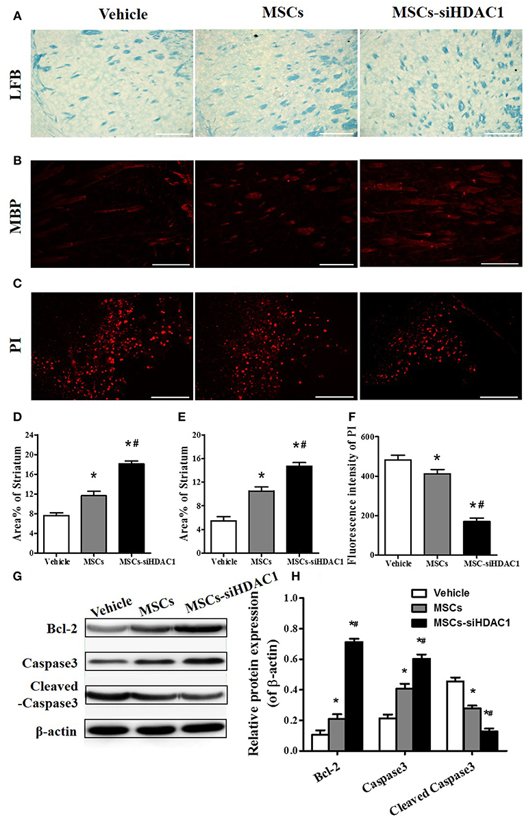
In the original article, there was a mistake in Figure 5C as published. The representative propidium iodide (PI) staining photo of the MSC-siHDAC1 group is incorrect. This photo was incorrectly chosen in the process of combining the figure.

Figure 5. HDAC1-silenced MSCs alleviated white matter injury and reduced cell death after TBI. (A) Representative images of luxol fast blue (LFB) staining. Scale bars = 100 μm. (B) Myelin basic protein (MBP) staining (red). Scale bars = 200 μm. (C) Representative propidium iodide (PI) staining (red) at 3 days after TBI. Scale bars = 100 μm. (D) Average area of LFB at 28 days after TBI. (E) Average area of MBP. (F) Quantitative analysis of PI fluorescence intensity in the injured cortex. (G) Western blotting and (H) densitometry measurement of Bcl-2, Caspase 3, and Cleaved caspase 3 in the lesion boundary zone of each group at 3 days post-injury. Data are presented as mean ± SEM. *p < 0.05 vs. Vehicle, #p < 0.05 vs. MSCs.
At the same time, we also reanalyzed the fluorescence intensity of PI. And, the results showed that MSCs-siHDAC1 more significantly decreased PI fluorescence intensity than that in the original article. The corrected Figure 5 appears below.
The authors apologize for this error and state that this does not change the scientific conclusions of the article in any way. The original article has been updated.
Keywords: histone deacetylase 1, human umbilical cord derived mesenchymal stem cells, traumatic brain injury, neuroprotection, PI3K/AKT
Citation: Xu L, Xing Q, Huang T, Zhou J, Liu T, Cui Y, Cheng T, Wang Y, Zhou X, Yang B, Yang GL, Zhang J, Zang X, Ma S and Guan F (2019) Corrigendum: HDAC1 Silence Promotes Neuroprotective Effects of Human Umbilical Cord-Derived Mesenchymal Stem Cells in a Mouse Model of Traumatic Brain Injury via PI3K/AKT Pathway. Front. Cell. Neurosci. 13:408. doi: 10.3389/fncel.2019.00408
Received: 12 June 2019; Accepted: 26 August 2019;
Published: 09 September 2019.
Edited by:
Thomas Fath, Macquarie University, AustraliaReviewed by:
Vladimir Sytnyk, University of New South Wales, AustraliaCopyright © 2019 Xu, Xing, Huang, Zhou, Liu, Cui, Cheng, Wang, Zhou, Yang, Yang, Zhang, Zang, Ma and Guan. This is an open-access article distributed under the terms of the Creative Commons Attribution License (CC BY). The use, distribution or reproduction in other forums is permitted, provided the original author(s) and the copyright owner(s) are credited and that the original publication in this journal is cited, in accordance with accepted academic practice. No use, distribution or reproduction is permitted which does not comply with these terms.
*Correspondence: Shanshan Ma, mashanshan84@163.com; Fangxia Guan, guanfangxia@126.com
†These authors have contributed equally to this work
 Ling Xu
 Qu Xing
 Tuanjie Huang
 Jiankang Zhou
 Tengfei Liu
 Yuanbo Cui
 Tian Cheng4
  Yaping Wang
 Xinkui Zhou
 Greta Luyuan Yang
 Xingxing Zang
 Shanshan Ma
 Fangxia Guan