 Maurizio Cundari
 Susanna Vestberg
 Peik Gustafsson
 Sorina Gorcenco
 Anders Rasmussen
- 1Department of Experimental Medical Science, Faculty of Medicine, Lund University, Lund, Sweden
- 2Unit of Neuropsychiatry, Hospital of Helsingborg, Helsingborg, Sweden
- 3Unit of Neurology, Hospital of Helsingborg, Helsingborg, Sweden
- 4Department of Psychology, Faculty of Social Science, Lund University, Lund, Sweden
- 5Child and Adolescent Psychiatry, Department of Clinical Sciences Lund, Medical Faculty, Lund University, Lund, Sweden
- 6Department for Clinical Sciences Lund, Neurology, Lund University, Skåne University Hospital, Lund, Sweden
The cerebellum plays a major role in balance, motor control and sensorimotor integration, but also in cognition, language, and emotional regulation. Several neuropsychiatric disorders such as attention deficit-hyperactivity disorder (ADHD), autism spectrum disorder (ASD), as well as neurological diseases such as spinocerebellar ataxia type 3 (SCA3) are associated with differences in cerebellar function. Morphological abnormalities in different cerebellar subregions produce distinct behavioral symptoms related to the functional disruption of specific cerebro-cerebellar circuits. The specific contribution of the cerebellum to typical development may therefore involve the optimization of the structure and function of cerebro-cerebellar circuits underlying skill acquisition in multiple domains. Here, we review cerebellar structural and functional differences between healthy and patients with ADHD, ASD, and SCA3, and explore how disruption of cerebellar networks affects the neurocognitive functions in these conditions. We discuss how cerebellar computations contribute to performance on cognitive and motor tasks and how cerebellar signals are interfaced with signals from other brain regions during normal and dysfunctional behavior. We conclude that the cerebellum plays a role in many cognitive functions. Still, more clinical studies with the support of neuroimaging are needed to clarify the cerebellum’s role in normal and dysfunctional behavior and cognitive functioning.
Introduction
Until recently, the cerebellum was regarded as a brain structure engaged mainly in motor control. Studies dating back to the early 19th century showed that cerebellar damage in pigeons resulted in an inability to fly due to uncoordinated wing movements (Flourens, 1824). After the cerebellum’s role in motor control was confirmed in studies on monkeys, it became a fundamental truth in clinical neurology and brain sciences (Russell and Horsley, 1894). Since then, numerous studies from the 20th and 21st centuries have reaffirmed the cerebellum as a structure that plays a vital role in motor control and motor learning (Manto et al., 2012). However, though the fields of cognitive neuroscience and experimental neuropsychology did not emerge until the early 20th century, other researchers in the 19th century reported emotional and intellectual impairment in patients with cerebellar agenesis (Fisher, 1839). Yet, it wasn’t until the 1990s that researchers refocused on the cerebellum’s non-motor functions (Schmahmann, 1991). Studies on cerebellar patients revealed significant impairments in executive function and attention (Stoodley and Schmahmann, 2010). Cerebellar abnormalities also have effects on other neurocognitive domains. These include language, visuospatial perception, visual construction ability, episodic memory, and social cognition (Hoche et al., 2016; Mariën and Borgatti, 2018; Slapik et al., 2019; Ray et al., 2020).
In line with these clinical findings, neuroimaging studies reveal that the cerebellum is anatomically and functionally interconnected with areas of the brain that are important for cognitive and emotional functions (Stoodley and Schmahmann, 2010). There is an abundance of mutual connections between the cerebellum and other areas of the brain, including the prefrontal cortex, the posterior parietal cortex, and the basal ganglia (Middleton and Strick, 1994; Kelly and Strick, 2003; Bostan et al., 2013).
This can explain why disorders affecting the cerebellum also lead to disrupted affective and cognitive functions, such as the cerebellar cognitive affective syndrome (CCAS) (Schmahmann and Sherman, 1998; Stoodley and Schmahmann, 2010; Ahmadian et al., 2019). Moreover, the profile and severity of CCAS symptoms varies in a predictable pattern depending on the pathological process and what part of the cerebellum is affected (Argyropoulos et al., 2020).
Here, we review literature on the anatomy and physiology of the cerebellum, and the neurocognitive functions of three conditions that have been linked to cerebellar function: SCA3, ADHD, and ASD. SCA3 is a neurological disease where cerebellar degeneration is clearly and robustly demonstrated, ADHD and ASD are neuropsychiatric disorders that have been associated with cerebellar abnormalities, but the extent of cerebellar involvement is not as pronounced as in the case of SCA3 (Riva et al., 2013; Wan et al., 2020; Duan et al., 2021; Stezin et al., 2021). We selected these three conditions to highlight the cerebellum’s role in cognition and how cognitive effects vary according to the severity of cerebellar abnormalities, more evident in SCA3 and lighter in ADHD and ASD.
We attempt to link these findings to contemporary theories and models of the cerebellum, and we discuss how symptoms may arise from interactions between the cerebellum and other brain parts that influence cognitive functions. We then discuss potential clinical implications from knowledge about the cerebellum’s involvement in cognitive functions. A cerebellar-dependent neuropsychological test battery could be a valuable tool in the diagnostic process when data from medical examinations such as biomarkers and neuroimaging data are missing or are unclear. This could lead to an improved understanding of the neurocognitive functions of these patient groups, and, ultimately, better treatment.
Methods
Search strategy and study selection
We performed a literature search in two electronic databases: PubMed and Embase. Through these searches, we identified articles supplying information on the neurocognitive profile, neurocognitive functions, and cerebellar functions of patients with three specific diagnoses ADHD, ASD, and SCA3.
Original articles and chapters reporting relevant studies on humans and animals were included. We used terms such as “ADHD,” “ASD,” and “SCA3” coupled with terms related to the cerebellum, cerebellar functions and to neurocognitive domains and neurocognitive profiles, such as “cognition,” “intelligence,” “visual perception/construction,” “processing speed,” “sensorimotor integration,” “executive function,” and “memory.”
Cerebellum
The cerebellum (literally “little brain” in Latin) contains 80,2% of all neurons in the human brain as well as 19% of all the non-neural cells in the brain (Azevedo et al., 2009). The anterior lobe of the cerebellum receives afferents from the spinal cord through the spinocerebellar tracts and from the cerebral cortex via corticopontine projections (Brodal, 1978; Hartmann-von Monakow et al., 1981; Schmahmann et al., 2004). The posterior lobe receives input mainly from the brainstem and cerebral cortex (Siegel and Sapru, 2006). The cerebellar vermis and fastigial nucleus are interconnected with the vestibular nuclei and other brainstem nuclei engaged in motor control, gait, and equilibrium, and with brainstem nuclei linked with limbic and paralimbic cortical and subcortical regions (Voogd, 2004).
The cerebellum is not only connected with structures concerned with motor control. Large parts of the cerebellum forms reciprocal connections with association areas of the cerebral cortex including the prefrontal cortex, posterior parietal cortex, superior temporal polymodal regions, cingulate gyrus, and posterior parahippocampal area (Strick et al., 2009; Bostan et al., 2013). The anatomical circuits linking the cerebellum with the cerebral cortex are organized in a two-stage feed-forward loop (the corticopontine and pontocerebellar projection) and a two-stage feedback loop (cerebellothalamic-thalamocortical projection). These loops are constituted by multiple parallel but partially overlapping subcircuits that link different areas of the cerebral cortex with specific regions of the cerebellum (Brodal, 1978; Voogd and Glickstein, 1998; Voogd, 2004; Apps and Garwicz, 2005). The cerebellum is also mutually interconnected with the basal ganglia via oligosynaptic loops (Hintzen et al., 2018). These anatomical connections hint that the cerebellum plays a role that goes beyond motor control. Indeed, as we will discuss below, evidence from recent decades suggests that the cerebellum is highly involved in a wide spectrum of cognitive functions.
Output from the cerebellar cortex goes via the cerebellar nuclei. The medial (fastigial) nucleus projects mainly to the brainstem and spinal cord; the interposed (comprising the emboliform and globose) nucleus targets mainly the midbrain and the thalamus; and the lateral (dentate) nucleus projects mainly to the thalamus. Some neurons in the cerebellar nuclei project directly to the red nucleus, which then projects to other nuclei that control muscles. This is how the cerebellum can elicit blink responses even in the absence of the cortex (Hesslow and Yeo, 2002). More recently, it was discovered that the cerebellar nuclei also contain neurons that project to the Purkinje cells forming the nucleo-cortical cerebellar pathway (Ankri et al., 2015). Apart from the inhibitory input from the Purkinje cells, the cerebellar nuclei also receive excitatory input from collaterals of both mossy fibers and climbing fibers (Ten Brinke et al., 2017).
Functions of the cerebellum
Knowledge of cerebellar functions has been derived mostly from studying natural and experimental ablative lesions (Fine et al., 2002). The cerebellum plays a crucial role in making predictions and creating feedforward models that guide our behavior (Wolpert et al., 1998; Welniarz et al., 2021). Consequently, cerebellar damage makes movements jerky, slow, and uncoordinated–as is evident in ataxia and dystonia (Therrien and Bastian, 2015). In line with the cerebellum’s role in motor coordination, disruption of the ionic homeostasis in cerebellar neurons has been shown to evoke motor impairments in mouse model studies (Hoxha et al., 2018). The impairments range from gross abnormalities to more specific impairments. For example, disruption of the high-voltage activated Ca2+-influx in Purkinje cells severely disrupts action potential firing and motor behavior in mouse model studies (Tara et al., 2018). Likewise, deletion of several chloride channels and transporters in mouse models has been associated with motor abnormalities (Poroca et al., 2017). For instance, knockout mouse models with deletion of GABAA receptors from Purkinje cells did not show ataxia, but they displayed significant impairments in several aspects of locomotion, motor performance and motor learning (Wulff et al., 2009).
Clinical research from the last three decades show that the cerebellum is not merely concerned with motor functions. The human cerebellum is linked with cerebral association areas concerned with intellect, social cognition, perception, recognition, forwarding and encoding of emotional information, and the regulation of emotional states in relation to cognitive, motor and social behaviors (Stoodley and Schmahmann, 2009a,b, Stoodley et al., 2010; Adamaszek et al., 2017). In line with the anatomical connections between the cerebellum, the basal ganglia (K B et al., 2022), and the cerebral cortex (Allen and Tsukahara, 1974; Holdefer et al., 2000; Strick et al., 2009), the cerebellum has been shown to play a role in many cognitive and emotional functions (Reeber et al., 2013). The limbic/cognitive cerebellum is identified in the cerebellar posterior lobe; but also, others areas of the cerebellum contribute to cognition like the sensorimotor cerebellum represented in the anterior lobe (Schmahmann, 2019). Furthermore, cerebellar damage is associated with lesions in the posterior lobe resulting in deficits in executive functions, visuospatial processing, language abilities and affect regulation (Karatekin et al., 2000; Grill et al., 2004; Manes et al., 2009). Other disorders linked to morphological abnormalities in the cerebellum, such as ASD, schizophrenia, and ADHD have also been linked with affective dysregulation (Bernard and Mittal, 2014; D’Mello et al., 2015; Stoodley, 2016; Kim et al., 2021). The cerebellum also contributes to visual cognition. Patients with degenerative cerebellar disorders showed impairments on mental rotation, an operation that requires continuous manipulation of a visual representation (McDougle et al., 2022). Further, the cerebellum is involved in face and emotion processing via its connections with limbic and cortical areas important for processing facial emotions perception (Fusar-Poli et al., 2009; Schutter et al., 2009). The cerebellum is involved as well in timing (Ivry and Keele, 1989).
Neuroimaging, cognitive and behavioral findings
Neuroimaging studies have verified that the cerebellum contributes to a broad range of cognitive functions, like perception, language, working memory, cognitive control, and social cognition (Diedrichsen et al., 2019). A review of clinical, experimental and neuroimaging studies shows that sensorimotor tasks have been linked to the anterior lobe, while higher function tasks, including language, executive function, and working memory, have been linked to the posterior lobe of the cerebellum. Processing of emotions was localized to vermal lobule VII and lateral posterior hemisphere (Stoodley and Schmahmann, 2009a). The involvement of the cerebellum in these processes is due to functional connections with frontal lobes via the cerebello-thalamo-cortical and cortico-ponto-cerebellar loops (Middleton and Strick, 1994; Kelly and Strick, 2003).
Functional magnetic resonance imaging (fMRI) shows cerebellar activation during many cognitive, affectional, and social tasks in healthy participants, although there is significant inter-participant variability in the pattern of topographical activation on a specific task (King et al., 2019). A meta-analysis concluded that specific parts of the cerebellum were activated during cognitive tasks and music elaboration/processing (E et al., 2014). One of the first positron emission tomography (PET) studies examining the cerebellum by Petersen et al. (1988) showed activation in the right lateral cerebellum when participants generated words. The cerebellum is also involved in lexical retrieval, syntax, aphasia, reading, writing and metalinguistic skills (De Smet et al., 2013). Other cognitive functions like working memory, social cognition, emotional memory, semantic memory and visuospatial abilities are also linked to the cerebellum (Ashida et al., 2019; Slapik et al., 2019; Van Overwalle et al., 2020; Gatti et al., 2021; Fastenrath et al., 2022). Another fMRI study showed that the dentate, the output nucleus of the cerebellum, was activated by cognitive processing, for example, during a realization of a puzzle that required multiple cognitive processes (Kim et al., 1994).
Petacchi et al. (2005) analyzed studies that used fMRI and PET techniques, their analysis shows that specific regions in the cerebellum are activated during auditory tasks and contribute to sensory auditory processing. Recent results demonstrated that the cerebellum shows increased activity during enhanced emotional memory encoding; the midline cerebellum seems to be activated during recall of emotional personal life episodes, remarking an aspect of this region in emotional memory retrieval (Fastenrath et al., 2022).
Neuroimaging studies have shown that explicit motor timing activates the cerebellum, the supplementary motor area, basal ganglia, right-inferior frontal, and parietal cortices (Avanzino et al., 2016). Timing ability affects sequential rhythmic movements, motor control, and many cognitive tasks used to assess executive functions (Homack et al., 2005). The cerebellum is essential in timing-dependent learning processes. It plays a role in finger tapping, in which the participant must maintain a rhythm, and eyeblink conditioning, a simple form of associative motor learning in which subjects learn to associate a neutral stimulus with a reflex-eliciting stimulus (Gerwig et al., 2007; Bracha et al., 2009). The cerebellum, together with other brain areas like the primary sensorimotor cortex, the supplementary motor area, and the occipital cortices, are necessary to complete the finger-tapping task in response to visual cues (Turesky et al., 2018). Previous studies have brought to light that the main neuronal plasticity responsible for generating the conditioned eyeblink response takes place in specific regions of the cerebellum (Steinmetz, 2000; Thürling et al., 2015).
Spinocerebellar ataxia type 3 (SCA3)
Ataxia derives from Greek and means “lack of order.” SCA is a rare inherited (autosomal dominant), progressive, neurodegenerative, and heterogeneous disease mainly affecting the cerebellum. SCA is the used nomenclature for Spinocerebellar Ataxia from type 1 to type 48 for autosomal dominant cerebellar ataxia a (Kuo, 2019). SCA3 is the world’s most common, dominantly inherited ataxia. Ataxia is characterized by decreased voluntary muscle coordination and impairment of movement control that affects gait stability, speech, and eye movement. Ataxia refers to the pattern of impaired movement and the group of diseases characterized by this specific symptom. The primary manifestation is losing balance while standing and walking (Ashizawa and Xia, 2016). Ataxia can be inherited, acquired, or idiopathic. Based on the inheritance mode, cerebellar ataxias are classified as Autosomal Dominant Cerebellar Ataxias, also known as SCA, Autosomal Recessive Cerebellar Ataxias, mitochondrial or X-linked ataxias (Durr, 2010; Klockgether, 2010). Acquired ataxias can be caused by many factors, such as strokes, tumors, multiple sclerosis (Kuo, 2019), alcoholism, peripheral neuropathy, metabolic disorders, or vitamin deficiencies (Klockgether, 2010). Cerebellar lesions associated with ataxia are often seen in neuroimaging (Kuo, 2019).
Due to the effects on the cerebellum, CCAS is present in SCA3 (Paulson, 2012). However, CCAS has been studied in SCA3 (Braga-Neto et al., 2012; Maas et al., 2021). Dystonia, depression, diplopia, sensorimotor polyneuropathy, restless legs syndrome, and parkinsonism are the recognized clinical symptoms of SCA3 patients (Kuo, 2019). Several genetic causes exist for ataxia; the CAG-repeat expansion is the leading cause (Durr, 2010). Besides the effect on the cerebellum (see below), the connectivity is impaired in both the striatal-cortical and cerebellar-cerebral networks (Lindsay and Storey, 2017). The striatal-cortical network, exactly the prefrontal striatal circuit, sustains a decisive role in higher-order cognitive functions (Alexander et al., 1986).
Cerebellum and ataxia
SCA3 influences the brain stem, cerebellum, and pyramidal tracts (D’Abreu et al., 2012; Wu et al., 2017). However, other studies also show an effect on other brain regions, such as basal ganglia, pallidum, and diencephalon (Arruda et al., 2020). Morphometric MRI has demonstrated typical patterns of atrophy or volume loss in the cerebellum and brainstem with ulterior lesions in some supratentorial areas (Wan et al., 2020; Stezin et al., 2021; van der Horn et al., 2022; Yap et al., 2022a). Some specific focal or regional atrophies noted in certain SCAs are present in SCA3, where studies show pontocerebellar atrophy with enlargement of the fourth ventricle (Bhandari et al., 2022). Animal models helped to clarify the pathogenesis of the cerebellar ataxias (Burright et al., 1995; Serra et al., 2006). Purkinje cell degeneration is one of the primary mechanisms in animal models of ataxia (Chen et al., 2008; Liu et al., 2009). Data from calcium imaging and electrophysiological studies demonstrate that Purkinje cells and their significant inputs are malfunctioning before the onset of degeneration in ataxia mouse models (Barnes et al., 2011; Hourez et al., 2011; Shakkottai et al., 2011; Kasumu et al., 2012; Hansen et al., 2013). Alterations of cerebellar function can be identified with motor learning paradigms that depend on the integrity of the olivocerebellar circuit, such as eyeblink. A recent study shows that this type of learning is impaired in preclinical carriers of a SCA3 mutation (van Gaalen et al., 2019).
The cerebellum has enduring subcortical and cortical connectivity, resulting in non-motor symptoms that are often neglected during clinical assessments in many neurodegenerative conditions, especially if clinical ataxia is not present (Schmahmann et al., 2004; Moro et al., 2019; Fastenrath et al., 2022). More than forty SCA have been recognized, SCA does not affect only the cerebellum, and SCA3 may involve other areas of the central nervous system, like pontine nuclei, spinal cord, cortex, peripheral nerves, and basal ganglia (Bhandari et al., 2022). Neural functions can be altered even if there are no visible imaging results, and these can be the primary cause of behavioral symptoms in cerebellar ataxia. Furthermore, cerebellar gray matter atrophy is found in several other neurodegenerative diseases such as amyotrophic lateral sclerosis, Alzheimer’s disease, frontotemporal dementia, Huntington’s disease, multiple systemic atrophy, Parkinson’s disease, and progressive supranuclear palsy (Gellersen et al., 2017). These studies together highlight the cerebellum’s role in both motor and cognitive functions, and they suggest that SCA affects motor function and cognition.
Neurocognitive functions in spinocerebellar ataxia type 3
Although previously thought to primarily impact motor functions, numerous studies that we review below highlight that SCA3 also significantly impacts multiple cognitive domains.
Executive functions
Previous studies show that SCA3 patients exhibit deficits on the Hayling Sentence Completion Test and the Stroop Color-Word Test–designed to assess inhibitory ability (Zawacki et al., 2002; Garrard et al., 2008; Moriarty et al., 2016). Feng et al. (2014) showed that the time required to complete Trail Making Test A did not differ significantly between SCA3 patients and controls, but it took significantly more time for SCA3 patients to finish Trail Making Test B. Results from Braga-Neto et al. (2012) showed that both Trail-Making Tests A and B were significantly more impaired in patients with SCA3 than controls. Patients with SCA3 have some deficits in auditory working memory, as measured by the Backward subtest of the Digit Span Test (Feng et al., 2014; Ma et al., 2014; Tamura et al., 2018). Several studies did, however, show average performance on Digit Span Test Forward, a measure of short-term memory that is more related to attention than to working memory (Zawacki et al., 2002; Bürk et al., 2003; Kawai et al., 2004; Braga-Neto et al., 2012; Lopes et al., 2013; Feng et al., 2014). Deficits in visuospatial working memory were also detected using Spatial Span and Corsi-Block tapping test (Braga-Neto et al., 2012; Lopes et al., 2013). The executive function impairments may demonstrate an interrupted cortico-striatal and cerebro-cerebellar network in patients affected by SCA3, coherent with similar cognitive difficulties highlighted in CCAS (Stoodley and Schmahmann, 2010; Lindsay and Storey, 2017).
Attention and processing speed
Contrasting results emerged for this cognitive domain. Yap et al. (2022b) described no impairment, while other studies show that SCA is associated with impaired visuospatial attention and processing speed (Zawacki et al., 2002; Braga-Neto et al., 2012; Tamura et al., 2018). The grade of ataxia severity, visuomotor search and cerebellar oculomotor problems may interfere with performance on neurocognitive tests (Braga-Neto et al., 2012; Tamura et al., 2018). On the other hand, auditory attention is not impaired in SCA3 (Garrard et al., 2008). The Symbol Digit Modalities Test and Elevator Counting subtest of the Test of Everyday Attention show impairment related to attention and process speed (Moriarty et al., 2016).
Language
Given that the cerebellum is also involved in language, it is perhaps unsurprising that SCA is associated with language problems (Mariën and Borgatti, 2018). One study on twenty SCA3 patients demonstrated that semantic fluency, phonemic fluency, and category switching were significantly lower in SCA3 patients compared to healthy controls (Maas et al., 2021); the same results were found in a longitudinal study (Moriarty et al., 2016). Verbal fluency was assessed in letter and category fluency tasks, it was also found to be impaired in multiple studies (Kawai et al., 2004; Klinke et al., 2010; Lopes et al., 2013; Feng et al., 2014; Ma et al., 2014; Tamura et al., 2018).
Besides, contrasting test results emerge for semantic verbal fluency with and without impairment (Bürk et al., 2003; Kawai et al., 2004; Garrard et al., 2008; Klinke et al., 2010; Braga-Neto et al., 2012; Feng et al., 2014; Tamura et al., 2018). According to some studies, the naming ability of SCA3 patients, an aspect of the semantic memory which requires retrieval capacity, is not impaired (Zawacki et al., 2002; Kawai et al., 2004; Lopes et al., 2013).
To some extent, impairment in executive functions may mediate the language deficits seen in patients with SCA. For example, patients may have trouble with word retrieval that implicates the cerebellum and its connections with the cerebrum (Mariën et al., 2014). Fundamental executive dysfunction in patients with SCA3 may interfere with the retrieval processes of verbal information from memory storage (Tamura et al., 2018). Divergent phonemic and semantic verbal fluency results indicate the cerebellum’s role in the motor and sequential speech mechanisms. Cerebellar impairment can affect verbal fluency by explicitly affecting the performance of phonemic rules. These results highlight the importance of computational properties in developing linguistic strategies by the cerebellum (Leggio et al., 2000).
Memory
Patients with SCA also exhibit memory problems. Several studies show that SCA3 patients have deficits with immediate auditory recall and delayed auditory recall (Bürk et al., 2003; Kawai et al., 2004; Garrard et al., 2008; Lopes et al., 2013; Feng et al., 2014; Ma et al., 2014; Tamura et al., 2018). A single study shows impairment of delayed recall but average immediate recall (Elyoseph et al., 2020). Deficits in both immediate and delayed recall in visual episodic memory were identified with the Visual Paired Associate test, and delayed recall in visual episodic memory was detected with the Visual Reproduction test. Scores were independent of ataxia severity even though motor coordination is necessary to complete this test (Kawai et al., 2004). Different results were obtained in another study that utilized the Visual Reproduction Test of Wechsler Memory Scale-III, where immediate visual recall was poorer while delayed visual recall was intact (Garrard et al., 2008).
SCA3 patients did not show any visual recall impairment measured with the Rey-Osterrieth Complex Figure test, a non-process-pure test that can measure more cognitive functions (Meyers and Kr, 1995; Bürk et al., 2003; Braga-Neto et al., 2012). Mainly, auditory and visual recognition memory was intact in several studies (Zawacki et al., 2002; Garrard et al., 2008; Klinke et al., 2010; Lopes et al., 2013; Elyoseph et al., 2020). A study that used the Recognition Memory Test showed that SCA3 patients have more impairment in face recognition than word recognition (Moriarty et al., 2016). Moreover, auditory and visual recognition is usually unaffected, indicating that encoding and consolidation work and memory difficulties result from impaired free recall. Problems with recall are due to impairments of executive functions. A study shows contrasting results for procedural learning, where SCA3 patients have impaired procedural learning of the sensory-motor sequence on the procedural Serial Reaction Time Test but regular learning on the procedural Weather Prediction Probabilistic Classification test (Elyoseph et al., 2020).
The results on sequence learning impairment are coherent with the present opinion on dysregulated striatal-cortical and cerebellar-cerebral networks in SCA3 (Lindsay and Storey, 2017). Parietal and prefrontal areas are activated during mindful learning and recall of sequence information mediated by working memory, where the cerebellum and motor-related areas are accountable for the habituation process (Eliassen et al., 2001). An impaired auditory and visual recall is present in SCA3 patients. Recognition memory is intact; cognitive impairments influence information retrieval, which is necessary for free recall test conditions. These study results do not prove a temporal lobe impairment (Lalonde and Botez-Marquard, 2000), where recall and recognition memory are significantly impaired. However, these results could confirm the involvement of a dysregulated prefrontal-striatal network (Lindsay and Storey, 2017). These results are coherent with the cognitive profile of the cerebellar cognitive affective syndrome, where the memory is altered as a consequence of impaired executive functions (Ray et al., 2020). In conclusion, several studies show that SCA3 is associated with memory deficits–especially for memory functions related to the frontal cortex.
Visuospatial perception and visuospatial ability
In contrast to the cognitive domains discussed so far, where there are evident deficits, at least in some sub-domains, the evidence for deficits in visuospatial perception and ability is relatively weak. Visual perception has been measured in SCA3 patients with the Incomplete Letters subtest of the Visual Object and Space Perception Battery. Results showed performances within the normal range (Garrard et al., 2008). No impairment was detected in studies that utilized the copy trial of the Rey–Osterrieth Complex Figure test, which measures visual-constructional ability and visual memory (Bürk et al., 2003; Braga-Neto et al., 2012). In contrast, the cube drawing test, which requires a three-dimensional perspective, showed impairments in this category of patients (Maas et al., 2021).
Social cognition
Theory of Mind is impaired in SCA3 patients (Moriarty et al., 2016). However, the ability to identify normative situations or violations of normative situations and attribute emotions like happiness, sadness, fear, anger or embarrassment is not impaired (Garrard et al., 2008; Sokolovsky et al., 2010). Difficulties emerged with the ability to describe, interpret, and justify the main character’s behavior during real social situations, presented in 14 stories. Social cognition has, however, rarely been examined in these patients, perhaps because SCA has always been treated as a disease that affects motor performance above other things. More research on the effects of cerebellar damage on social cognition is needed.
General intelligence
Several studies have investigated the impact of SCA3 on general intelligence, with contradictory results. A recent review by Yap et al. (2022b) concluded that general intelligence and neurocognitive functions such as verbal reasoning, semantic language, recognition, visuospatial perception, visuoconstructive ability, attention, and processing speed are relatively preserved in SCA3 patients (Yap et al., 2022b). Some studies reported the preservation of general intelligence, as measured by a composite score that includes both non-verbal and verbal reasoning (Zawacki et al., 2002; Bürk et al., 2003; Kawai et al., 2004; Kamphaus, 2005; Klinke et al., 2010), while one study underlined lower intelligence level compare to the control group (Garrard et al., 2008). However, one study reported lower general intelligence scores in SCA3 patients compared to the average population (Garrard et al., 2008). On the other hand, verbal reasoning appeared to be preserved, with average scores found on the Verbal Comprehension subtest of the Wechsler Adult Intelligence Scale (Zawacki et al., 2002; Bürk et al., 2003; Garrard et al., 2008; Klinke et al., 2010; Braga-Neto et al., 2012; Lopes et al., 2013). The results for non-verbal reasoning were less consistent with some studies demonstrating apparent impairment on the Perceptual Reasoning subtest of the WAIS (Kawai et al., 2004; Garrard et al., 2008; Braga-Neto et al., 2012), while others did not find any significant impairment (Zawacki et al., 2002; Bürk et al., 2003; Lopes et al., 2013). Overall, the literature suggests that general intelligence is, at most, only mildly impacted in SCA3 patients.
Summary
In conclusion, multiple studies on SCA3 patients with cerebellar damage have shown impairments in various cognitive domains, particularly in executive functions, and secondary effects in other areas. The neurocognitive functions are illustrated in Table 1. The cognitive profile of SCA3 shows impairment in the executive functions of inhibition, cognitive flexibility, and visual and verbal working memory (Zawacki et al., 2002; Braga-Neto et al., 2012; Feng et al., 2014).
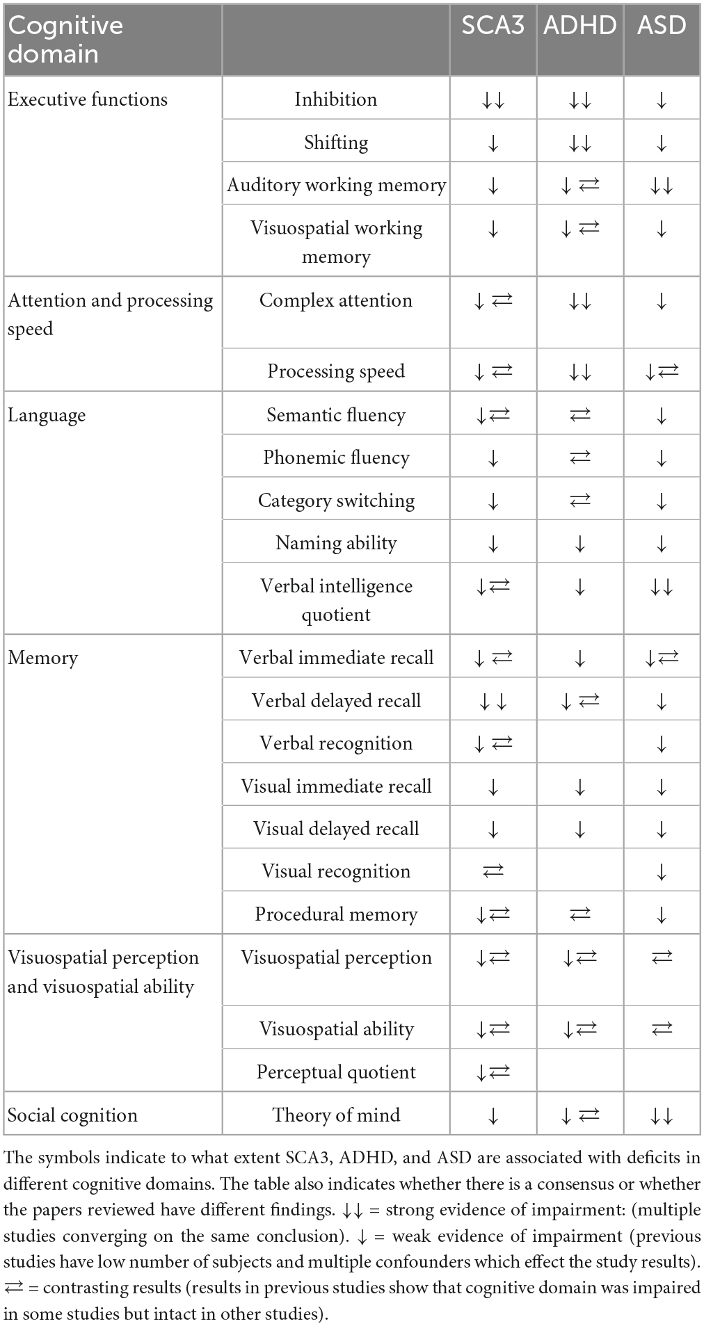
Table 1. Summarizes the evidence in the papers reviewed.
Previous results showed a link between activity in the cerebro-ponto-cerebello-thalamo-cortical loop and executive functions in SCAs (Lindsay and Storey, 2017). This is consistent with the previously reported core impairment of CCAS (Stoodley and Schmahmann, 2010; Lindsay and Storey, 2017).
Executive dysfunctions also affect verbal functions like semantic and phonemic fluency as well as fluency while switching between categories (Kawai et al., 2004; Moriarty et al., 2016; Maas et al., 2021). These are all functions that demand retrieval from long-term semantic memory. Semantic memory seems to be unaffected in these patients (Zawacki et al., 2002; Kawai et al., 2004; Lopes et al., 2013). Executive dysfunctions also impact episodic memory, with results showing evident deficits in information retrieval in visual and verbal episodic memory, while recognition and consolidation of information are partially intact (Bürk et al., 2003; Garrard et al., 2008; Braga-Neto et al., 2012; Moriarty et al., 2016). Visuospatial attention, visuospatial ability, and processing speed are also impaired (Zawacki et al., 2002; Moriarty et al., 2016; Tamura et al., 2018).
While social cognition is not well-studied in patients with SCA, some findings suggest that the Theory of Mind may be affected. Psychiatric conditions such as anxiety and depression, common among SCA3 patients, may also harm cognition. Language and cultural differences may also confound psychological studies, including those we have reviewed here. The severity of ataxia, the duration of the disease, and visual, articulatory, or upper limb motor difficulties can also influence cognition and hence the performance on neuropsychological assessments. However, because of the small sample sizes of previous studies, different confounders, and different neuropsychological methods that have been used, the neurocognitive profile of SCA3 needs to be better characterized and needs to be investigated further.
Attention deficit hyperactivity disorder
ADHD is a neuropsychiatric disorder present in approximately 5% of all children and in approximately 2.5% of the adult population (Regier et al., 2013; Austerman, 2015). It is estimated that about 15% of patients in adult psychiatry have ADHD (Deberdt et al., 2015). ADHD is defined based on clinical symptoms indicating dysregulation of attention and or activity/impulse control. When ADHD is suspected in adulthood, the investigation can be supplemented with the Wender Utah Rating Scale and Adult ADHD Self-Report Scale (Anbarasan et al., 2020).
Adults with ADHD show emotional dysregulation and executive function deficits (Barkley, 2010; Kessler et al., 2010; Biederman et al., 2019). While adults with ADHD often find ways to deal with attention problems and hyperactivity, the demands on executive abilities increase. Most jobs require complex planning and organizational skills, causing difficulties for adults with ADHD. Organizational problems often cause frustration and despair that lead to anxiety, sleep disorders, fatigue, depression or other conditions (Brattberg, 2006; Dirks et al., 2017; Schiweck et al., 2021). ADHD symptoms are normally distributed in the population, meaning they are present to varying degrees in many people. It is only when the symptoms are prominent and create a significant functional impairment that an ADHD diagnosis is justified. To receive the diagnosis, manifestations of the main symptoms of inattention and/or hyperactivity and impulsivity, along with clear functional problems due to ADHD symptoms, should be present before the age of twelve (Doernberg and Hollander, 2016). Patients with ADHD do not just look for treatment and assistance for what is classically perceived as ADHD, but for various psychiatric symptoms and functional problems, such as depression, fluctuating mood, fatigue, and anxiety (Brattberg, 2006). In summary, to diagnose ADHD is still challenging. This heterogeneous neuropsychiatric condition has many comorbidities with similar phenotypes like anxiety disorders, mood disorders, bipolar disorder, hypomania, substance use disorders, sleep disorders, Tourette syndrome, ASD and other behavioral disorders (Hegerl et al., 2010; El Malhany et al., 2015; Doernberg and Hollander, 2016; Fadeuilhe et al., 2021; Schiweck et al., 2021). Even if ADHD is considered a neurodevelopmental disorder, ADHD symptoms usually persist in late adulthood and is often confounded with mild cognitive impairment (Callahan et al., 2017).
Cerebellum and ADHD
The brain of patients with ADHD has been the subject of extensive research. One of the most robust findings is the abnormal structure and function of the cerebellum in patients diagnosed with ADHD. Abnormal cerebellar development has been implicated in ADHD (Vloet et al., 2006; Vaidya, 2011; Stoodley, 2016; Bruchhage et al., 2018). Functional imaging studies point to abnormalities in the cerebellum and other brain structures. Cerebellar and cerebral volume is ∼4% smaller in ADHD patients compared to controls (Castellanos et al., 2002; Vaidya, 2011). One study which used diffusion tensor imaging showed that children with ADHD have thinner, less myelinated, and less consistently organized fibers in the right cerebral peduncle, left middle cerebellar peduncle, and left cerebellum (Ashtari et al., 2005). Previous studies using MRI in children with ADHD have observed structural changes in cerebellum and specifically in the vermis (Castellanos et al., 1996; Berquin et al., 1998; Mackie et al., 2007). A section of the executive/non-motor portion of the cerebellum, the left Crus I result has been shown to have a lower grey-matter volume in ADHD children compared to healthy controls (Fernandez et al., 2022).
However, the brain changes associated with ADHD extend beyond the cerebellum. Structural brain imaging studies have revealed that patients with ADHD display smaller brain volumes in multiple regions, including the cerebellum, dorsolateral prefrontal cortex, caudate, pallidum, and corpus callosum (Seidman et al., 2005; Luders et al., 2009). Moreover, morphological and functional imaging studies in patients with ADHD have identified abnormal white-matter structure of neural pathways connecting prefrontal and parieto-occipital areas with the striatum and the cerebellum (Silk et al., 2009).
This evidence of ADHD being linked to cerebellar abnormalities is further supported by studies revealing that children with ADHD perform poorly on tasks that demand cerebellar involvement. Children with ADHD exhibit abnormal tapping responses and abnormal synchronized oscillatory mechanisms (Rivkin et al., 2003; Ben-Pazi et al., 2006). Children with focal cerebellar disorders and ADHD showed assorted impairments of eyeblink conditioning (Frings et al., 2010; Reeb-Sutherland and Fox, 2015). Ample research shows that ADHD is associated with changes in cerebellar morphology and cerebellar function (Buderath et al., 2009; Frings et al., 2010; Bledsoe et al., 2011). The role of the vermis in ADHD is still not clear but some researchers have pointed out its possible role. Bledsoe et al. (2011) highlighted that a smaller volume of the posterior inferior vermis is connected to behavioral outcomes like hyperactivity, attention and impulsivity reported by parents of children with ADHD.
Increased cerebro-cerebellar functional connectivity in the superior temporal gyrus within the somatomotor network could underlie impairments in cognitive control and somatic motor function in ADHD. Furthermore, increasing cerebro-cerebellar functional connectivity in older participants with ADHD suggests that enhanced cerebellar involvement may compensate for dysfunctions of the cerebral cortex in ADHD (Wang et al., 2022). Moreover, research also suggests that ADHD is also associated with changes in how the cerebellum interacts with other brain regions, including the cerebral cortex (Qiu et al., 2011). This may in turn explain some of the changes in cognition seen in cerebellar patients.
In summary, abnormal cerebellar development, morphological differences of the cerebellum and cerebellar connectivity, and results from behavioral studies where the cerebellum is involved prove that this structure of the brain is clearly involved with ADHD.
Neurocognitive functions in ADHD
In the following section, we analyze studies that examine the neurocognitive functions of patients with ADHD. The research indicates that patients with ADHD experience deficits across all cognitive domains, although the specific subdomains affected differ from those observed in the other disorders reviewed.
Executive functions
Studies have shown differences in neurocognitive function between patients with ADHD and controls, as evidenced by results from the 3-dimensional computerized Tower of London Test. This test measures both planning latencies and accuracy and has demonstrated that patients with ADHD exhibit impairments in both areas. This trend of impairment is thought to stem from difficulties in inhibiting responses when faced with problem-solving tasks, resulting in decreased planning activity (Young et al., 2007). The working memory capacity, which is also important for planning ability of patients with ADHD was evaluated using the Tower of London-Freiburg Version, and the results showed no significant differences between the ADHD patients and healthy controls (Mohamed et al., 2021). The Stroop Test, which measures patient’s ability to shift attention and inhibit interfering information (MacLeod, 1991), was conducted to assess the inhibitory ability of ADHD patients.
Results showed that patients with ADHD exhibit impaired inhibition compared to healthy controls, as reported in various studies (Fabio and Caprì, 2017; Guo et al., 2021; Callahan et al., 2022). However, it should also be noted that some studies have found no differences in executive control and inhibitory ability between ADHD patients and healthy controls, as indicated by similar error rates (Aycicegi-Dinn et al., 2011). While some tests suggest impairments in cognitive flexibility, attentional switching, and working memory, others find no significant differences between ADHD patients and healthy controls. For example, the Trail Making Test-Langensteinbacher Version showed no impairments in cognitive flexibility and attentional switching in ADHD patients (Mohamed et al., 2021). In contrast, adults with ADHD performed worse on Trial Making Test Part B used to measure visual attention and task switching (Tatar and Cansız, 2022). Cognitive flexibility and reaction time were measured with the Cognitive Flexibility Inventory and the Iowa Gambling Task, results were significantly lower in the ADHD patients compared to the control group (Roshani et al., 2020). Studies evaluating working memory using various tests, including the 2-Back Test, the Vienna Test System (Schellig and Schuri, 2009), and the Tower of Hanoi Test (Fabio and Caprì, 2017), also produced contradictory results. While some studies found impairments in working memory function in ADHD patients (Fabio and Caprì, 2017; Nikolas et al., 2019), others found no significant differences (Mohamed et al., 2021). The Letter/Number Sequencing Task (Gold et al., 1997) was used to assess auditory working memory, and results showed that ADHD patients had significantly lower scores compared to healthy controls (Aycicegi-Dinn et al., 2011). In contrast, visuospatial working memory, which does not require auditory processing, was found to be similar between ADHD patients and controls (Aycicegi-Dinn et al., 2011). Studies have linked deficits in executive function in ADHD patients specifically to the cerebellum. In an fMRI study, Depue et al. (2010) showed that ADHD patients with executive dysfunction had reduced cerebellar activation and decreased cerebellar volume, plus decreased activation of the frontal cortex and basal ganglia compared to healthy controls.
Another fMRI study by Hirose et al. (2014), showed changes in cerebro-cerebellar interaction during response inhibition in task performances. This suggests that the involvement of the cerebellum in the functional enhancement of inhibitory control may be sustained by modifications of the prefrontal-cerebellar interactions.
Attention and processing speed
Patients with ADHD exhibit difficulties with processing speed, distractibility, and selective attention, leading to an increased number of omission errors compared to healthy controls on tests such as the Vigilance and Sustained Attention Test and Selective Attention Test, two subtests of the Vienna Test System (Sturm, 2017; Mohamed et al., 2021). Similarly, ADHD patients showed a clear impairment in the Continuous performance test which is used to measure sustained attention and response inhibition (Tatar and Cansız, 2022). Performance deficits in patients with ADHD were seen for errors of omission, correct responses, late responses, and errors of commission compared to healthy controls. Low scores emerged using the non-conflict block of the Go/No-Go task and the Stroop Color-Word Task to assess processing speed. Taken together these results have shown that ADHD patients have slower response times compared to healthy controls (Aycicegi-Dinn et al., 2011). Furthermore, research has consistently demonstrated that sustained attention, reaction time, inattention, impulsivity, response speed and variability of reaction time are significantly impaired in patients with ADHD (Tinius, 2003; Nikolas et al., 2019; Guo et al., 2021). Like with executive functions, deficits in attention and processing speed have been linked to the cerebellum. For example, structural neuroimaging studies have shown reduced cerebellar volumes in ADHD which in turn correlates with attentional problem (Petacchi et al., 2005; Kucyi et al., 2015).
Language
The results of verbal fluency tests in patients with ADHD are inconsistent. While some studies have found no difference between healthy controls and ADHD patients using the Resensburg Word Fluency Test (Aycicegi-Dinn et al., 2011; Mohamed et al., 2021), others have reported impaired verbal fluency when different testing methods were used (Hervey et al., 2004; Boonstra et al., 2005; Tucha et al., 2005; Guo et al., 2021). The naming ability of patients with ADHD, which is related to semantic memory retrieval capacity, has not yet been fully explored in terms of its contribution to language in this neuropsychiatric disorder.
Children with ADHD showed slower speed response than typically developed children on a naming test (Wang et al., 2017a). Furthermore, ADHD children show lower accuracy, higher number of errors and slower responses in figure naming compared to healthy controls (Koltermann et al., 2020). A previous study by Takeda et al. (2020) attempted to identify differences in Wechsler Adult Intelligence Scale profiles between ADHD patients and healthy controls, but no significant group differences were found in Verbal intelligence quotient. However, a more recent study by Doi et al. (2022) found that female ADHD patients had a lower Verbal intelligence quotient compared to males when characterizing the cognitive profile of ADHD and ASD.
The contribution of the cerebellum, together with other brain areas are necessary for language processing. Verbal fluency and lexical retrieval tasks are associated with cerebellar activation. For example, the contralateral cerebellar hemisphere, together with Broca’s area, was actively involved in the production of semantically related verbs in response to visually presented nouns. This is an example how these actions are due to non-motor cognitive processes subserving semantic word association (Petersen et al., 1988, 1989; Stoodley and Schmahmann, 2009a; De Smet et al., 2013). Further reinforcing the cerebellums role in language, Arasanz et al. (2012) demonstrated that continuous cerebellar theta burst stimulation (cTBS) using Transcranial Magnetic Stimulation (TMS) interfered with performance on phonemic and semantic fluency tasks.
Memory
ADHD patients have shown lower scores in delayed recall of verbal memory compared to healthy controls as tested by the Verbal Learning and Memory Test (Mohamed et al., 2021). Likewise, immediate and delayed verbal recall, tested using the Logical Memory Subtest of the Wechsler Memory Scale, have been found to be clearly impaired in ADHD patients (Aycicegi-Dinn et al., 2011; Callahan et al., 2022). Further, the immediate and delayed visual recall tested using the Rey-Osterrieth Complex Figure test showed that ADHD patients performed worse than healthy controls (Aycicegi-Dinn et al., 2011). On the other hand, Dinn and colleagues found that patients with ADHD do not have a memory loss once the information is encoded (Aycicegi-Dinn et al., 2011).
A meta-analysis investigating the presence of procedural sequence learning found that ADHD patients do not exhibit significant deficits and that procedural sequence learning appears to be intact in ADHD (Sanjeevan et al., 2020). However, another study reported impaired results for procedural memory (Butzbach et al., 2019).
The cerebellum also contributes to memory functions, for example, Fastenrath et al. (2022) show that primarily the amygdala and successively the cerebellum is part of a network implicate in emotional enhancement of episodic memory and as well the right cerebellum is involved in semantic memory task (Gatti et al., 2021).
Visuospatial perception and visuospatial ability
Visuospatial ability, tested with the Rey-Osterrieth Complex Figure test, did not show significant differences between healthy controls and ADHD patients (Aycicegi-Dinn et al., 2011; Callahan et al., 2022). Schreiber et al. (1999) suggested that lower results on Rey-Osterrieth Complex Figure test copy trial was due to executive dysfunction in adults with ADHD and Rey-Osterrieth Complex Figure test can be a sensitive test to detect these deficits.
Social cognition
Social cognition is still poorly explored in patients with ADHD, in particular regarding adults (Morellini et al., 2022). Contrasting results in the literature emerge for this cognitive domain. Some studies demonstrated that emotion recognition and processing, emotional prosody and empathy are impaired in ADHD patients. Control groups performed better than ADHD patients on the Cambridge Behavior Scale, the Tübingen Affect Battery and Reading mind from the eyes test (Uekermann et al., 2010; Kis et al., 2017; Tatar and Cansız, 2022). In contrast, healthy controls and ADHD patients did not differ on theory of mind task like the revised version of Reading the Mind in the Eyes Test and the Object Alternation Test (Aycicegi-Dinn et al., 2011).
Cerebellum plays a fundamental role in identifying sequences in movements that are important to understand the intentions of others. For example, mirroring, attribute mental states in others, mentalizing, and making predictions regarding social behavior (Van Overwalle et al., 2020). Interestingly, the same functions are impaired in patients with ASD diagnosis (see below).
General intelligence
Intelligence is not impaired in patients with ADHD. Researchers who used the Cattell’s Culture Fair Intelligence Test found no difference between healthy controls and adult ADHD patients (Aycicegi-Dinn et al., 2011). Likewise, Takeda et al. (2020) showed that intelligence quotient was not impaired in adults with ADHD, although working memory and processing speed indices of Wechsler Memory Scale-III were significantly lower than healthy controls.
Summary
Adults with ADHD exhibit clear impairments in multiple cognitive domains that impact their daily functioning. The cognitive heterogeneity among adults with ADHD may be due to variations in comorbid disorders, medication status, and mood disorders within the ADHD group (Cumyn et al., 2009; Chen et al., 2018).
An overview of the neurocognitive functions and how they are affected in ADHD according to this review are illustrated in Table 1. Executive functions, such as the ability to inhibit, cognitive flexibility, as well as the processing of auditory information, are particularly compromised in these patients. On the other hand, visuospatial working memory is less impacted than auditory working memory as it does not require additional auditory processing (Aycicegi-Dinn et al., 2011).
Several deficits were identified under the attention cognitive domain and processing speed as well ias verbal fluency tasks (Hervey et al., 2004; Boonstra et al., 2005; Tucha et al., 2005; Guo et al., 2021). Visual episodic memory appears to be less affected compared to verbal episodic memory in patients with ADHD. However, impairments in verbal delayed recall and visual immediate and delayed recall have been observed, but recognition ability appears to be preserved irrespective of modality (Aycicegi-Dinn et al., 2011; Callahan et al., 2017; Butzbach et al., 2019; Sanjeevan et al., 2020; Mohamed et al., 2021). Hence, the ability to store (transfer?) information to the declarative long-term memory is not affected, but rather the retrieval of information.
The results of social cognition studies in patients with ADHD are contradictory, and the presence of undiagnosed comorbid ASD could be a confounding factor that negatively impacts their social cognition abilities (Uekermann et al., 2010; Aycicegi-Dinn et al., 2011; Morellini et al., 2022).
Mental rigidity, a common feature in patients with ADHD and ASD, could also be contributing to the varying results. The presence of disinhibition, difficulties with planning, and lack of motivation, as well as other dysexecutive symptoms characteristic of frontal lobe syndrome, have been reported among patients diagnosed with ADHD. These symptoms can impact other cognitive domains, and self-report questionnaires have shown high scores for executive dysfunction, indicating deficits in neuropsychological assessments (Aycicegi-Dinn et al., 2011).
When it comes to ADHD, the presence of undiagnosed comorbidities and other psychiatric disorders can significantly impact the results of neurocognitive assessments. Additionally, the consumption of psychotropic substances and multiple medications by some ADHD patients can greatly impact their cognitive and executive function performance (Vik et al., 2004). For instance, in a study done by Crunelle et al. (2013), ADHD patients that used cocaine had higher levels of motor and cognitive impulsivity compared to healthy controls.
Collectively, the evidence from behavioral and neuroimaging studies, indicates that ADHD is associated with cerebellar abnormalities and that these cerebellar abnormalities impact cognitive functions. This suggests that the symptoms associated with ADHD are partly due to changes in cerebellar function. However, it is also clear that ADHD is a disorder that affects multiple brain regions, and more research is required to determine the precise nature of the relationship between these deficits and the extent to which they are due to cerebellar dysfunction or problems with the interplay between various brain regions, including the cerebellum. Identifying the specific contribution of cerebellar impairment in the context of ADHD is crucial to advance our understanding of this disorder and inform the development of effective interventions to improve cognitive and motor functions.
Autism spectrum disorder
Autism spectrum disorder is characterized by difficulties with social interaction, communication, limited interests, and repetitive and restrictive behavior (Hodges et al., 2020). These problems can be of varying degrees and be associated with disorders of language development, general intelligence, emotional regulation, sensory sensitivity, and executive abilities. Sometimes intelligence and general cognitive functioning are preserved (Robertson and Baron-Cohen, 2017; Cai et al., 2018; Demetriou et al., 2018). There is no pharmacological treatment for ASD. However, early diagnosis and intervention coupled with remedial services can benefit patients (Baio et al., 2018).
The most common suggested causes of ASD are metabolic and physiological disorders, immunological dysfunction, oxidative stress, and mitochondrial dysfunction (Nardone and Elliott, 2016). Altered sensory processing and perception have been one of the characteristics of ASD (Cheung and Lau, 2020). Patients with ASD are extra sensitive to the sensory input of all modalities. This sensitivity reflects differences in sensory and cognitive processing as well as differences in low-level processing in sensory-devoted areas (Robertson and Baron-Cohen, 2017; Cai et al., 2018).
Autism spectrum disorder has many comorbidities, including morphological abnormalities like macrocephaly, cerebral palsy, physiological impairments like gastrointestinal, sleep problems, psychiatric disorders like anxiety, ADHD, obsessive-compulsive disorder, tic disorders, Tourette syndrome, epilepsy, and Rett syndrome (Sharma et al., 2018; Al-Beltagi, 2021). Patients who have shown delayed development, such as late language development, lack of interest in social interaction with other children, weak motor ability, and abnormal eye contact, have an increased risk of having ASD (Miniscalco et al., 2006). In severe cases, the child does not develop normal language, lacks social interaction, or has deviant behavior such as repetitive movements or compulsive attachment to particular objects. Patients with ASD show stereotypes, rituals, and mental rigidity (Esbensen et al., 2009). Occasionally hallucinations, delinquency and self-harming behavior emerge (Lai et al., 2014). Patients with ASD and normal language development can go undetected into adulthood (Gilchrist et al., 2001). Patients with ASD also often have social and emotional problems and may exhibit intense periods of motor anxiety and irritability typically triggered by changes in routines and the environment or sensory overload (Yuan et al., 2022). For them, intuitive thinking is often altered, and they often have great difficulties reading body language, understanding irony, and interpreting non-verbal signals (Noens and van Berckelaer-Onnes, 2005).
Executive dysfunction can contribute to the symptoms associated with ASD (Demetriou et al., 2018; Jones et al., 2018). The prevalence of autistic traits is normally distributed in the population. For a diagnosis, the symptoms and problems must have been consistent since childhood, be clear in all or most contexts, and lead to a significant disability in several functional domains (Cai et al., 2018; Styles et al., 2020).
Oristaglio et al. (2013) tested eyeblink conditioning on children with ASD, and they showed abnormally timed conditioned blink responses compared to the control group. Different diagnostic subgroups of ASD like Asperger’s syndrome, pervasive developmental disorder, and typical ASD, displayed differences in the timing of conditioned responses during trace and delay eyeblink conditioning (Oristaglio et al., 2013; Welsh and Oristaglio, 2016). Differences have also been observed in finger tapping–another task that has been linked to cerebellar functioning. Compared to controls, patients with ASD showed altered motor performance and temporal processing instability (Morimoto et al., 2018). In line with the cerebellum importance of finger tapping, children with ASD showed a reduced activation of the ipsilateral cerebellum while performing the task (Mostofsky et al., 2009).
Cerebellum and ASD
Post-mortem and structural MRI studies have highlighted that the frontal lobes, amygdala and cerebellum are abnormal in ASD (Amaral et al., 2008). Structural brain differences have been reported in terms of increased total brain volume, especially in young children with ASD, and regional gray/white matter differences in both adults and children with ASD. However, the results are not totally clear because of variations in diagnostic set-up, inclusion criteria, age, and IQ of participants. Voxel-based morphometry has revealed gray matter increases in brain areas implicated in social cognition, communication, and stereotyped behaviors–the core atypical features of ASD (Hyde et al., 2010). A number of changes has also been reported in the cerebellum in patients with ASD, including abnormal development of the cortex, reduced numbers of Purkinje cells, and hypoplasia without cogent gliosis (Bauman and Kemper, 2003; Whitney et al., 2008). These changes may in part be caused by an increment of interleukin 6 that acts as both a pro-inflammatory cytokine and an anti-inflammatory myokine and causes functional deficits of cerebellar circuits (Wei et al., 2011). Other studies support the hypothesis that a reduced release of dopamine in the frontal cortex is due to alteration in the cerebellum and with a decrease in the functional connectivity between the cerebellum and the cerebral cortex (Mittleman et al., 2008).
In addition to these morphological differences, there are many studies showing that the performance of patients with ASD differs from controls on tasks related to the cerebellum. For example, performance on eyeblink conditioning, a simple form of learning that is dependent on the cerebellum, differs between patients with ASD and controls (Sears et al., 1994; Welsh and Oristaglio, 2016). Eye-tracking to assess cerebellar integrity and assessment of an eventual comorbid epilepsy may allow for better sub-phenotyping within the heterogeneous populations that comprise ASD (Bozzi et al., 2018; Freedman and Foxe, 2018). Freedman and Foxe (2018) reported in their review a reduced size of the cerebellar vermis in ASD, which could critically impact visuo-sensorimotor development in early ages, and disrupt visual orienting, communication, and social interaction. ASD patients showed lower saccades movement and less peak speeds than controls on volitional saccade task (Stanley-Cary et al., 2011). Differences in velocity and shift in saccade dynamics typically occurs during adaptation in ASD participants (Johnson et al., 2013). Behavioral studies on mice with distinct degrees of developmental Purkinje cell loss show a correlation between the number of Purkinje cells and the degree of behavioral rigidity on learning tasks designed to assess autistic behavior in mice (Dickson et al., 2010; Martin et al., 2010). Damage to the cerebellum at the beginning of development can have long-term effects on movement, cognition, and emotional regulation. These effects may in part reflect disruption of interaction between the cerebellum and other brain regions (Wang et al., 2014). Abnormalities in different cerebellar subregions produce specific behavioral symptoms related to the functional disruption of specific cerebro-cerebellar circuits and influence brain structural development (Stoodley, 2014, 2016).
Taken together, ample evidence shows cerebellar impairment in patients and mouse models with ASD. Cerebellar abnormalities cause disruption of functions related to the cerebellum, but they may also negatively affect the ability to interpret external stimuli and organize internal processes. This doesn’t mean that the fundamental cause of ASD is to be found within the cerebellum, but rather that whatever neural alterations lead to ASD, the cerebellum likely plays an important role.
Neurocognitive functions in ASD
Executive functions
Rumsey (1985) started to explore executive functions deficits in ASD. He demonstrated that patients with a clear ASD diagnosis compared to healthy controls displayed an increased number of perseverative responses on the Wisconsin Card Sorting Test, used to measure abstract reasoning capacity and the ability to shift cognitive strategies. Perseverative responses can be due, partially to difficulties with response inhibition and to cognitive flexibility, which can be observed in patients with ASD in everyday resistance to change, and difficulty with shifts between activities. Russo et al. (2007) summed up and analyzed several studies in order to clarify inhibition’s ability, working memory and cognitive flexibility in ASD patients; results concerning these functions were variable but, however, impaired. ASD patients also have significant deficits with control of interference, motor timing and ability to inhibit an automatic response (Hlavatá et al., 2018). In another study motor inhibition ability was also impaired (Hughes et al., 1994).
Neuropsychological studies demonstrated deficits in working memory and other executive dysfunctions for patients affected by ASD compared to healthy controls (Hill, 2004; Kercood et al., 2014; Marinopoulou et al., 2016). Results of neuropsychological assessments were significantly lower concerning cognitive flexibility, planning ability, auditory working memory and visuospatial working memory in ASD patients compared to controls (Geurts et al., 2004; Goldberg et al., 2005; Landa and Goldberg, 2005). Wilson et al. (2014) used the Non-word Repetition task to assess phonological working memory, speech perception, phonological assembly, and short-term memory. Test results did not show significant differences between healthy controls and patients with ASD.
There is growing evidence that the changes in executive functions observed in ASD patients are linked to changes in the connectivity between the cerebellum and the cerebrum (Gilbert et al., 2008). For example, Bednarz and Kana (2019) highlighted cerebellar contribution to altered network connectivity in ASD. Intrinsic connectivity networks such as the executive control network, the default mode network, and salience networks are particularly important for cognitive processing.
Attention and processing speed
Patients with ASD scored significantly lower than healthy controls on the Purdue Pegboard Test, a psychomotor test of manual dexterity and bimanual coordination also used to measure executive functions and attention (Wilson et al., 2014). ASD patients demonstrate clear impairments compared to healthy controls for attentional set-shifting and planning measured, respectively, with Intra-Dimensional Extra-Dimensional set shifting task and Tower of London test (Hughes et al., 1994). Adults with ASD have been shown to have evident deficits in the processing speed subtests of the WAIS-III and WAIS-IV (Marinopoulou et al., 2016; Braconnier and Siper, 2021). In contrast, previous studies did not find any impairment in this cognitive domain (Minshew et al., 1997).
Language
Evident abnormalities were previously identified in the domain of the verbal functions. Difficulties with social interaction, verbal and non-verbal communication, and imaginative activities consistently occurred together with language difficulties (Wing and Gould, 1979). Adults with ASD have been shown to have deficits in verbal reasoning subtests of the WAIS-IV, the total Verbal Intelligence Quotient of ASD patients were found to be much lower than other cognitive quotients and this could be used as a possible ASD predictor (Braconnier and Siper, 2021; Johnson et al., 2021). Previous clinical research showed no impairment in verbal fluency that was tested with the FAS task on ASD patients (Wilson et al., 2014). The use of complex language and reasoning domains showed impairments in ASD patients (Minshew et al., 1997). The naming ability of patients with ASD, which is related to semantic memory retrieval capacity, has not yet been fully explored in terms of its contribution to language. Recent results in a neuropsychological study on children with ASD demonstrate that their results did not differ from healthy controls on naming accuracy and naming speed (Fletcher et al., 2020).
Memory
Williams et al. (2017) tested memory functions on children and adults with ASD using the Logical Memory subtest of the Wechsler Memory Scale-III and the Wide Range Assessment of Memory and Learning. Their study results for adults with ASD showed that on the WMS recall score for story details there was a significant main effect for immediate, short-term delay and long-term delay conditions. For adult thematic scores there was a relatively small discrepancy between the ASD and healthy controls on immediate recall but there was a clear discrepancy on long-term memory. Results for children and adolescent with ASD indicated that they did not differ from healthy controls in the percentages of patients with relative impairment at the immediate or 30-min delay intervals; but the ASD group had a significantly higher percentage of patients with relative impairment at the 2-day delay interval (Williams et al., 2017). Decreased story themes reflect the use of underlying organizational strategies.
In a recent study Griffin et al. (2022) focused on evaluating episodic memory in ASD; they showed that immediate recall, delayed recall and recognition are impaired in both auditory and visual modalities. General deficits were identified in the prospective memory in ASD, performance was impaired in time-based tasks and event-based cues of non-focality or low salience, that require high cognitive functioning and elevated attention (Sheppard et al., 2018).
Visuospatial perception and visuospatial ability
In a previous study there were no significant performance differences between healthy controls and ASD patients on the Embedded Figures Test that measure local/global perceptual style, central coherence and perception ability (Wilson et al., 2014). According to previous studies visuospatial abilities seem not be impaired (Minshew et al., 1997). Adults with ASD have been shown to have deficits with the visuospatial perception task of the WAIS-IV (Braconnier and Siper, 2021). Visual detection, motion perception, and face processing are altered in ASD, where attention deficits and social brain network impairment influence these three brain functions (Chung and Son, 2020). Guy et al. (2019) studied visuospatial perception on patients with ASD using the Navon task. The results did not differ for reaction times between controls and ASD patients, but the effect of interference was significantly higher in participants with ASD. Other clinical researches which were focused to clarify visuoperceptual functions did not demonstrate any impairment but instead that patients with ASD have an enhanced ability to process local or detailed stimuli (Mottron and Burack, 2001; Behrmann et al., 2006; Mottron et al., 2006).
Social cognition
Social cognition seems to be one of the most impaired cognitive domains for patients affected by ASD. Many studies demonstrated a strong impairment with social cognition in ASD patients, where, respectively, the theory of mind, emotion processing and social orientation were deficient (Happé et al., 2006; Enticott et al., 2014; Wilson et al., 2014; Griffiths et al., 2019). Patients with ASD performed more poorly than healthy controls on Frith-Happé Animations Test, on Karolinska Directed Emotional Faces test and on “Reading the Mind in the Eyes” Task used for emotion recognition (Wilson et al., 2014). Social skill and social motivation are robustly impaired in ASD patients compared to non-ASD controls (Morrison et al., 2020). In a previous study the Awareness of Social Inference Test Part 2A has been used to assess explicit theory of mind and social perception; results showed that patients with ASD had deficits with regard to complex social perception compared to their matched controls (Veddum et al., 2019). Executive dysfunctions also negatively influence social cognition in ASD (Pellicano, 2007; Freeman et al., 2017). Low cognitive abilities and in particular executive dysfunctions affect adequate responses to the environment and the ability to learn from social interactions (Sarovic, 2021).
General intelligence
Social, cognitive, and adaptive skill deficits characterize intellectual disabilities, which are present in this neuropsychiatric and neurodevelopmental disorder (Downs et al., 2008; Matson and Shoemaker, 2009). The normal population shows higher cognitive ability in terms of intelligence level and executive functions (La Malfa et al., 2004; Russo et al., 2007; Wang et al., 2017b). Around 45% to 75% of patients with ASD have an intellectual disability and from 20% to 40% of patients with intellectual disability turn out to have ASD (La Malfa et al., 2004; Saemundsen et al., 2010; Lai et al., 2014). Patients with ASD have their cognitive strengths and weaknesses as well as variable intelligence levels. Dawson et al. (2007) tested ASD children with the Raven’s Progressive Matrices to measure crystallized intelligence, performances on this test were spread on the lower and higher parts of the average from 30th percentiles to 70th percentiles (Dawson et al., 2007). Within the entire ASD, there are patients with a low intelligence quotient and also over-talented patients. However, the average intelligence quotient is significantly lower than in the general population, probably around 80 rather than 100. The diagnosis of ASD is generally associated with an intellectual quotient below 80 (Gillberg and Rasmussen, 2011). Thus, it could be more appropriate to distinguish ASD patients with cognitive and intellectual disability and ASD patients with a normative or above average IQ.
Summary
The neurocognitive functions in ASD are summarized in Table 1. Multiple cognitive functions in ASD are impaired like abstract reasoning capacity, cognitive flexibility, inhibition ability, working memory, planning ability, attention, verbal functions, process speed, emotion processing, social cognition, immediate recall, delayed recall, and recognition in both visual and verbal modalities (Hughes et al., 1994; Geurts et al., 2004; Hill, 2004; Goldberg et al., 2005; Russo et al., 2007; Wilson et al., 2014; Hlavatá et al., 2018; Sheppard et al., 2018; Griffin et al., 2022). Executive dysfunctions significantly decrease the life quality of patients affected by ASD (de Vries and Geurts, 2015). A major challenge when studying ASD is that ASD is a heterogenic developmental disorder, resulting in high variability on cognitive tests (Newschaffer et al., 2002). ASD often co-occurs with ADHD and patients with both diagnoses often have a different pattern of symptoms (Leitner, 2014; Ashwood et al., 2015). Other medical conditions like hyperkinetic disorders, obsessive-compulsive disorder, Tourette’s syndrome, language delay, epilepsy, depression, anxiety, schizophrenia, bipolar disorder, personality disorder, fragile X syndrome, tuberous sclerosis, or general learning disability can be critical confounders that affect performance on neuropsychological assessments. Attention deficits often cause fatigue and reduce the patient’s cognitive performance (Zwick, 2017). Repetitive behavior and rigid mentality have been associated with caudate abnormality in ASD (Sears et al., 1999). The neurocognitive profile of adults with ASD is, however, still partially unclear. As for ADHD, evidence suggests that ASD is associated with changes in multiple brain regions, including the cerebellum. Combined with studies showing that ASD is associated with deficits in tasks related to cerebellar function, there is convincing evidence that the symptoms associated with ASD are partly due to cerebellar abnormalities. Again, future studies need to address the extent to which these deficits are linked directly to the cerebellum versus the interplay between the cerebellum and other parts of the brain.
Discussion
This review highlights differences in neurocognitive functions, illustrated in Table 1, of two disorders and one disease affecting the cerebellum – ADHD, ASD, and SCA3. Multiple cognitive impairments are evident in all three conditions (Barnard et al., 2008; Guo et al., 2021; Yap et al., 2022b). The cognitive impairments associated can exacerbate functional deficits, leading to significant negative impacts on daily life, socioeconomic status, and personal relationships.
These three conditions are all associated with clear cerebellar abnormalities, which may in turn affect cognitive functions, directly or indirectly. Although progress has been made in identifying cognitive difficulties, further research is needed to fully understand the disorders and their effects on other cognitive functions. Currently, many of the cognitive difficulties are not sufficiently acknowledged, resulting in limited recognition and understanding in society. A key goal of future research should be to determine if these cognitive deficits are directly caused by the disorders, by other comorbid conditions, or a combination of both.
Other conditions such as low or high intelligence quotient, epilepsy, tics, depression, anxiety, eating disorders, obsessive-compulsive disorder, and burnout syndrome can impact the performance of patients with SCA3, ADHD, and ASD on neuropsychological assessments. The presence of these conditions can be confounding factors that clinicians must take into account when interpreting test results and associated cognitive functions. Other problems with earlier studies include small sample sizes, poor neurocognitive task selection, cultural differences and patient heterogeneity; which may also contribute to the variations in performances revealed in this review.
There are several reasons why it is important to assess neurocognitive function in ADHD, ASD, and SCA3. First, identifying cognitive deficits that arise during the early stages of these conditions could help prevent or alleviate further cognitive impairments. Second, understanding the cognitive challenges facing these patients can help caregivers as well as the patients themselves to facilitate their daily routines. Third, the identification of cognitive deficits can be a starting point for personalized work and educational rehabilitation programs. Failure to detect cognitive impairments, on the other hand, can have severe effects. Cognitive deficits can trigger high levels of psychosocial stress at school or work which can lead to depression, anxiety, and ultimately self-harm, violence, and increased risk of suicide.
Neuropsychological assessments can contribute to the discrimination between many disorders and diseases, for example, Alzheimer’s disease, frontotemporal dementia, and post-traumatic stress syndrome. However, for ADHD, SCA3, and ASD there are few neuropsychological assessments used in clinical practice. The neurocognitive functions have not been extensively evaluated and clarified in SCA3. Future studies need to clarify how the damage to the cerebellum affects cognitive functions with the support of neuroimaging, how cerebellar damage in SCA3 affects the neurocognitive domains, and if it is possible to detect a mild cognitive impairment which often leads to cognitive diseases or dementia (Teive and Arruda, 2009; Lindsay and Storey, 2017).
For ADHD the Integrated Visual and Auditory continuous performance test is commonly used by researchers and clinicians (Tinius, 2003; Arble et al., 2014). This test has good specificity and sensitivity for categorizing ADHD type. However, a disadvantage of this continuous performance test is its relatively long duration of 15 min without interruption, resulting in many patients not completing the test. Therefore, other tests are needed in clinical evaluations that can measure the same functions but without these disadvantages. Neuroimaging has shown promise as a quantitative and non-invasive method for the assessment of structural and functional brain alterations in clinical SCA3 patients as well as preclinical carriers. Neuropsychological assessments can as well contribute to identifying a possible hidden mild cognitive impairment, when there are no clear biomarkers but light cognitive symptoms that clinicians often miss (Stezin et al., 2021). Furthermore, neuropsychological assessments can contribute to giving a comprehensive diagnostic, functional description, and evaluate the severity of the symptoms, supported by cognitive profiles.
However, for neuropsychiatric disorders like ADHD and ASD neuroimaging results have been less clear, perhaps because of the heterogeneity of these disorders (Anagnostou and Taylor, 2011; Cortese and Castellanos, 2012; Wolff et al., 2018; Giedd, 2019; Pereira-Sanchez and Castellanos, 2021). ADHD and ASD are diagnosed on the basis of behavioral symptoms and clinical history but cognitive abilities may also be useful in characterizing patients with ADHD and ASD.
Future directions
Future studies are needed to further explain the role of the cerebellum in both normal and pathological behavior, mood regulation, impulse control and how it contributes to executive cognitive functioning. Further, new clinical studies investigating the cognitive profiles of ADHD, ASD, and SCA3, are necessary to gain insight into the nuances of the disorders which in turn can improve the differential diagnosis and the potential to identify sub-cognitive phenotypes of patients with ADHD and ASD, which in turn may help us develop better treatments and strategies. Non-pharmacological strategies could also help the large group of patients with neuropsychiatric symptoms who do not fulfill all the diagnostics criteria to obtain a formal ADHD or ASD diagnosis. Given that most of the studies on ASD have focused on children or adolescents, further studies are needed to clarify the cognitive functions of adults with ADHD and ASD.
To fully understand how the cerebellum works we must study how it contributes to different psychological functions and neurocognitive domains. Considering the rich cerebro-cerebellar connections, we should expect that changes in the cerebellum can affect cognitive functions such as verbal functions, cognitive flexibility, impulse control, task initiation, efficiency, self-monitoring, planning, and prioritizing. The findings from neuroimaging and neuropsychology reviewed here show cogent evidence that the human cerebellum plays a pivotal role in many cognitive functions. Accordingly, ADHD, ASD, and SCA3–three conditions associated with abnormalities in cerebellar morphology–all affect various cognitive functions, although more research is needed to uncover the causal relationships involved. Future clinical studies on these patient groups and other patients in which the cerebellum is affected can help us uncover the cerebellum’s role in cognition and lead to an improved understanding of the difficulties facing patients with cerebellar alterations.
Author contributions
MC: wrote the first draft, conception, disposition, and organization of the work. SV: reviewed and critiqued the parts inherent to cognition, neurocognitive impairments, and neuropsychology. PG: reviewed and critiqued the parts inherent to ADHD and ASD. SG: reviewed and critiqued the parts inherent to ataxia and SCA3. AR: wrote the final draft, supervised the main work, conception, financing, and organization of the work. All authors contributed to the article and approved the submitted version.
Funding
This study was supported by grants to AR from the Swedish Research Council (2020-01468), the Crafoord Foundation (20180704, 20200729, and 20220796), Per-Eric och Ulla Schybergs stiftelse (42630), the Segerfalk Foundation (2019-2246), Åke-Wibergs foundation (M18-0070, M19-0375, and M20-0008), Fredrik and Ingrid Thurings foundation (2018-00366 and 2019-00516), Pia Ståhls Foundation (20012), Magnus Bergvalls Foundation (2020-03788), and the Anna-Lisa Rosenberg Foundation. The funders had no role in study design, data collection, analysis, publication decision, or manuscript preparation.
Conflict of interest
The authors declare that the research was conducted in the absence of any commercial or financial relationships that could be construed as a potential conflict of interest.
Publisher’s note
All claims expressed in this article are solely those of the authors and do not necessarily represent those of their affiliated organizations, or those of the publisher, the editors and the reviewers. Any product that may be evaluated in this article, or claim that may be made by its manufacturer, is not guaranteed or endorsed by the publisher.
References
Adamaszek, M., D’Agata, F., Ferrucci, R., Habas, C., Keulen, S., Kirkby, K. C., et al. (2017). Consensus paper: Cerebellum and emotion. Cerebellum 16, 552–576. doi: 10.1007/s12311-016-0815-8
Ahmadian, N., van Baarsen, K., van Zandvoort, M., and Robe, P. A. (2019). The cerebellar cognitive affective syndrome-a meta-analysis. Cerebellum 18, 941–950. doi: 10.1007/s12311-019-01060-2
Al-Beltagi, M. (2021). Autism medical comorbidities. World J. Clin. Pediatr. 10, 15–28. doi: 10.5409/wjcp.v10.i3.15
Alexander, G. E., DeLong, M. R., and Strick, P. L. (1986). Parallel organization of functionally segregated circuits linking basal ganglia and cortex. Annu. Rev. Neurosci. 9, 357–381. doi: 10.1146/annurev.ne.09.030186.002041
Allen, G. I., and Tsukahara, N. (1974). Cerebrocerebellar communication systems. Physiol. Rev. 54, 957–1006. doi: 10.1152/physrev.1974.54.4.957
Amaral, D. G., Schumann, C. M., and Nordahl, C. W. (2008). Neuroanatomy of autism. Trends Neurosci. 31, 137–145. doi: 10.1016/j.tins.2007.12.005
Anagnostou, E., and Taylor, M. J. (2011). Review of neuroimaging in autism spectrum disorders: what have we learned and where we go from here. Mol. Autism 2:4. doi: 10.1186/2040-2392-2-4
Anbarasan, D., Kitchin, M., and Adler, L. A. (2020). Screening for Adult ADHD. Curr. Psychiatry Rep. 22:72. doi: 10.1007/s11920-020-01194-9
Ankri, L., Husson, Z., Pietrajtis, K., Proville, R., Léna, C., Yarom, Y., et al. (2015). A novel inhibitory nucleo-cortical circuit controls cerebellar Golgi cell activity. eLife 4:e06262. doi: 10.7554/eLife.06262
Apps, R., and Garwicz, M. (2005). Anatomical and physiological foundations of cerebellar information processing. Nat. Rev. Neurosci. 6, 297–311. doi: 10.1038/nrn1646
Arasanz, C. P., Staines, W. R., Roy, E. A., and Schweizer, T. A. (2012). The cerebellum and its role in word generation: a cTBS study. Cortex 48, 718–724. doi: 10.1016/j.cortex.2011.02.021
Arble, E., Kuentzel, J., and Barnett, D. (2014). Convergent validity of the Integrated Visual and Auditory Continuous Performance Test (IVA+Plus): associations with working memory, processing speed, and behavioral ratings. Arch. Clin. Neuropsychol. 29, 300–312. doi: 10.1093/arclin/acu006
Argyropoulos, G. P. D., van Dun, K., Adamaszek, M., Leggio, M., Manto, M., Masciullo, M., et al. (2020). The cerebellar cognitive affective/schmahmann syndrome: A task force paper. Cerebellum 19, 102–125. doi: 10.1007/s12311-019-01068-8
Arruda, W. O., Meira, A. T., Ono, S. E., de Carvalho Neto, A., Betting, L. E. G. G., Raskin, S., et al. (2020). Volumetric MRI Changes in Spinocerebellar Ataxia (SCA3 and SCA10) Patients. Cerebellum 19, 536–543. doi: 10.1007/s12311-020-01137-3
Ashida, R., Cerminara, N. L., Edwards, R. J., Apps, R., and Brooks, J. C. W. (2019). Sensorimotor, language, and working memory representation within the human cerebellum. Hum. Brain Mapp. 40, 4732–4747. doi: 10.1002/hbm.24733
Ashizawa, T., and Xia, G. (2016). Ataxia. Continuum 22, 1208–1226. doi: 10.1212/CON.0000000000000362
Ashtari, M., Kumra, S., Bhaskar, S. L., Clarke, T., Thaden, E., Cervellione, K. L., et al. (2005). Attention-deficit/hyperactivity disorder: a preliminary diffusion tensor imaging study. Biol. Psychiatry 57, 448–455. doi: 10.1016/j.biopsych.2004.11.047
Ashwood, K. L., Tye, C., Azadi, B., Cartwright, S., Asherson, P., and Bolton, P. (2015). Brief Report: Adaptive functioning in children with ASD, ADHD and ASD ADHD. J. Autism Dev. Disord. 45, 2235–2242. doi: 10.1007/s10803-014-2352-y
Austerman, J. (2015). ADHD and behavioral disorders: Assessment, management, and an update from DSM-5. Cleve. Clin. J. Med. 82, S2–S7. doi: 10.3949/ccjm.82.s1.01
Avanzino, L., Pelosin, E., Vicario, C. M., Lagravinese, G., Abbruzzese, G., and Martino, D. (2016). Time processing and motor control in movement disorders. Front. Hum. Neurosci. 10:631. doi: 10.3389/fnhum.2016.00631
Aycicegi-Dinn, A., Dervent-Ozbek, S., Yazgan, Y., Bicer, D., and Dinn, W. M. (2011). Neurocognitive correlates of adult attention-deficit/hyperactivity disorder in a Turkish sample. Atten. Defic. Hyperact. Disord. 3, 41–52. doi: 10.1007/s12402-010-0050-y
Azevedo, F. A. C., Carvalho, L. R. B., Grinberg, L. T., Farfel, J. M., Ferretti, R. E. L., Leite, R. E. P., et al. (2009). Equal numbers of neuronal and nonneuronal cells make the human brain an isometrically scaled-up primate brain. J. Comp. Neurol. 513, 532–541. doi: 10.1002/cne.21974
Baio, J., Wiggins, L., Christensen, D. L., Maenner, M. J., Daniels, J., Warren, Z., et al. (2018). Prevalence of Autism spectrum disorder among children aged 8 Years - Autism and developmental disabilities monitoring network, 11 Sites. United States, 2014. MMWR Surveill. Summ. 67, 1–23. doi: 10.15585/mmwr.ss6706a1
Barkley, R. A. (2010). Differential diagnosis of adults with ADHD: the role of executive function and self-regulation. J. Clin. Psychiatry 71:e17. doi: 10.4088/JCP.9066tx1c
Barnard, L., Muldoon, K., Hasan, R., O’Brien, G., and Stewart, M. (2008). Profiling executive dysfunction in adults with autism and comorbid learning disability. Autism 12, 125–141. doi: 10.1177/1362361307088486
Barnes, J. A., Ebner, B. A., Duvick, L. A., and Gao, W. (2011). Abnormalities in the climbing fiber-Purkinje cell circuitry contribute to neuronal dysfunction in ATXN1 [82Q] mice. J. Neurosci. 31, 12778–12789. doi: 10.1523/JNEUROSCI.2579-11.2011
Bauman, M. L., and Kemper, T. L. (2003). The neuropathology of the autism spectrum disorders: what have we learned? Novartis Found. Symp. 251, 281–297.
Bednarz, H. M., and Kana, R. K. (2019). Patterns of Cerebellar Connectivity with Intrinsic Connectivity Networks in Autism Spectrum Disorders. J. Autism Dev. Disord. 49, 4498–4514. doi: 10.1007/s10803-019-04168-w
Behrmann, M., Thomas, C., and Humphreys, K. (2006). Seeing it differently: visual processing in autism. Trends Cogn. Sci. 10, 258–264. doi: 10.1016/j.tics.2006.05.001
Ben-Pazi, H., Shalev, R. S., Gross-Tsur, V., and Bergman, H. (2006). Age and medication effects on rhythmic responses in ADHD: possible oscillatory mechanisms? Neuropsychologia 44, 412–416. doi: 10.1016/j.neuropsychologia.2005.05.022
Bernard, J. A., and Mittal, V. A. (2014). Cerebellar-motor dysfunction in schizophrenia and psychosis-risk: the importance of regional cerebellar analysis approaches. Front. Psychiatry 5:160. doi: 10.3389/fpsyt.2014.00160
Berquin, P. C., Giedd, J. N., Jacobsen, L. K., Hamburger, S. D., Krain, A. L., Rapoport, J. L., et al. (1998). Cerebellum in attention-deficit hyperactivity disorder: a morphometric MRI study. Neurology 50, 1087–1093. doi: 10.1212/WNL.50.4.1087
Bhandari, J., Thada, P. K., and Samanta, D. (2022). “Spinocerebellar Ataxia,” in StatPearls. Treasure Island, FL: StatPearls Publishing.
Biederman, J., Lanier, J., DiSalvo, M., Noyes, E., Fried, R., Woodworth, K. Y., et al. (2019). Clinical correlates of mind wandering in adults with ADHD. J. Psychiatr. Res. 117, 15–23. doi: 10.1016/j.jpsychires.2019.06.012
Bledsoe, J. C., Semrud-Clikeman, M., and Pliszka, S. R. (2011). Neuroanatomical and neuropsychological correlates of the cerebellum in children with attention-deficit/hyperactivity disorder–combined type. J. Am. Acad. Child Adolesc. Psychiatry 50, 593–601. doi: 10.1016/j.jaac.2011.02.014
Boonstra, A. M., Oosterlaan, J., Sergeant, J. A., and Buitelaar, J. K. (2005). Executive functioning in adult ADHD: a meta-analytic review. Psychol. Med. 35, 1097–1108. doi: 10.1017/S003329170500499X
Bostan, A. C., Dum, R. P., and Strick, P. L. (2013). Cerebellar networks with the cerebral cortex and basal ganglia. Trends Cogn. Sci. 17, 241–254. doi: 10.1016/j.tics.2013.03.003
Bozzi, Y., Provenzano, G., and Casarosa, S. (2018). Neurobiological bases of autism-epilepsy comorbidity: a focus on excitation/inhibition imbalance. Eur. J. Neurosci. 47, 534–548. doi: 10.1111/ejn.13595
Bracha, V., Zbarska, S., Parker, K., Carrel, A., Zenitsky, G., and Bloedel, J. R. (2009). The cerebellum and eye-blink conditioning: learning versus network performance hypotheses. Neuroscience 162, 787–796. doi: 10.1016/j.neuroscience.2008.12.042
Braconnier, M. L., and Siper, P. M. (2021). Neuropsychological Assessment in Autism Spectrum Disorder. Curr. Psychiatry Rep. 23:63. doi: 10.1007/s11920-021-01277-1
Braga-Neto, P., Pedroso, J. L., Alessi, H., Dutra, L. A., Felício, A. C., Minett, T., et al. (2012). Cerebellar cognitive affective syndrome in Machado Joseph disease: core clinical features. Cerebellum 11, 549–556. doi: 10.1007/s12311-011-0318-6
Brattberg, G. (2006). PTSD and ADHD: underlying factors in many cases of burnout. Stress Health 22, 305–313. doi: 10.1002/smi.1112
Brodal, P. (1978). Principles of organization of the monkey corticopontine projection. Brain Res. 148, 214–218. doi: 10.1016/0006-8993(78)90392-X
Bruchhage, M. M. K., Bucci, M.-P., and Becker, E. B. E. (2018). Cerebellar involvement in autism and ADHD. Handb. Clin. Neurol. 155, 61–72. doi: 10.1016/B978-0-444-64189-2.00004-4
Buderath, P., Gärtner, K., Frings, M., Christiansen, H., Schoch, B., Konczak, J., et al. (2009). Postural and gait performance in children with attention deficit/hyperactivity disorder. Gait Posture 29, 249–254. doi: 10.1016/j.gaitpost.2008.08.016
Bürk, K., Globas, C., Bösch, S., Klockgether, T., Zühlke, C., Daum, I., et al. (2003). Cognitive deficits in spinocerebellar ataxia type 1, 2, and 3. J. Neurol. 250, 207–211. doi: 10.1007/s00415-003-0976-5
Burright, E. N., Clark, H. B., Servadio, A., Matilla, T., Feddersen, R. M., Yunis, W. S., et al. (1995). SCA1 transgenic mice: a model for neurodegeneration caused by an expanded CAG trinucleotide repeat. Cell 82, 937–948. doi: 10.1016/0092-8674(95)90273-2
Butzbach, M., Fuermaier, A. B. M., Aschenbrenner, S., Weisbrod, M., Tucha, L., and Tucha, O. (2019). Basic processes as foundations of cognitive impairment in adult ADHD. J. Neural Transm. 126, 1347–1362. doi: 10.1007/s00702-019-02049-1
Cai, R. Y., Richdale, A. L., Uljarević, M., Dissanayake, C., and Samson, A. C. (2018). Emotion regulation in autism spectrum disorder: Where we are and where we need to go. Autism Res. 11, 962–978. doi: 10.1002/aur.1968
Callahan, B. L., Bierstone, D., Stuss, D. T., and Black, S. E. (2017). Adult ADHD: Risk Factor for Dementia or Phenotypic Mimic? Front. Aging Neurosci. 9:260. doi: 10.3389/fnagi.2017.00260
Callahan, B. L., Ramakrishnan, N., Shammi, P., Bierstone, D., Taylor, R., Ozzoude, M., et al. (2022). Cognitive and neuroimaging profiles of older adults with attention deficit/hyperactivity disorder presenting to a memory clinic. J. Atten. Disord. 26, 1118–1129. doi: 10.1177/10870547211060546
Castellanos, F. X., Giedd, J. N., Marsh, W. L., Hamburger, S. D., Vaituzis, A. C., Dickstein, D. P., et al. (1996). Quantitative brain magnetic resonance imaging in attention-deficit hyperactivity disorder. Arch. Gen. Psychiatry 53, 607–616. doi: 10.1001/archpsyc.1996.01830070053009
Castellanos, F. X., Lee, P. P., Sharp, W., Jeffries, N. O., Greenstein, D. K., Clasen, L. S., et al. (2002). Developmental trajectories of brain volume abnormalities in children and adolescents with attention-deficit/hyperactivity disorder. JAMA 288, 1740–1748. doi: 10.1001/jama.288.14.1740
Chen, Q., Hartman, C. A., Haavik, J., Harro, J., Klungsøyr, K., Hegvik, T.-A., et al. (2018). Common psychiatric and metabolic comorbidity of adult attention-deficit/hyperactivity disorder: A population-based cross-sectional study. PLoS One 13:e0204516. doi: 10.1371/journal.pone.0204516
Chen, X., Tang, T.-S., Tu, H., Nelson, O., Pook, M., Hammer, R., et al. (2008). Deranged calcium signaling and neurodegeneration in spinocerebellar ataxia type 3. J. Neurosci. 28, 12713–12724. doi: 10.1523/JNEUROSCI.3909-08.2008
Cheung, P. P. P., and Lau, B. W. M. (2020). Neurobiology of sensory processing in autism spectrum disorder. Prog. Mol. Biol. Transl. Sci. 173, 161–181. doi: 10.1016/bs.pmbts.2020.04.020
Chung, S., and Son, J.-W. (2020). Visual perception in Autism Spectrum Disorder: A review of neuroimaging studies. Soa Chongs. Chong. Uihak 31, 105–120. doi: 10.5765/jkacap.200018
Cortese, S., and Castellanos, F. X. (2012). Neuroimaging of attention-deficit/hyperactivity disorder: current neuroscience-informed perspectives for clinicians. Curr. Psychiatry Rep. 14, 568–578. doi: 10.1007/s11920-012-0310-y
Crunelle, C. L., Veltman, D. J., van Emmerik-van Oortmerssen, K., Booij, J., and van den Brink, W. (2013). Impulsivity in adult ADHD patients with and without cocaine dependence. Drug Alcohol Depend. 129, 18–24. doi: 10.1016/j.drugalcdep.2012.09.006
Cumyn, L., French, L., and Hechtman, L. (2009). Comorbidity in adults with attention-deficit hyperactivity disorder. Can. J. Psychiatry 54, 673–683. doi: 10.1177/070674370905401004
D’Abreu, A., França, M. C. Jr., Yasuda, C. L., Campos, B. A. G., Lopes-Cendes, I., and Cendes, F. (2012). Neocortical atrophy in Machado-Joseph disease: a longitudinal neuroimaging study. J. Neuroimaging 22, 285–291. doi: 10.1111/j.1552-6569.2011.00614.x
Dawson, M., Soulières, I., Gernsbacher, M. A., and Mottron, L. (2007). The level and nature of autistic intelligence. Psychol. Sci. 18, 657–662. doi: 10.1111/j.1467-9280.2007.01954.x
De Smet, H. J., Paquier, P., Verhoeven, J., and Mariën, P. (2013). The cerebellum: its role in language and related cognitive and affective functions. Brain Lang. 127, 334–342. doi: 10.1016/j.bandl.2012.11.001
de Vries, M., and Geurts, H. (2015). Influence of Autism traits and executive functioning on quality of life in children with an Autism spectrum disorder. J. Autism Dev. Disord. 45, 2734–2743. doi: 10.1007/s10803-015-2438-1
Deberdt, W., Thome, J., Lebrec, J., Kraemer, S., Fregenal, I., Ramos-Quiroga, J. A., et al. (2015). Prevalence of ADHD in nonpsychotic adult psychiatric care (ADPSYC): A multinational cross-sectional study in Europe. BMC Psychiatry 15:242. doi: 10.1186/s12888-015-0624-5
Demetriou, E. A., Lampit, A., Quintana, D. S., Naismith, S. L., Song, Y. J. C., Pye, J. E., et al. (2018). Autism spectrum disorders: a meta-analysis of executive function. Mol. Psychiatry 23, 1198–1204. doi: 10.1038/mp.2017.75
Depue, B. E., Burgess, G. C., Willcutt, E. G., Bidwell, L. C., Ruzic, L., and Banich, M. T. (2010). Symptom-correlated brain regions in young adults with combined-type ADHD: their organization, variability, and relation to behavioral performance. Psychiatry Res. 182, 96–102. doi: 10.1016/j.pscychresns.2009.11.011
Dickson, P. E., Rogers, T. D., Del Mar, N., Martin, L. A., Heck, D., Blaha, C. D., et al. (2010). Behavioral flexibility in a mouse model of developmental cerebellar Purkinje cell loss. Neurobiol. Learn. Mem. 94, 220–228. doi: 10.1016/j.nlm.2010.05.010
Diedrichsen, J., King, M., Hernandez-Castillo, C., Sereno, M., and Ivry, R. B. (2019). Universal transform or multiple functionality? Understanding the contribution of the human cerebellum across task domains. Neuron 102, 918–928. doi: 10.1016/j.neuron.2019.04.021
Dirks, H., Scherbaum, N., Kis, B., and Mette, C. (2017). ADHD in Adults and Comorbid Substance Use Disorder: Prevalence, Clinical Diagnostics and Integrated Therapy. Fortschr. Neurol. Psychiatr. 85, 336–344. doi: 10.1055/s-0043-100763
D’Mello, A. M., Crocetti, D., Mostofsky, S. H., and Stoodley, C. J. (2015). Cerebellar gray matter and lobular volumes correlate with core autism symptoms. NeuroImage 7, 631–639. doi: 10.1016/j.nicl.2015.02.007
Doernberg, E., and Hollander, E. (2016). Neurodevelopmental Disorders (ASD and ADHD): DSM-5, ICD-10, and ICD-11. CNS Spectr. 21, 295–299. doi: 10.1017/S1092852916000262
Doi, H., Kanai, C., and Ohta, H. (2022). Transdiagnostic and sex differences in cognitive profiles of autism spectrum disorder and attention-deficit/hyperactivity disorder. Autism Res. 15, 1130–1141. doi: 10.1002/aur.2712
Downs, A., Downs, R. C., and Rau, K. (2008). Effects of training and feedback on Discrete Trial Teaching skills and student performance. Res. Dev. Disabil. 29, 235–246. doi: 10.1016/j.ridd.2007.05.001
Duan, K., Jiang, W., Rootes-Murdy, K., Schoenmacker, G. H., Arias-Vasquez, A., Buitelaar, J. K., et al. (2021). Gray matter networks associated with attention and working memory deficit in ADHD across adolescence and adulthood. Transl. Psychiatry 11:184. doi: 10.1038/s41398-021-01301-1
Durr, A. (2010). Autosomal dominant cerebellar ataxias: polyglutamine expansions and beyond. Lancet Neurol. 9, 885–894. doi: 10.1016/S1474-4422(10)70183-6
E, K.-H., Chen, S.-H. A., Ho, M.-H. R., and Desmond, J. E. (2014). A meta-analysis of cerebellar contributions to higher cognition from PET and fMRI studies. Hum. Brain Mapp. 35, 593–615. doi: 10.1002/hbm.22194
El Malhany, N., Gulisano, M., Rizzo, R., and Curatolo, P. (2015). Tourette syndrome and comorbid ADHD: causes and consequences. Eur. J. Pediatr. 174, 279–288. doi: 10.1007/s00431-014-2417-0
Eliassen, J. C., Souza, T., and Sanes, J. N. (2001). Human brain activation accompanying explicitly directed movement sequence learning. Exp. Brain Res. 141, 269–280. doi: 10.1007/s002210100822
Elyoseph, Z., Mintz, M., Vakil, E., Zaltzman, R., and Gordon, C. R. (2020). Selective Procedural Memory Impairment but Preserved Declarative Memory in Spinocerebellar Ataxia Type 3. Cerebellum 19, 226–234. doi: 10.1007/s12311-019-01101-w
Enticott, P. G., Kennedy, H. A., Johnston, P. J., Rinehart, N. J., Tonge, B. J., Taffe, J. R., et al. (2014). Emotion recognition of static and dynamic faces in autism spectrum disorder. Cogn. Emot. 28, 1110–1118. doi: 10.1080/02699931.2013.867832
Esbensen, A. J., Seltzer, M. M., Lam, K. S. L., and Bodfish, J. W. (2009). Age-related differences in restricted repetitive behaviors in autism spectrum disorders. J. Autism Dev. Disord. 39, 57–66. doi: 10.1007/s10803-008-0599-x
Fabio, R. A., and Caprì, T. (2017). The executive functions in a sample of Italian adults with ADHD: attention, response inhibition and planning/organization. Mediter. J. Clin. Psychology 5, 1–17.
Fadeuilhe, C., Daigre, C., Richarte, V., Grau-López, L., Palma-Álvarez, R. F., Corrales, M., et al. (2021). Insomnia Disorder in Adult Attention-Deficit/Hyperactivity Disorder Patients: Clinical. Comorbidity, and Treatment Correlates. Front. Psychiatry 12:663889. doi: 10.3389/fpsyt.2021.663889
Fastenrath, M., Spalek, K., Coynel, D., Loos, E., Milnik, A., Egli, T., et al. (2022). Human cerebellum and corticocerebellar connections involved in emotional memory enhancement. Proc. Natl. Acad. Sci. U. S. A. 119:e2204900119. doi: 10.1073/pnas.2204900119
Feng, L., Chen, D. B., Hou, L., Huang, L. H., Lu, S. Y., Liang, X. L., et al. (2014). Cognitive impairment in native Chinese with spinocerebellar ataxia type 3. Eur. Neurol. 71, 262–270. doi: 10.1159/000357404
Fernandez, L., Burmester, A., Duque, J. D., Silk, T. J., Hyde, C. E., Kirkovski, M., et al. (2022). Examination of Cerebellar Grey-Matter Volume in Children with Neurodevelopmental Disorders: a Coordinated Analysis Using the ACAPULCO Algorithm. Cerebellum [Epub ahead of print]. doi: 10.1007/s12311-022-01503-3
Fine, E. J., Ionita, C. C., and Lohr, L. (2002). The history of the development of the cerebellar examination. Semin. Neurol. 22, 375–384. doi: 10.1055/s-2002-36759
Fisher, J. D. T. (1839). ART. VII. Contributions Illustrative of the Functions of the Cerebellum. Am. J. Med. Sci. 23:1827.
Fletcher, F. E., Knowland, V., Walker, S., Gaskell, M. G., Norbury, C., and Henderson, L. M. (2020). Atypicalities in sleep and semantic consolidation in autism. Dev. Sci. 23:e12906. doi: 10.1111/desc.12906
Flourens, P. (1824). Experimental research on the properties and functions of the nervous system in vertebrate animals. Paris: Crevot.
Freedman, E. G., and Foxe, J. J. (2018). Eye movements, sensorimotor adaptation and cerebellar-dependent learning in autism: toward potential biomarkers and subphenotypes. Eur. J. Neurosci. 47, 549–555. doi: 10.1111/ejn.13625
Freeman, L. M., Locke, J., Rotheram-Fuller, E., and Mandell, D. (2017). Brief Report: Examining Executive and Social Functioning in Elementary-Aged Children with Autism. J. Autism Dev. Disord. 47, 1890–1895. doi: 10.1007/s10803-017-3079-3
Frings, M., Gaertner, K., Buderath, P., Gerwig, M., Christiansen, H., Schoch, B., et al. (2010). Timing of conditioned eyeblink responses is impaired in children with attention-deficit/hyperactivity disorder. Exp. Brain Res. 201, 167–176. doi: 10.1007/s00221-009-2020-1
Fusar-Poli, P., Placentino, A., Carletti, F., Landi, P., Allen, P., Surguladze, S., et al. (2009). Functional atlas of emotional faces processing: a voxel-based meta-analysis of 105 functional magnetic resonance imaging studies. J. Psychiatry Neurosci. 34, 418–432.
Garrard, P., Martin, N. H., Giunti, P., and Cipolotti, L. (2008). Cognitive and social cognitive functioning in spinocerebellar ataxia: a preliminary characterization. J. Neurol. 255, 398–405. doi: 10.1007/s00415-008-0680-6
Gatti, D., Vecchi, T., and Mazzoni, G. (2021). Cerebellum and semantic memory: A TMS study using the DRM paradigm. Cortex 135, 78–91. doi: 10.1016/j.cortex.2020.11.017
Gellersen, H. M., Guo, C. C., O’Callaghan, C., Tan, R. H., Sami, S., and Hornberger, M. (2017). Cerebellar atrophy in neurodegeneration-a meta-analysis. J. Neurol. Neurosurg. Psychiatry 88, 780–788. doi: 10.1136/jnnp-2017-315607
Gerwig, M., Kolb, F. P., and Timmann, D. (2007). The involvement of the human cerebellum in eyeblink conditioning. Cerebellum 6, 38–57. doi: 10.1080/14734220701225904
Geurts, H. M., Verté, S., Oosterlaan, J., Roeyers, H., and Sergeant, J. A. (2004). How specific are executive functioning deficits in attention deficit hyperactivity disorder and autism? J. Child Psychol. Psychiatry 45, 836–854. doi: 10.1111/j.1469-7610.2004.00276.x
Giedd, J. N. (2019). The Enigma of Neuroimaging in ADHD. Am. J. Psychiatry 176, 503–504. doi: 10.1176/appi.ajp.2019.19050540
Gilbert, S. J., Bird, G., Brindley, R., Frith, C. D., and Burgess, P. W. (2008). Atypical recruitment of medial prefrontal cortex in autism spectrum disorders: an fMRI study of two executive function tasks. Neuropsychologia 46, 2281–2291. doi: 10.1016/j.neuropsychologia.2008.03.025
Gilchrist, A., Green, J., Cox, A., Burton, D., Rutter, M., and Le Couteur, A. (2001). Development and current functioning in adolescents with Asperger syndrome: a comparative study. J. Child Psychol. Psychiatry 42, 227–240. doi: 10.1111/1469-7610.00714
Gillberg, C., and Rasmussen, P. (2011). “Neuropsykiatriska funktionsnedsättningar med debut i tidig barndom,” in Kognitiv Medicin, ed. C. N. A. A. W. Lars-Olof Wahlund (Swedish: Norstedts), 160–182.
Gold, J. M., Carpenter, C., Randolph, C., Goldberg, T. E., and Weinberger, D. R. (1997). Auditory working memory and Wisconsin Card Sorting Test performance in schizophrenia. Arch. Gen. Psychiatry 54, 159–165. doi: 10.1001/archpsyc.1997.01830140071013
Goldberg, M. C., Mostofsky, S. H., Cutting, L. E., Mahone, E. M., Astor, B. C., Denckla, M. B., et al. (2005). Subtle executive impairment in children with autism and children with ADHD. J. Autism Dev. Disord. 35, 279–293. doi: 10.1007/s10803-005-3291-4
Griffin, J. W., Bauer, R., and Gavett, B. E. (2022). The Episodic Memory Profile in Autism Spectrum Disorder: A Bayesian Meta-Analysis. Neuropsychol. Rev. 32, 316–351. doi: 10.1007/s11065-021-09493-5
Griffiths, S., Jarrold, C., Penton-Voak, I. S., Woods, A. T., Skinner, A. L., and Munafò, M. R. (2019). Impaired Recognition of Basic Emotions from Facial Expressions in Young People with Autism Spectrum Disorder: Assessing the Importance of Expression Intensity. J. Autism Dev. Disord. 49, 2768–2778. doi: 10.1007/s10803-017-3091-7
Grill, J., Viguier, D., Kieffer, V., Bulteau, C., Sainte-Rose, C., Hartmann, O., et al. (2004). Critical risk factors for intellectual impairment in children with posterior fossa tumors: the role of cerebellar damage. J. Neurosurg. 101, 152–158. doi: 10.3171/ped.2004.101.2.0152
Guo, N., Fuermaier, A. B. M., Koerts, J., Mueller, B. W., Diers, K., Mroß, A., et al. (2021). Neuropsychological functioning of individuals at clinical evaluation of adult ADHD. J. Neural Transm. 128, 877–891. doi: 10.1007/s00702-020-02281-0
Guy, J., Mottron, L., Berthiaume, C., and Bertone, A. (2019). A Developmental Perspective of Global and Local Visual Perception in Autism Spectrum Disorder. J. Autism Dev. Disord. 49, 2706–2720. doi: 10.1007/s10803-016-2834-1
Hansen, S. T., Meera, P., Otis, T. S., and Pulst, S. M. (2013). Changes in Purkinje cell firing and gene expression precede behavioral pathology in a mouse model of SCA2. Hum. Mol. Genet. 22, 271–283. doi: 10.1093/hmg/dds427
Happé, F., Ronald, A., and Plomin, R. (2006). Time to give up on a single explanation for autism. Nat. Neurosci. 9, 1218–1220. doi: 10.1038/nn1770
Hartmann-von Monakow, K., Akert, K., and Künzle, H. (1981). Projection of precentral, premotor and prefrontal cortex to the basilar pontine grey and to nucleus reticularis tegmenti pontis in the monkey (Macaca fascicularis). Schweiz. Arch. Neurol. Neurochir. Psychiatr. 129, 189–208.
Hegerl, U., Himmerich, H., Engmann, B., and Hensch, T. (2010). Mania and attention-deficit/hyperactivity disorder: common symptomatology, common pathophysiology and common treatment? Curr. Opin. Psychiatry 23, 1–7. doi: 10.1097/YCO.0b013e328331f694
Hervey, A. S., Epstein, J. N., and Curry, J. F. (2004). Neuropsychology of adults with attention-deficit/hyperactivity disorder: a meta-analytic review. Neuropsychology 18, 485–503. doi: 10.1037/0894-4105.18.3.485
Hesslow, G., and Yeo, C. H. (2002). “The Functional Anatomy of Skeletal Conditioning,” in A Neuroscientist’s Guide to Classical Conditioning, ed. J. W. Moore (New York, NY: Springer New York), 86–146. doi: 10.1007/978-1-4419-8558-3_4
Hill, E. L. (2004). Executive dysfunction in autism. Trends Cogn. Sci. 8, 26–32. doi: 10.1016/j.tics.2003.11.003
Hintzen, A., Pelzer, E. A., and Tittgemeyer, M. (2018). Thalamic interactions of cerebellum and basal ganglia. Brain Struct. Funct. 223, 569–587. doi: 10.1007/s00429-017-1584-y
Hirose, S., Jimura, K., Kunimatsu, A., Abe, O., Ohtomo, K., Miyashita, Y., et al. (2014). Changes in cerebro-cerebellar interaction during response inhibition after performance improvement. Neuroimage 99, 142–148. doi: 10.1016/j.neuroimage.2014.05.007
Hlavatá, P., Kašpárek, T., Linhartová, P., Ošlejšková, H., and Bareš, M. (2018). Autism, impulsivity and inhibition a review of the literature. Basal Ganglia 14, 44–53. doi: 10.1016/j.baga.2018.10.002
Hoche, F., Guell, X., Sherman, J. C., Vangel, M. G., and Schmahmann, J. D. (2016). Cerebellar Contribution to Social Cognition. Cerebellum 15, 732–743. doi: 10.1007/s12311-015-0746-9
Hodges, H., Fealko, C., and Soares, N. (2020). Autism spectrum disorder: definition, epidemiology, causes, and clinical evaluation. Transl. Pediatr. 9, S55–S65. doi: 10.21037/tp.2019.09.09
Holdefer, R. N., Miller, L. E., Chen, L. L., and Houk, J. C. (2000). Functional connectivity between cerebellum and primary motor cortex in the awake monkey. J. Neurophysiol. 84, 585–590. doi: 10.1152/jn.2000.84.1.585
Homack, S., Lee, D., and Riccio, C. A. (2005). Test review: Delis-Kaplan executive function system. J. Clin. Exp. Neuropsychol. 27, 599–609. doi: 10.1080/13803390490918444
Hourez, R., Servais, L., Orduz, D., Gall, D., Millard, I., de Kerchove, et al. (2011). Aminopyridines correct early dysfunction and delay neurodegeneration in a mouse model of spinocerebellar ataxia type 1. J. Neurosci. 31, 11795–11807. doi: 10.1523/JNEUROSCI.0905-11.2011
Hoxha, E., Balbo, I., Miniaci, M. C., and Tempia, F. (2018). Purkinje Cell Signaling Deficits in Animal Models of Ataxia. Front. Synaptic Neurosci. 10:6. doi: 10.3389/fnsyn.2018.00006
Hughes, C., Russell, J., and Robbins, T. W. (1994). Evidence for executive dysfunction in autism. Neuropsychologia 32, 477–492. doi: 10.1016/0028-3932(94)90092-2
Hyde, K. L., Samson, F., Evans, A. C., and Mottron, L. (2010). Neuroanatomical differences in brain areas implicated in perceptual and other core features of autism revealed by cortical thickness analysis and voxel-based morphometry. Hum. Brain Mapp. 31, 556–566. doi: 10.1002/hbm.20887
Ivry, R. B., and Keele, S. W. (1989). Timing functions of the cerebellum. J. Cogn. Neurosci. 1, 136–152. doi: 10.1162/jocn.1989.1.2.136
Johnson, B. P., Rinehart, N. J., White, O., Millist, L., and Fielding, J. (2013). Saccade adaptation in autism and Asperger’s disorder. Neuroscience 243, 76–87. doi: 10.1016/j.neuroscience.2013.03.051
Johnson, C. N., Ramphal, B., Koe, E., Raudales, A., Goldsmith, J., and Margolis, A. E. (2021). Cognitive correlates of autism spectrum disorder symptoms. Autism Res. 14, 2405–2411. doi: 10.1002/aur.2577
Jones, C. R. G., Simonoff, E., Baird, G., Pickles, A., Marsden, A. J. S., Tregay, J., et al. (2018). The association between theory of mind, executive function, and the symptoms of autism spectrum disorder. Autism Res. 11, 95–109. doi: 10.1002/aur.1873
K B, R., Krzyspiak, J., and Khodakhah, K. (2022). Subthalamic nucleus modulation of the pontine nuclei and its targeting of the cerebellar cortex. J. Neurosci. [Epub ahead of print]. doi: 10.1523/JNEUROSCI.2388-19.2022
Kamphaus, R. W. (2005). Clinical Assessment of Child and Adolescent Intelligence. New York, NY: Springer. doi: 10.1007/978-0-387-29149-9_11
Karatekin, C., Lazareff, J. A., and Asarnow, R. F. (2000). Relevance of the cerebellar hemispheres for executive functions. Pediatr. Neurol. 22, 106–112. doi: 10.1016/S0887-8994(99)00128-9
Kasumu, A. W., Liang, X., Egorova, P., Vorontsova, D., and Bezprozvanny, I. (2012). Chronic suppression of inositol 1,4,5-triphosphate receptor-mediated calcium signaling in cerebellar purkinje cells alleviates pathological phenotype in spinocerebellar ataxia 2 mice. J. Neurosci. 32, 12786–12796. doi: 10.1523/JNEUROSCI.1643-12.2012
Kawai, Y., Takeda, A., Abe, Y., Washimi, Y., Tanaka, F., and Sobue, G. (2004). Cognitive impairments in Machado-Joseph disease. Arch. Neurol. 61, 1757–1760. doi: 10.1001/archneur.61.11.1757
Kelly, R. M., and Strick, P. L. (2003). Cerebellar loops with motor cortex and prefrontal cortex of a nonhuman primate. J. Neurosci. 23, 8432–8444. doi: 10.1523/JNEUROSCI.23-23-08432.2003
Kercood, S., Grskovic, J. A., Banda, D., and Begeske, J. (2014). Working memory and autism: A review of literature. Res. Autism Spectr. Disord. 8, 1316–1332. doi: 10.1016/j.rasd.2014.06.011
Kessler, R. C., Green, J. G., Adler, L. A., Barkley, R. A., Chatterji, S., Faraone, S. V., et al. (2010). Structure and diagnosis of adult attention-deficit/hyperactivity disorder: analysis of expanded symptom criteria from the Adult ADHD Clinical Diagnostic Scale. Arch. Gen. Psychiatry 67, 1168–1178. doi: 10.1001/archgenpsychiatry.2010.146
Kim, S. E., Jung, S., Sung, G., Bang, M., and Lee, S.-H. (2021). Impaired cerebro-cerebellar white matter connectivity and its associations with cognitive function in patients with schizophrenia. npj Schizophrenia 7, 1–7. doi: 10.1038/s41537-021-00169-w
Kim, S. G., Uğurbil, K., and Strick, P. L. (1994). Activation of a cerebellar output nucleus during cognitive processing. Science 265, 949–951. doi: 10.1126/science.8052851
King, M., Hernandez-Castillo, C. R., Poldrack, R. A., Ivry, R. B., and Diedrichsen, J. (2019). Functional boundaries in the human cerebellum revealed by a multi-domain task battery. Nat. Neurosci. 22, 1371–1378. doi: 10.1038/s41593-019-0436-x
Kis, B., Guberina, N., Kraemer, M., Niklewski, F., Dziobek, I., Wiltfang, J., et al. (2017). Perception of emotional prosody in adults with attention deficit hyperactivity disorder. Acta Psychiatr. Scand. 135, 506–514. doi: 10.1111/acps.12719
Klinke, I., Minnerop, M., Schmitz-Hübsch, T., Hendriks, M., Klockgether, T., Wüllner, U., et al. (2010). Neuropsychological features of patients with spinocerebellar ataxia (SCA) types 1, 2, 3, and 6. Cerebellum 9, 433–442. doi: 10.1007/s12311-010-0183-8
Klockgether, T. (2010). Sporadic ataxia with adult onset: classification and diagnostic criteria. Lancet Neurol. 9, 94–104. doi: 10.1016/S1474-4422(09)70305-9
Koltermann, G., Becker, N., Wauke, A. P. T., de Oliveira, C. P., Gomides, M. R., de, A., et al. (2020). Intragroup differences and similarities in performance on rapid automatized naming tasks in children with ADHD symptoms, children with reading disabilities, and controls. Trends Psychiatry Psychother 42, 190–194. doi: 10.1590/2237-6089-2019-0014
Kucyi, A., Hove, M. J., Biederman, J., Van Dijk, K. R. A., and Valera, E. M. (2015). Disrupted functional connectivity of cerebellar default network areas in attention-deficit/hyperactivity disorder. Hum. Brain Mapp. 36, 3373–3386. doi: 10.1002/hbm.22850
La Malfa, G., Lassi, S., Bertelli, M., Salvini, R., and Placidi, G. F. (2004). Autism and intellectual disability: a study of prevalence on a sample of the Italian population. J. Intellect. Disabil. Res. 48, 262–267. doi: 10.1111/j.1365-2788.2003.00567.x
Lai, M.-C., Lombardo, M. V., and Baron-Cohen, S. (2014). Autism. The Lancet 383, 896–910. doi: 10.1016/S0140-6736(13)61539-1
Lalonde, R., and Botez-Marquard, T. (2000). Neuropsychological deficits of patients with chronic or acute cerebellar lesions. J. Neurolinguistics 13, 117–128. doi: 10.1016/S0911-6044(00)00007-5
Landa, R. J., and Goldberg, M. C. (2005). Language, social, and executive functions in high functioning autism: a continuum of performance. J. Autism Dev. Disord. 35, 557–573. doi: 10.1007/s10803-005-0001-1
Leggio, M. G., Silveri, M. C., Petrosini, L., and Molinari, M. (2000). Phonological grouping is specifically affected in cerebellar patients: a verbal fluency study. J. Neurol. Neurosurg. Psychiatry 69, 102–106. doi: 10.1136/jnnp.69.1.102
Leitner, Y. (2014). The co-occurrence of autism and attention deficit hyperactivity disorder in children - What Do We Know? Front. Hum. Neurosci. 8:268. doi: 10.3389/fnhum.2014.00268
Lindsay, E., and Storey, E. (2017). Cognitive Changes in the Spinocerebellar Ataxias Due to Expanded Polyglutamine Tracts: A Survey of the Literature. Brain Sci. 7:83. doi: 10.3390/brainsci7070083
Liu, J., Tang, T.-S., Tu, H., Nelson, O., Herndon, E., Huynh, D. P., et al. (2009). Deranged calcium signaling and neurodegeneration in spinocerebellar ataxia type 2. J. Neurosci. 29, 9148–9162. doi: 10.1523/JNEUROSCI.0660-09.2009
Lopes, T. M., D’Abreu, A., França, M. C. Jr., Yasuda, C. L., Betting, L. E., Samara, A. B., et al. (2013). Widespread neuronal damage and cognitive dysfunction in spinocerebellar ataxia type 3. J. Neurol. 260, 2370–2379. doi: 10.1007/s00415-013-6998-8
Luders, E., Narr, K. L., Hamilton, L. S., Phillips, O. R., Thompson, P. M., Valle, J. S., et al. (2009). Decreased callosal thickness in attention-deficit/hyperactivity disorder. Biol. Psychiatry 65, 84–88. doi: 10.1016/j.biopsych.2008.08.027
Ma, J., Wu, C., Lei, J., and Zhang, X. (2014). Cognitive impairments in patients with spinocerebellar ataxia types 1, 2 and 3 are positively correlated to the clinical severity of ataxia symptoms. Int. J. Clin. Exp. Med. 7, 5765–5771.
Maas, R. P. P. W. M., Killaars, S., van de Warrenburg, B. P. C., and Schutter, D. J. L. G. (2021). The cerebellar cognitive affective syndrome scale reveals early neuropsychological deficits in SCA3 patients. J. Neurol. 268, 3456–3466. doi: 10.1007/s00415-021-10516-7
Mackie, S., Shaw, P., Lenroot, R., Pierson, R., Greenstein, D. K., Nugent, T. F., et al. (2007). Cerebellar development and clinical outcome in attention deficit hyperactivity disorder. AJP 164, 647–655. doi: 10.1176/ajp.2007.164.4.647
MacLeod, C. M. (1991). Half a century of research on the Stroop effect: an integrative review. Psychol. Bull. 109, 163–203. doi: 10.1037/0033-2909.109.2.163
Manes, F., Villamil, A. R., Ameriso, S., Roca, M., and Torralva, T. (2009). “Real life” executive deficits in patients with focal vascular lesions affecting the cerebellum. J. Neurol. Sci. 283, 95–98. doi: 10.1016/j.jns.2009.02.316
Manto, M., Bower, J. M., Conforto, A. B., Delgado-García, J. M., da Guarda, S. N. F., Gerwig, M., et al. (2012). Consensus Paper: Roles of the Cerebellum in Motor Control—The Diversity of Ideas on Cerebellar Involvement in Movement. Cerebellum 11, 457–487. doi: 10.1007/s12311-011-0331-9
Mariën, P., Ackermann, H., Adamaszek, M., Barwood, C. H. S., Beaton, A., Desmond, J., et al. (2014). Consensus paper: Language and the cerebellum: an ongoing enigma. Cerebellum 13, 386–410. doi: 10.1007/s12311-013-0540-5
Mariën, P., and Borgatti, R. (2018). Language and the cerebellum. Handb. Clin. Neurol. 154, 181–202. doi: 10.1016/B978-0-444-63956-1.00011-4
Marinopoulou, M., Lugnegård, T., Hallerbäck, M. U., Gillberg, C., and Billstedt, E. (2016). Asperger Syndrome and Schizophrenia: A Comparative Neuropsychological Study. J. Autism Dev. Disord. 46, 2292–2304. doi: 10.1007/s10803-016-2758-9
Martin, L. A., Goldowitz, D., and Mittleman, G. (2010). Repetitive behavior and increased activity in mice with Purkinje cell loss: a model for understanding the role of cerebellar pathology in autism. Eur. J. Neurosci. 31, 544–555. doi: 10.1111/j.1460-9568.2009.07073.x
Matson, J. L., and Shoemaker, M. (2009). Intellectual disability and its relationship to autism spectrum disorders. Res. Dev. Disabil. 30, 1107–1114. doi: 10.1016/j.ridd.2009.06.003
McDougle, S. D., Tsay, J. S., Pitt, B., King, M., Saban, W., Taylor, J. A., et al. (2022). Continuous manipulation of mental representations is compromised in cerebellar degeneration. Brain [Epub ahead of print]. doi: 10.1093/brain/awac072
Meyers, J. E., and Kr, M. (1995). Rey Complex Figure Test and Recognition Trial. Odessa, TX: Psychological Assessment Resources.
Middleton, F. A., and Strick, P. L. (1994). Anatomical evidence for cerebellar and basal ganglia involvement in higher cognitive function. Science 266, 458–461. doi: 10.1126/science.7939688
Miniscalco, C., Nygren, G., Hagberg, B., Kadesjö, B., and Gillberg, C. (2006). Neuropsychiatric and neurodevelopmental outcome of children at age 6 and 7 years who screened positive for language problems at 30 months. Dev. Med. Child Neurol. 48, 361–366. doi: 10.1017/S0012162206000788
Minshew, N. J., Goldstein, G., and Siegel, D. J. (1997). Neuropsychologic functioning in autism: profile of a complex information processing disorder. J. Int. Neuropsychol. Soc. 3, 303–316. doi: 10.1017/S1355617797003032
Mittleman, G., Goldowitz, D., Heck, D. H., and Blaha, C. D. (2008). Cerebellar modulation of frontal cortex dopamine efflux in mice: relevance to autism and schizophrenia. Synapse 62, 544–550. doi: 10.1002/syn.20525
Mohamed, S. M. H., Butzbach, M., Fuermaier, A. B. M., Weisbrod, M., Aschenbrenner, S., Tucha, L., et al. (2021). Basic and complex cognitive functions in Adult ADHD. PLoS One 16:e0256228. doi: 10.1371/journal.pone.0256228
Morellini, L., Ceroni, M., Rossi, S., Zerboni, G., Rege-Colet, L., Biglia, E., et al. (2022). Social Cognition in Adult ADHD: A Systematic Review. Front. Psychol. 13:940445. doi: 10.3389/fpsyg.2022.940445
Moriarty, A., Cook, A., Hunt, H., Adams, M. E., Cipolotti, L., and Giunti, P. (2016). A longitudinal investigation into cognition and disease progression in spinocerebellar ataxia types 1, 2, 3, 6, and 7. Orphanet J. Rare Dis. 11:82. doi: 10.1186/s13023-016-0447-6
Morimoto, C., Hida, E., Shima, K., and Okamura, H. (2018). Temporal processing instability with millisecond accuracy is a cardinal feature of sensorimotor impairments in Autism spectrum disorder: analysis using the synchronized finger-tapping task. J. Autism Dev. Disord. 48, 351–360. doi: 10.1007/s10803-017-3334-7
Moro, A., Moscovich, M., Farah, M., Camargo, C. H. F., Teive, H. A. G., and Munhoz, R. P. (2019). Nonmotor symptoms in spinocerebellar ataxias (SCAs). Cerebel. Atax. 6:12. doi: 10.1186/s40673-019-0106-5
Morrison, K. E., DeBrabander, K. M., Jones, D. R., Ackerman, R. A., and Sasson, N. J. (2020). Social Cognition, Social Skill, and Social Motivation Minimally Predict Social Interaction Outcomes for Autistic and Non-Autistic Adults. Front. Psychol. 11:591100. doi: 10.3389/fpsyg.2020.591100
Mostofsky, S. H., Powell, S. K., Simmonds, D. J., Goldberg, M. C., Caffo, B., and Pekar, J. J. (2009). Decreased connectivity and cerebellar activity in autism during motor task performance. Brain 132, 2413–2425. doi: 10.1093/brain/awp088
Mottron, L., and Burack, J. A. (2001). “Enhanced perceptual functioning in the development of autism,” in The development of autism: Perspectives from theory and research, ed. J. A. Burack (Mahwah, NJ: Lawrence Erlbaum Associates Publishers), 131–148.
Mottron, L., Dawson, M., Soulières, I., Hubert, B., and Burack, J. (2006). Enhanced perceptual functioning in autism: an update, and eight principles of autistic perception. J. Autism Dev. Disord. 36, 27–43. doi: 10.1007/s10803-005-0040-7
Nardone, S., and Elliott, E. (2016). The Interaction between the Immune System and Epigenetics in the Etiology of Autism Spectrum Disorders. Front. Neurosci. 10:329. doi: 10.3389/fnins.2016.00329
Newschaffer, C. J., Fallin, D., and Lee, N. L. (2002). Heritable and nonheritable risk factors for autism spectrum disorders. Epidemiol. Rev. 24, 137–153. doi: 10.1093/epirev/mxf010
Nikolas, M. A., Marshall, P., and Hoelzle, J. B. (2019). The role of neurocognitive tests in the assessment of adult attention-deficit/hyperactivity disorder. Psychol. Assess. 31, 685–698. doi: 10.1037/pas0000688
Noens, I. L. J., and van Berckelaer-Onnes, I. A. (2005). Captured by details: sense-making, language and communication in autism. J. Commun. Disord. 38, 123–141. doi: 10.1016/j.jcomdis.2004.06.002
Oristaglio, J., Hyman West, S., Ghaffari, M., Lech, M. S., Verma, B. R., Harvey, J. A., et al. (2013). Children with autism spectrum disorders show abnormal conditioned response timing on delay, but not trace, eyeblink conditioning. Neuroscience 248, 708–718. doi: 10.1016/j.neuroscience.2013.06.007
Paulson, H. (2012). “Machado–Joseph disease/spinocerebellar ataxia type 3,” in Handbook of Clinical Neurology, eds S. H. Subramony and A. Dürr (Berlin: Elsevier), 437–449. doi: 10.1016/B978-0-444-51892-7.00027-9
Pellicano, E. (2007). Links between theory of mind and executive function in young children with autism: clues to developmental primacy. Dev. Psychol. 43, 974–990. doi: 10.1037/0012-1649.43.4.974
Pereira-Sanchez, V., and Castellanos, F. X. (2021). Neuroimaging in attention-deficit/hyperactivity disorder. Curr. Opin. Psychiatry 34, 105–111. doi: 10.1097/YCO.0000000000000669
Petacchi, A., Laird, A. R., Fox, P. T., and Bower, J. M. (2005). Cerebellum and auditory function: an ALE meta-analysis of functional neuroimaging studies. Hum. Brain Mapp. 25, 118–128. doi: 10.1002/hbm.20137
Petersen, S. E., Fox, P. T., Posner, M. I., Mintun, M., and Raichle, M. E. (1988). Positron emission tomographic studies of the cortical anatomy of single-word processing. Nature 331, 585–589. doi: 10.1038/331585a0
Petersen, S. E., Fox, P. T., Posner, M. I., Mintun, M., and Raichle, M. E. (1989). Positron emission tomographic studies of the processing of singe words. J. Cogn. Neurosci. 1, 153–170. doi: 10.1162/jocn.1989.1.2.153
Poroca, D. R., Pelis, R. M., and Chappe, V. M. (2017). ClC Channels and Transporters: Structure, Physiological Functions, and Implications in Human Chloride Channelopathies. Front. Pharmacol. 8:151. doi: 10.3389/fphar.2017.00151
Qiu, M.-G., Ye, Z., Li, Q.-Y., Liu, G.-J., Xie, B., and Wang, J. (2011). Changes of brain structure and function in ADHD children. Brain Topogr. 24, 243–252. doi: 10.1007/s10548-010-0168-4
Ray, K. L., Ragland, J. D., MacDonald, A. W., Gold, J. M., Silverstein, S. M., Barch, D. M., et al. (2020). Dynamic reorganization of the frontal parietal network during cognitive control and episodic memory. Cogn. Affect. Behav. Neurosci. 20, 76–90. doi: 10.3758/s13415-019-00753-9
Reeber, S. L., Otis, T. S., and Sillitoe, R. V. (2013). New roles for the cerebellum in health and disease. Front. Syst. Neurosci. 7:83. doi: 10.3389/fnsys.2013.00083
Reeb-Sutherland, B. C., and Fox, N. A. (2015). Eyeblink conditioning: a non-invasive biomarker for neurodevelopmental disorders. J. Autism Dev. Disord. 45, 376–394. doi: 10.1007/s10803-013-1905-9
Regier, D. A., Kuhl, E. A., and Kupfer, D. J. (2013). The DSM-5: Classification and criteria changes. World Psychiatry 12, 92–98. doi: 10.1002/wps.20050
Riva, D., Annunziata, S., Contarino, V., Erbetta, A., Aquino, D., and Bulgheroni, S. (2013). Gray matter reduction in the vermis and CRUS-II is associated with social and interaction deficits in low-functioning children with autistic spectrum disorders: a VBM-DARTEL Study. Cerebellum 12, 676–685. doi: 10.1007/s12311-013-0469-8
Rivkin, M. J., Vajapeyam, S., Hutton, C., Weiler, M. L., Hall, E. K., Wolraich, D. A., et al. (2003). A functional magnetic resonance imaging study of paced finger tapping in children. Pediatr. Neurol. 28, 89–95. doi: 10.1016/S0887-8994(02)00492-7
Robertson, C. E., and Baron-Cohen, S. (2017). Sensory perception in autism. Nat. Rev. Neurosci. 18, 671–684. doi: 10.1038/nrn.2017.112
Roshani, F., Piri, R., Malek, A., Michel, T. M., and Vafaee, M. S. (2020). Comparison of cognitive flexibility, appropriate risk-taking and reaction time in individuals with and without adult ADHD. Psychiatry Res. 284:112494. doi: 10.1016/j.psychres.2019.112494
Rumsey, J. M. (1985). Conceptual problem-solving in highly verbal, nonretarded autistic men. J. Autism Dev. Disord. 15, 23–36. doi: 10.1007/BF01837896
Russell, J. S. R., and Horsley, V. A. H. (1894). XVIII. Experimental researches into the functions of the cerebellum. Philos. Trans. R. Soc. Lond. 185, 819–861. doi: 10.1098/rstb.1894.0018
Russo, N., Flanagan, T., Iarocci, G., Berringer, D., Zelazo, P. D., and Burack, J. A. (2007). Deconstructing executive deficits among persons with autism: implications for cognitive neuroscience. Brain Cogn. 65, 77–86. doi: 10.1016/j.bandc.2006.04.007
Saemundsen, E., Juliusson, H., Hjaltested, S., Gunnarsdottir, T., Halldorsdottir, T., Hreidarsson, S., et al. (2010). Prevalence of autism in an urban population of adults with severe intellectual disabilities–a preliminary study. J. Intellect. Disabil. Res. 54, 727–735. doi: 10.1111/j.1365-2788.2010.01300.x
Sanjeevan, T., Cardy, R. E., and Anagnostou, E. (2020). Procedural Sequence Learning in Attention Deficit Hyperactivity Disorder: A Meta-Analysis. Front. Psychol. 11:560064. doi: 10.3389/fpsyg.2020.560064
Sarovic, D. (2021). A Unifying Theory for Autism: The Pathogenetic Triad as a Theoretical Framework. Front. Psychiatry 12:767075. doi: 10.3389/fpsyt.2021.767075
Schiweck, C., Arteaga-Henriquez, G., Aichholzer, M., Edwin Thanarajah, S., Vargas-Cáceres, S., Matura, S., et al. (2021). Comorbidity of ADHD and adult bipolar disorder: A systematic review and meta-analysis. Neurosci. Biobehav. Rev. 124, 100–123. doi: 10.1016/j.neubiorev.2021.01.017
Schmahmann, J. D. (1991). An emerging concept. The cerebellar contribution to higher function. Arch. Neurol. 48, 1178–1187. doi: 10.1001/archneur.1991.00530230086029
Schmahmann, J. D. (2019). The cerebellum and cognition. Neurosci. Lett. 688, 62–75. doi: 10.1016/j.neulet.2018.07.005
Schmahmann, J. D., Ko, R., and MacMore, J. (2004). The human basis pontis: motor syndromes and topographic organization. Brain 127, 1269–1291. doi: 10.1093/brain/awh138
Schmahmann, J. D., and Sherman, J. C. (1998). The cerebellar cognitive affective syndrome. Brain 121, 561–579. doi: 10.1093/brain/121.4.561
Schreiber, H. E., Javorsky, D. J., Robinson, J. E., and Stern, R. A. (1999). Rey-Osterrieth Complex Figure performance in adults with attention deficit hyperactivity disorder: a validation study of the Boston Qualitative Scoring System. Clin. Neuropsychol. 13, 509–520. doi: 10.1076/1385-4046(199911)13:04;1-Y;FT509
Schutter, D. J. L. G., Enter, D., and Hoppenbrouwers, S. S. (2009). High-frequency repetitive transcranial magnetic stimulation to the cerebellum and implicit processing of happy facial expressions. J. Psychiatry Neurosci. 34, 60–65.
Sears, L. L., Finn, P. R., and Steinmetz, J. E. (1994). Abnormal classical eye-blink conditioning in autism. J. Autism Dev. Disord. 24, 737–751. doi: 10.1007/BF02172283
Sears, L. L., Vest, C., Mohamed, S., Bailey, J., Ranson, B. J., and Piven, J. (1999). An MRI study of the basal ganglia in autism. Prog. Neuropsychopharmacol. Biol. Psychiatry 23, 613–624. doi: 10.1016/S0278-5846(99)00020-2
Seidman, L. J., Valera, E. M., and Makris, N. (2005). Structural brain imaging of attention-deficit/hyperactivity disorder. Biol. Psychiatry 57, 1263–1272. doi: 10.1016/j.biopsych.2004.11.019
Serra, H. G., Duvick, L., Zu, T., Carlson, K., Stevens, S., Jorgensen, N., et al. (2006). RORalpha-mediated Purkinje cell development determines disease severity in adult SCA1 mice. Cell 127, 697–708. doi: 10.1016/j.cell.2006.09.036
Shakkottai, V. G., do Carmo, Costa, M., Dell’Orco, J. M., Sankaranarayanan, A., Wulff, H., et al. (2011). Early changes in cerebellar physiology accompany motor dysfunction in the polyglutamine disease spinocerebellar ataxia type 3. J. Neurosci. 31, 13002–13014. doi: 10.1523/JNEUROSCI.2789-11.2011
Sharma, S. R., Gonda, X., and Tarazi, F. I. (2018). Autism Spectrum Disorder: Classification, diagnosis and therapy. Pharmacol. Ther. 190, 91–104. doi: 10.1016/j.pharmthera.2018.05.007
Sheppard, D. P., Bruineberg, J. P., Kretschmer-Trendowicz, A., and Altgassen, M. (2018). Prospective memory in autism: theory and literature review. Clin. Neuropsychol. 32, 748–782. doi: 10.1080/13854046.2018.1435823
Siegel, A., and Sapru, H. N. (2006). Essential Neuroscience. Philadelphia, PA: Lippincott Williams & Wilkins.
Silk, T. J., Vance, A., Rinehart, N., Bradshaw, J. L., and Cunnington, R. (2009). White-matter abnormalities in attention deficit hyperactivity disorder: a diffusion tensor imaging study. Hum. Brain Mapp. 30, 2757–2765. doi: 10.1002/hbm.20703
Slapik, M., Kronemer, S. I., Morgan, O., Bloes, R., Lieberman, S., Mandel, J., et al. (2019). Visuospatial Organization and Recall in Cerebellar Ataxia. Cerebellum 18, 33–46. doi: 10.1007/s12311-018-0948-z
Sokolovsky, N., Cook, A., Hunt, H., Giunti, P., and Cipolotti, L. (2010). A preliminary characterisation of cognition and social cognition in spinocerebellar ataxia types 2, 1, and 7. Behav. Neurol. 23, 17–29. doi: 10.1155/2010/395045
Stanley-Cary, C., Rinehart, N., Tonge, B., White, O., and Fielding, J. (2011). Greater disruption to control of voluntary saccades in autistic disorder than Asperger’s disorder: evidence for greater cerebellar involvement in autism? Cerebellum 10, 70–80. doi: 10.1007/s12311-010-0229-y
Steinmetz, J. E. (2000). Brain substrates of classical eyeblink conditioning: a highly localized but also distributed system. Behav. Brain Res. 110, 13–24. doi: 10.1016/S0166-4328(99)00181-3
Stezin, A., Bhardwaj, S., Hegde, S., Jain, S., Bharath, R. D., Saini, J., et al. (2021). Cognitive impairment and its neuroimaging correlates in spinocerebellar ataxia 2. Parkinsonism Relat. Disord. 85, 78–83. doi: 10.1016/j.parkreldis.2021.02.028
Stoodley, C. J. (2014). Distinct regions of the cerebellum show gray matter decreases in autism, ADHD, and developmental dyslexia. Front. Syst. Neurosci. 8:92. doi: 10.3389/fnsys.2014.00092
Stoodley, C. J. (2016). The cerebellum and neurodevelopmental disorders. Cerebellum 15, 34–37. doi: 10.1007/s12311-015-0715-3
Stoodley, C. J., and Schmahmann, J. D. (2009b). The cerebellum and language: evidence from patients with cerebellar degeneration. Brain Lang. 110, 149–153. doi: 10.1016/j.bandl.2009.07.006
Stoodley, C. J., and Schmahmann, J. D. (2009a). Functional topography in the human cerebellum: a meta-analysis of neuroimaging studies. Neuroimage 44, 489–501. doi: 10.1016/j.neuroimage.2008.08.039
Stoodley, C. J., and Schmahmann, J. D. (2010). Evidence for topographic organization in the cerebellum of motor control versus cognitive and affective processing. Cortex 46, 831–844. doi: 10.1016/j.cortex.2009.11.008
Stoodley, C. J., Valera, E. M., and Schmahmann, J. D. (2010). An fMRI study of intra-individual functional topography in the human cerebellum. Behav. Neurol. 23, 65–79. doi: 10.1155/2010/840942
Strick, P. L., Dum, R. P., and Fiez, J. A. (2009). Cerebellum and nonmotor function. Annu. Rev. Neurosci. 32, 413–434. doi: 10.1146/annurev.neuro.31.060407.125606
Styles, M., Alsharshani, D., Samara, M., Alsharshani, M., Khattab, A., Qoronfleh, M. W., et al. (2020). Risk factors, diagnosis, prognosis and treatment of autism. Front. Biosci. 25, 1682–1717. doi: 10.2741/4873
Takeda, T., Nakashima, Y., and Tsuji, Y. (2020). Discrepancies in Wechsler Adult Intelligent Scale III profile in adult with and without attention-deficit hyperactivity disorder. Neuropsychopharmacol. Rep. 40, 166–174. doi: 10.1002/npr2.12106
Tamura, I., Takei, A., Hamada, S., Soma, H., Nonaka, M., Homma, S., et al. (2018). Executive dysfunction in patients with spinocerebellar ataxia type 3. J. Neurol. 265, 1563–1572. doi: 10.1007/s00415-018-8883-y
Tara, E., Vitenzon, A., Hess, E., and Khodakhah, K. (2018). Aberrant cerebellar Purkinje cell activity as the cause of motor attacks in a mouse model of episodic ataxia type 2. Dis. Model. Mech. 11:34181. doi: 10.1242/dmm.034181
Tatar, Z. B., and Cansız, A. (2022). Executive function deficits contribute to poor theory of mind abilities in adults with ADHD. Appl. Neuropsychol. Adult 29, 244–251. doi: 10.1080/23279095.2020.1736074
Teive, H. A. G., and Arruda, W. O. (2009). Cognitive dysfunction in spinocerebellar ataxias. Dement. Neuropsychol. 3, 180–187. doi: 10.1590/S1980-57642009DN30300002
Ten Brinke, M. M., Heiney, S. A., Wang, X., Proietti-Onori, M., Boele, H.-J., Bakermans, J., et al. (2017). Dynamic modulation of activity in cerebellar nuclei neurons during pavlovian eyeblink conditioning in mice. eLife 6:e28132. doi: 10.7554/eLife.28132
Therrien, A. S., and Bastian, A. J. (2015). Cerebellar damage impairs internal predictions for sensory and motor function. Curr. Opin. Neurobiol. 33, 127–133. doi: 10.1016/j.conb.2015.03.013
Thürling, M., Kahl, F., Maderwald, S., Stefanescu, R. M., Schlamann, M., Boele, H.-J., et al. (2015). Cerebellar cortex and cerebellar nuclei are concomitantly activated during eyeblink conditioning: a 7T fMRI study in humans. J. Neurosci. 35, 1228–1239. doi: 10.1523/JNEUROSCI.2492-14.2015
Tinius, T. P. (2003). The Integrated Visual and Auditory Continuous Performance Test as a neuropsychological measure. Arch. Clin. Neuropsychol. 18, 439–454. doi: 10.1093/arclin/18.5.439
Tucha, O., Mecklinger, L., Laufkötter, R., Kaunzinger, I., Paul, G. M., Klein, H. E., et al. (2005). Clustering and switching on verbal and figural fluency functions in adults with attention deficit hyperactivity disorder. Cogn. Neuropsychiatry 10, 231–248. doi: 10.1080/13546800444000047
Turesky, T. K., Olulade, O. A., Luetje, M. M., and Eden, G. F. (2018). An fMRI study of finger tapping in children and adults. Hum. Brain Mapp. 39, 3203–3215. doi: 10.1002/hbm.24070
Uekermann, J., Kraemer, M., Abdel-Hamid, M., Schimmelmann, B. G., Hebebrand, J., Daum, I., et al. (2010). Social cognition in attention-deficit hyperactivity disorder (ADHD). Neurosci. Biobehav. Rev. 34, 734–743. doi: 10.1016/j.neubiorev.2009.10.009
Vaidya, C. J. (2011). Neurodevelopmental Abnormalities in ADHD. Behav. Neurosci. Attent. Deficit Hyperact. Disord. Treat. 9, 49–66. doi: 10.1007/7854_2011_138
van der Horn, H. J., Meles, S. K., Kok, J. G., Vergara, V. M., Qi, S., Calhoun, V. D., et al. (2022). A resting-state fMRI pattern of spinocerebellar ataxia type 3 and comparison with 18F-FDG PET. Neuroimage Clin. 34:103023. doi: 10.1016/j.nicl.2022.103023
van Gaalen, J., Maas, R. P. P. W. M., Ippel, E. F., Elting, M. W., van Spaendonck-Zwarts, K. Y., Vermeer, S., et al. (2019). Abnormal eyeblink conditioning is an early marker of cerebellar dysfunction in preclinical SCA3 mutation carriers. Exp. Brain Res. 237, 427–433. doi: 10.1007/s00221-018-5424-y
Van Overwalle, F., Manto, M., Cattaneo, Z., Clausi, S., Ferrari, C., Gabrieli, J. D. E., et al. (2020). Consensus Paper: Cerebellum and Social Cognition. Cerebellum 19, 833–868. doi: 10.1007/s12311-020-01155-1
Veddum, L., Pedersen, H. L., Landert, A.-S. L., and Bliksted, V. (2019). Do patients with high-functioning autism have similar social cognitive deficits as patients with a chronic cause of schizophrenia? Nord. J. Psychiatry 73, 44–50. doi: 10.1080/08039488.2018.1554697
Vik, P. W., Cellucci, T., Jarchow, A., and Hedt, J. (2004). Cognitive impairment in substance abuse. Psychiatr. Clin. North Am. 27, 97–109. doi: 10.1016/S0193-953X(03)00110-2
Vloet, T. D., Neufang, S., Herpertz-Dahlmann, B., and Konrad, K. (2006). [Neuroimaging data of ADHD, tic-disorder and obsessive-compulsive-disorder in children and adolescents]. Z. Kinder Jugendpsychiatr. Psychother. 34, 343–355. doi: 10.1024/1422-4917.34.5.343
Voogd, J. (2004). “Cerebellum and Precerebellar Nuclei,” in The Human Nervous System, eds G. Paxinos and J. Mai (San Diego, CA: Elsevier), doi: 10.1016/B978-012547626-3/50012-0
Voogd, J., and Glickstein, M. (1998). The anatomy of the cerebellum. Trends Neurosci. 21, 370–375. doi: 10.1016/S0166-2236(98)01318-6
Wan, N., Chen, Z., Wan, L., Tang, B., and Jiang, H. (2020). MR Imaging of SCA3/MJD. Front. Neurosci. 14:749. doi: 10.3389/fnins.2020.00749
Wang, E., Sun, M., Tao, Y., Gao, X., Guo, J., Zhao, C., et al. (2017a). Attentional selection predicts rapid automatized naming ability in Chinese-speaking children with ADHD. Sci. Rep. 7:939. doi: 10.1038/s41598-017-01075-x
Wang, Y., Zhang, Y.-B., Liu, L.-L., Cui, J.-F., Wang, J., Shum, D. H. K., et al. (2017b). A Meta-Analysis of Working Memory Impairments in Autism Spectrum Disorders. Neuropsychol. Rev. 27, 46–61. doi: 10.1007/s11065-016-9336-y
Wang, P., Wang, J., Jiang, Y., Wang, Z., Meng, C., Castellanos, F. X., et al. (2022). Cerebro-cerebellar Dysconnectivity in Children and Adolescents With Attention-Deficit/Hyperactivity Disorder. J. Am. Acad. Child Adolesc. Psychiatry 61, 1372–1384. doi: 10.1016/j.jaac.2022.03.035
Wang, S. S.-H., Kloth, A. D., and Badura, A. (2014). The cerebellum, sensitive periods, and autism. Neuron 83, 518–532. doi: 10.1016/j.neuron.2014.07.016
Wei, H., Zou, H., Sheikh, A. M., Malik, M., Dobkin, C., Brown, W. T., et al. (2011). IL-6 is increased in the cerebellum of autistic brain and alters neural cell adhesion, migration and synaptic formation. J. Neuroinflammation 8:52. doi: 10.1186/1742-2094-8-52
Welniarz, Q., Worbe, Y., and Gallea, C. (2021). The Forward Model: A Unifying Theory for the Role of the Cerebellum in Motor Control and Sense of Agency. Front. Syst. Neurosci. 15:644059. doi: 10.3389/fnsys.2021.644059
Welsh, J. P., and Oristaglio, J. T. (2016). Autism and classical eyeblink conditioning: performance changes of the conditioned response related to autism spectrum disorder diagnosis. Front. Psychiatry 7:137. doi: 10.3389/fpsyt.2016.00137
Whitney, E. R., Kemper, T. L., Bauman, M. L., Rosene, D. L., and Blatt, G. J. (2008). Cerebellar Purkinje cells are reduced in a subpopulation of autistic brains: a stereological experiment using calbindin-D28k. Cerebellum 7, 406–416. doi: 10.1007/s12311-008-0043-y
Williams, D. L., Minshew, N. J., Goldstein, G., and Mazefsky, C. A. (2017). Long-term memory in older children/adolescents and adults with autism spectrum disorder. Autism Res. 10, 1523–1532. doi: 10.1002/aur.1801
Wilson, C. E., Happé, F., Wheelwright, S. J., Ecker, C., Lombardo, M. V., Johnston, P., et al. (2014). The neuropsychology of male adults with high-functioning autism or asperger syndrome. Autism Res. 7, 568–581. doi: 10.1002/aur.1394
Wing, L., and Gould, J. (1979). Severe impairments of social interaction and associated abnormalities in children: epidemiology and classification. J. Autism Dev. Disord. 9, 11–29. doi: 10.1007/BF01531288
Wolff, J. J., Jacob, S., and Elison, J. T. (2018). The journey to autism: Insights from neuroimaging studies of infants and toddlers. Dev. Psychopathol. 30, 479–495. doi: 10.1017/S0954579417000980
Wolpert, D. M., Miall, R. C., and Kawato, M. (1998). Internal models in the cerebellum. Trends Cogn. Sci. 2, 338–347. doi: 10.1016/S1364-6613(98)01221-2
Wu, X., Liao, X., Zhan, Y., Cheng, C., Shen, W., Huang, M., et al. (2017). Microstructural Alterations in Asymptomatic and Symptomatic Patients with Spinocerebellar Ataxia Type 3: A Tract-Based Spatial Statistics Study. Front. Neurol. 8:714. doi: 10.3389/fneur.2017.00714
Wulff, P., Schonewille, M., Renzi, M., Viltono, L., Sassoè-Pognetto, M., Badura, A., et al. (2009). Synaptic inhibition of Purkinje cells mediates consolidation of vestibulo-cerebellar motor learning. Nat. Neurosci. 12, 1042–1049. doi: 10.1038/nn.2348
Yap, K. H., Abdul Manan, H., Yahya, N., Azmin, S., Mohamed Mukari, S. A., and Mohamed Ibrahim, N. (2022a). Magnetic Resonance Imaging and Its Clinical Correlation in Spinocerebellar Ataxia Type 3: A Systematic Review. Front. Neurosci. 16:859651. doi: 10.3389/fnins.2022.859651
Yap, K. H., Kessels, R. P. C., Azmin, S., van de Warrenburg, B., and Mohamed Ibrahim, N. (2022b). Neurocognitive Changes in Spinocerebellar Ataxia Type 3: A Systematic Review with a Narrative Design. Cerebellum 21, 314–327. doi: 10.1007/s12311-021-01282-3
Young, S., Morris, R., Toone, B., and Tyson, C. (2007). Planning ability in adults with attention-deficit/hyperactivity disorder. Neuropsychology 21, 581–589. doi: 10.1037/0894-4105.21.5.581
Yuan, H.-L., Lai, C. Y. Y., Wong, M. N. K., Kwong, T. C., Choy, Y. S., Mung, S. W. Y., et al. (2022). Interventions for Sensory Over-Responsivity in Individuals with Autism Spectrum Disorder: A Narrative Review. Children 9:101584. doi: 10.3390/children9101584
Zawacki, T.M., Grace, J., Friedman, J. H., and Sudarsky, L. (2002). Executive and emotional dysfunction in Machado-Joseph disease. Mov. Disord. 17, 1004–1010. doi: 10.1002/mds.10033
Keywords: cerebellum, cognition, neuropsychology, neurology, neuropsychiatry, spinocerebellar ataxia (SCA), attention deficit hyperactivity disorder (ADHD), autism spectrum disorder (ASD)
Citation: Cundari M, Vestberg S, Gustafsson P, Gorcenco S and Rasmussen A (2023) Neurocognitive and cerebellar function in ADHD, autism and spinocerebellar ataxia. Front. Syst. Neurosci. 17:1168666. doi: 10.3389/fnsys.2023.1168666
Received: 17 February 2023; Accepted: 06 June 2023;
Published: 21 June 2023.
Edited by:
Lisa Mapelli, University of Pavia, ItalyReviewed by:
Anila Maria D’Mello, University of Texas Southwestern Medical Center, United StatesGiusy Olivito, Sapienza University of Rome, Italy
Copyright © 2023 Cundari, Vestberg, Gustafsson, Gorcenco and Rasmussen. This is an open-access article distributed under the terms of the Creative Commons Attribution License (CC BY). The use, distribution or reproduction in other forums is permitted, provided the original author(s) and the copyright owner(s) are credited and that the original publication in this journal is cited, in accordance with accepted academic practice. No use, distribution or reproduction is permitted which does not comply with these terms.
*Correspondence: Maurizio Cundari, TWF1cml6aW8uY3VuZGFyaUBtZWQubHUuc2U=