Performance and overview of clinically relevant areas of application of saliva testing in the cat
 Maike Schroers
 Andrea Meyer-Lindenberg
- Clinic of Small Animal Surgery and Reproduction, Veterinary Faculty, Ludwig-Maximilians-University Munich, Munich, Germany
Introduction: The cat represents an important model in order to investigate basic physiological knowledge of salivary secretion as well as pharmacokinetics of active substances.
Objective: The aim of the study was to review in which diagnostic application areas saliva testing is routinely used and in which areas it could be further explored in the future.
Materials and methods: Literature relevant to the research question was collected in March 2022 using the Pubmed database.
Results: The diagnosis of infectious diseases in cat saliva is one of the most important fields of application. Saliva diagnostics may also indicate dental diseases, allergies or kidney and other metabolic diseases. Sexual and stress hormones can also be measured in cat saliva. A number of clinically relevant allergens in cat saliva that may cause allergies in humans has been investigated and described, in addition to infectious agents that can be transmitted from cats to humans.
Conclusions: Saliva testing in cats can be useful in many areas, including the detection of infectious diseases, allergies and dental disease. However, it is far from being used to its full potential within veterinary medicine.
1 Introduction
As the knowledge of common human and animal diseases increases, so do the possibilities for diagnosis and treatment. In the field of laboratory diagnostics, saliva diagnostics is becoming increasingly important alongside the examination of blood, cerebrospinal fluid, urine and other body fluids. Saliva is produced in the cat's oral cavity by large glands, the parotid gland (Glandula parotis) and the mandibular gland (Glandula mandibularis), as well as other small glands (1). It is 99.5% water, the other components being mucins (called glycoproteins), proteins and digestive enzymes (2). The salivary glands are very well-vascularized and innervated (3). Through the salivary ducts, saliva enters the oral cavity through their endings, called acini. The acinar cells are surrounded by an extracellular matrix, myoepithelial cells, myofibroblasts, endothelial cells, stromal cells, immune cells and nerve fibers (3). Naturally, saliva is rich in immunoglobulins (4, 5). In addition to providing digestive enzymes, saliva is primarily responsible for maintaining and preserving oral tissue.
This review article critically examines the areas of application of feline salivary testing in order to identify areas for further investigation in order to further improve feline health. Literature relevant to the research question was collected in March 2022 using the Pubmed database. The terms “cat” and “saliva” were searched. This was followed by the filtering process, before the articles were sorted thematically and the content was recorded. The first filtering excluded papers that did not fit the research question based on their title or content. The second filtering excluded articles that were not directly related to salivary diagnostics. Review articles were also excluded. After the filtering process, the relevant articles were sorted by topic and the content, if available, was entered into an Excel spreadsheet based on the abstract or full text. The articles were sorted by topic into different spreadsheets. A total of 432 articles were found. After filtering, 225 articles could be included in the study (Figure 1). When sorting the articles by topic, a distinction was made between studies relating to the cat and studies investigating cat saliva in relation to human diseases (Figure 2). Experimental studies investigating the physiology of salivary secretion in a feline model were also included.
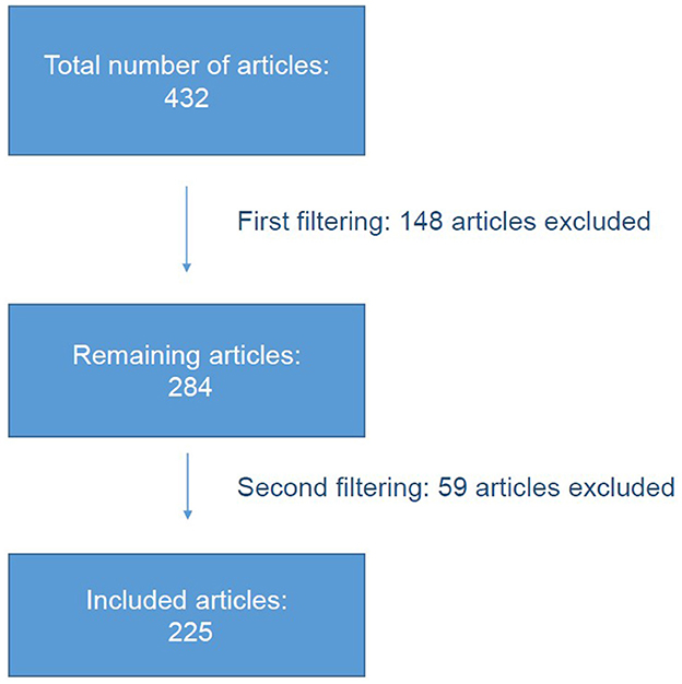
Figure 1. Filtering of the articles.
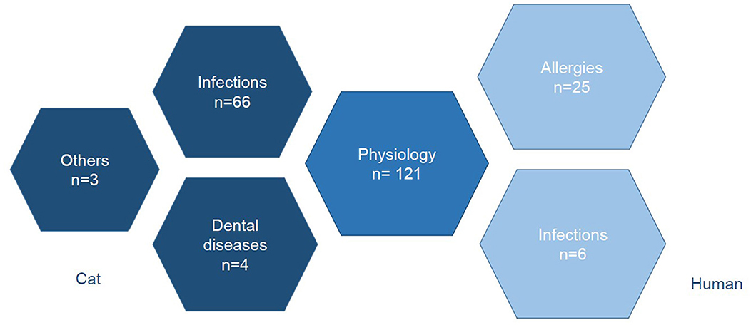
Figure 2. Sorting the articles into different subject areas.
2 The cat as a model for basic salivary physiology research
The cat is an important model for basic research on salivary secretion. In particular, the influence of the autonomic nervous system on salivary secretion has been investigated in many studies (6–9). The roles of the sympathetic and parasympathetic nervous systems have been studied by severing (10) or stimulation of the corresponding nerves in the model in experimental studies (11). For example, the parasympathetic fibers of the chorda tympani of the facial nerve have been shown to significantly stimulate salivary gland secretion (12). Acetylcholine can be used to experimentally induce changes in electrolyte balance, such as intracellular Na+ and K+ concentrations, in a perfused salivary gland (13–17). The role of hexosamines, calcium and various proteins that may affect secretion were also discussed (18). The endogenous hormone kallikrein, which occurs as a serine protease in the salivary gland, also has a vasodilatory effect and therefore appears to influence salivary secretion (19–22). In addition to basic research on salivary secretion in the feline model, salivary diagnostics appears to be useful for assessing the pharmacokinetics of drugs such as pradofloxacin and doxycycline in the cat (23).
3 Infectious diseases of the cat
Infections such as Feline Immunodeficiency virus (FIV) (24–26), Feline leukemia virus (FeLV) (27–29), phlebovirus (30), rabies (31, 32), feline coronavirus (33), feline herpesvirus (26, 34–36), calicivirus (36–38), anellovirus (39, 40), picornavirus (35), feline foamy virus (41), west nile virus (42), hendra virus (43), bartonella (44–46), mycoplasma (47–51), helicobacter pylori (49–51), staphylococci (52), anaplasma (53), ehrlichia (53) and rickettsia can be detected by special saliva swab tests (53). Detection can be by polymerase chain reaction (PCR) (30, 46), enzyme-linked immunosorbent assay (ELISA) (54, 55) or immunofluorescence assay (IFA) (56). For diagnostic purposes, rapid saliva test kits, e.g., for antigen detection of FeLV, have also been evaluated (56, 57).
In addition to direct pathogen detection and pathogenesis studies, the prevalence of diseases in specific countries such as bartonella in Korea (44), FeLV in New Zealand (54) or in Switzerland (58) and of FIV in Italy has been determined (25). Also of interest was the occurrence of different diseases such as feline foamy virus and FeLV (41) as well as the influence of diseases like FIV on the course of a calicivirus infection (36). Mechanisms of the acute and chronic course of calicivirus infection have been investigated with a view to possible vaccination (38). There are strains of the virus that are resistant to vaccination and may contribute to a chronic course.
4 Dental diseases of the cat
Elevated serum and salivary immunoglobulin concentrations were also measured by ELISA in cats with gingivostomatitis (59) or periodontal disease in general (60).
5 Further studies on cat saliva
A novel saliva-based test for the diagnosis of food allergy in cats has been studied. It detected antibodies in saliva to certain feed ingredients, including lamb, beef, pork, turkey, fish, wheat, potato, millet, and rice (61).
In a study on blood grouping of cats based on saliva, the authors could not prove any helpful parameters (62).
In a study of blood grouping in cats based on saliva, the authors did not find any useful parameters (63).
6 Infections of humans by cat saliva
The diagnosis of infectious agents with zoonotic potential in cat saliva is also of great scientific interest. These include primarily bacteria such as Pasteurella multocida (64), which can be transmitted to humans via cat saliva. In addition to the transmission of other pathogens such as Enterobacteriaceae (65, 66), streptococci and staphylococci (64) and bartonella (67), the zoonotic potential of the bacterium Capnocytophaga canimorsus is discussed, which can cause symptoms such as sepsis and meningitis has been discussed (68, 69).
7 Allergies of humans due to cat saliva
Allergens shed by cats that may cause allergic reactions in humans have been investigated in several studies (70, 71). The most important allergen is Fel d1 (72–77). Saliva tests have been performed by ELISA (61, 72, 73, 77), radioimmonoassay (RIA) (76), high performance liquid chromatography (HPLC) (78), immunoelectrophoresis (IEP) (79), a quantitative assay for the detection of monoclonal antibodies, or radioallergosorbent test (RAST), which is also used to detect antibodies to a specific antigen (80, 81). In one study the authors claimed that cat fur was the main source of cat allergens, while another study showed that saliva was the main reservoir (82). The authors of another study considered the skin to be the main source of allergens (74). Thus, the main source of allergens responsible for cat allergy in humans has been controversial for many years. Overall, allergens have been detected in cat excreta such as fur, saliva, skin particles and urine (74, 77, 83, 84).
Recently, new approaches have emerged to investigate Fel d1 blocking antibodies that may reduce cat allergen shedding (75). For example, a clinical trial has already shown that feeding cats a special diet containing these blocking antibodies reduces the rate of antigen excretion (85). From a human health point of view, such a diet therefore appears to be a promising future option for reducing the risk of allergy in the owner.
8 Discussion
The aim of this study was to provide an overview through a systematic review of the literature and to identify areas of underuse in cats.
The physiology of salivary secretion in the cat model represents the majority of articles found, 121 out of 225. This is basic research on animal models, with the aim of drawing conclusions about the physiology of other species or humans. These studies can be used in translational medicine, which is why the research interest seems to be particularly high.
With 104 articles, there is still a large number of articles on clinical applications of feline salivary diagnostics. Another major topic was the diagnosis of feline infectious diseases such as FIV and FeLV. This can be explained by the fact that these diseases are very widespread throughout the world (54). The advantage of saliva diagnostics is the high sensitivity for many viruses such as FIV and FeLV, so saliva sampling can be considered a simple alternative to blood sampling in this area of pathogen detection (54).
It should be noted that for some other pathogens, diagnostic results should be interpreted with caution. For example, in a study of Candidatus Mycoplasma haemominutum in cats after natural infection, the pathogen was detected in the blood but not in feces and saliva (48). Candidatus Mycoplasma turicensis was also detected in cat saliva only in the early phase of infection, not in the late phase (48). The superiority of a saliva test over a blood test depends on the pathogen and must be considered on a case-by-case basis.
In the case of zoonoses transmitted from cats to humans, it should be borne in mind that they are particularly dangerous for young, elderly, pregnant and immunocompromised, e.g., asplenic people. In addition to general hygiene measures such as regular hand washing, vector prophylaxis to prevent communicable diseases should not be neglected. For example, cats should receive regular ectoparasite prophylaxis to minimize the risk of transmission of e.g., Bartonella henselae via fleas (86). The transmission of disease from cats to humans through bites is particularly dangerous. These are often underestimated, but can lead to serious consequences and even death (87–89). The most common pathogens include Pasteurella multocida, but Staphylococcus aureus and a wide range of aerobic and anaerobic pathogens are also commonly found in bite wounds, often requiring appropriate wound revision and antibiotic treatment (89).
Saliva testing can also be useful in feline dentistry, although there are only a few articles on this subject (60, 90). However, it should be noted that infectious diseases such as calici or feline foamy virus can contribute to diseases such as feline gingivostomatitis (91, 92). Fungal diseases can also contribute to gingivostomatitis (93). Although salivary diagnostics are not yet routinely used in veterinary practice, parameters such as oxidative stress have been found to be indicative of tooth resorption and periodontitis (94). Elevated levels of salivary immunoglobulins may indicate feline gingivostomatitis (59). An altered microbiome may also be associated with an increased prevalence of diseases such as gingivostomatitis (95, 96). The pH of saliva and the presence of bacteria such as Streptococcus mutans may explain why the prevalence of dental disease such as carious lesions differs between humans and cats. In human medicine, indications of caries can be found by examining the microflora of the saliva (97). Parameters such as alanine asparate aminotransferase may also indicate parodontitis (98). In addition, oral lichen planus disease can be diagnosed based on the microflora and cytokines in the mouth, which has not been directly described in cats (99). Several studies have been conducted to diagnose oral squamous cell carcinoma using saliva (100, 101).
A common disease in small animal medicine is food allergy (63, 87). In a 2018 review article on this topic, the authors concluded that elimination diets are the most important component of diagnosis and treatment (102). However, a year later, a study was published on a novel saliva-based test for the diagnosis of food allergy in cats, which could simplify the diagnosis (61). This test has already been described for dogs. As an elimination diet is often a lengthy and cumbersome procedure, saliva diagnostics could be a faster and less invasive alternative.
It was also possible to find studies in which the test results contradicted the authors' expectations. In a study of AB blood groups, no substances were found in cat saliva that could be used to determine the blood group of the cat (62). Saliva diagnostics is therefore an area of ongoing research.
A comparison of the areas of application for dogs and cats shows that there is still a long way to go in feline medicine. In addition to infectious diseases and allergens (103, 104), stress hormones such as cortisol (105) and vasopressin (106) are measured in dogs. In the present literature search, only one study was found in which salivary cortisol concentrations were measured in cats after administration of alfaxalon or propofol (63). Stress hormones are useful biomarkers of stress and pain, and further studies could be conducted in this area.
Renal values such as salivary urea and creatinine have already been investigated in a pilot study in dogs with chronic kidney disease (107), but no studies have yet been conducted in cats. Inflammatory markers such as C-reactive protein have also been measured in dog saliva (108). In addition, inflammatory parameters such as tumor necrosis factor or various interleukins have already been measured in the saliva of dogs with diabetes mellitus (109). Studies in cats are also lacking in this area. Other known applications in human medicine include genetic analysis (110) and the determination of sex hormones, such as testosterone (111) and progesterone (112) which have not yet been used in feline medicine. There are also studies on the diagnosis of cystic fibrosis (113), Sjögren's syndrome (114), or Prader-Willi syndrome (115) but also tumor diseases [such as breast cancer (116)], pancreatitis (117, 118), diabetes mellitus (119, 120), or sepsis (121–123). Due to the large number of studies in human medicine, only examples can be given here. However, it is clear that the potential of saliva diagnostics in veterinary medicine has not yet been fully exploited.
The advantage of saliva diagnostics is that it may be possible to avoid taking blood from the animal if, for example, the aforementioned infectious diseases are to be detected. In general, salivettes (Figure 1) are often used for saliva diagnostics. In some cases, they need to remain in the animal's mouth for several seconds to collect enough saliva for measurement. However, not only in studies on the detection of infectious agents, but also in studies on stress hormones such as cortisol, a conventional swab was sufficient to collect enough saliva (63). After transferring the salivettes to the appropriate tubes (Figure 3), they are centrifuged and the saliva can then be pipetted and stored until analysis. It should be noted that there is no standardized protocol for salivary diagnostics in cats. The amount of saliva will depend on the parameter being assessed. Saliva samples may be collected in the awake state or under sedation, although certain medications may stimulate salivary secretion. The extent to which stimulation of salivary secretion may affect the test result must be considered for each parameter. The concentrations of the parameter to be determined are usually much lower in saliva than in blood. This is also a disadvantage of saliva diagnostics.
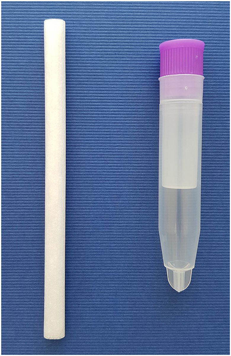
Figure 3. Salivette and corresponding tube for saliva collection.
In the present study, the search was carried out using the Pubmed database only. Databases such as Scopus, Web of Science and Google Scholar were not searched, which is a limitation of the work. Nevertheless, a total of 225 articles provided an overview of the relevant areas of application of salivary diagnostics in cats. With 148 articles, a large number of studies had to be excluded. Due to the unspecific search for “cat saliva”, for example, articles on “catfish” and “wild cats” were found, which did not fit the topic. Articles on saliva studies of people with cat allergy and saliva studies of ticks, which can transmit infectious agents to cats, were also excluded (124). Articles were also excluded if they assessed the epidemiology of, for example, pasteurella or rabies in different countries, without examining cat saliva (125). Similarly, articles sialoceles (126), adenocarcinoma of the salivary gland (127) and scintigraphy for thyroid function had to be excluded as well, because saliva was not examined (128). One article on the pancreas described physiological processes that could also be found in other exocrine glands, such as the salivary glands (129), but as salivary diagnostics was not the primary concern, these articles were also excluded.
Due to the large number of search results, the topics could only be summarized and illustrated with examples in this paper. The focus of the studies presented was on frequently investigated areas of application that are of great scientific interest, even though by far not all the possibilities are fully exploited in feline medicine, such as the measurement of stress hormones to assess stress and pain, as is already done in dogs. Overall, salivary testing in cats can be useful in many areas, and in some cases can even save the patient from having to take a blood sample.
Author contributions
MS: Conceptualization, Data curation, Formal analysis, Investigation, Methodology, Project administration, Software, Writing—original draft. AM-L: Supervision, Writing—review & editing.
Funding
The author(s) declare that no financial support was received for the research, authorship, and/or publication of this article.
Conflict of interest
The authors declare that the research was conducted in the absence of any commercial or financial relationships that could be construed as a potential conflict of interest.
Publisher's note
All claims expressed in this article are solely those of the authors and do not necessarily represent those of their affiliated organizations, or those of the publisher, the editors and the reviewers. Any product that may be evaluated in this article, or claim that may be made by its manufacturer, is not guaranteed or endorsed by the publisher.
References
1. Fromme V, Kohler C, Piesnack S, Oechtering G, Ludewig E. Computed tomographic anatomy of the salivary glands in the cat. Tierarztl Prax Ausg K Kleintiere Heimtiere. (2016) 44:16–25. doi: 10.15654/TPK-150097
2. Pasha S, Inui T, Chapple I, Harris S, Holcombe L, Grant MM. The saliva proteome of dogs: variations within and between breeds and between species. Proteomics. (2018) 18:1700293. doi: 10.1002/pmic.201700293
3. Holmberg KV, Hoffman MP. Anatomy, biogenesis and regeneration of salivary glands. Monogr Oral Sci. (2014) 24:1–13. doi: 10.1159/000358776
4. Harvey CE. Oral diseases and veterinary dentistry. Tijdschr Diergeneeskd. (1987) 112 (Suppl. 1):20S−5S.
5. Schultz RD, Scott FW, Duncan JR, Gillespie JH. Feline immunoglobulins. Infect Immun. (1974) 9:391–3. doi: 10.1128/iai.9.2.391-393.1974
6. Emmelin N. Parotid secretion after cutting the auriculotemporal nerve at different levels. J Physiol. (1967) 188:44P–5P.
7. Karn RC. The mouse salivary androgen-binding protein (ABP) alpha subunit closely resembles chain 1 of the cat allergen Fel dl. Biochem Genet. (1994) 32:271–7. doi: 10.1007/BF00555830
8. Dische Z, Kahn N, Rothschild C, Danilchenko A, Licking J, Wang SC. Glycoproteins of submaxillary saliva of the cat: differences in composition produced by sympathetic and parasympathetic nerve stimulation. J Neurochem. (1970) 17:649–58. doi: 10.1111/j.1471-4159.1970.tb00544.x
9. Gutkin VI, Kuptsov SA. Effect of postganglionic parasympathetic and sympathetic denervation of the submandibular gland on the electrolyte composition of its secretion. Fiziol Zh SSSR Im I M Sechenova. (1973) 59:133–9.
10. Edwards AV, Garrett JR. Submandibular responses to stimulation of the sympathetic innervation following parasympathetic denervation in cats. J Physiol. (1988) 397:421–31. doi: 10.1113/jphysiol.1988.sp017009
11. Garrett JR, Kidd A. Effects of secretory nerve stimulation on acid phosphatase and peroxidase in submandibular saliva and acini in cats. Histochem J. (1977) 9:435–51. doi: 10.1007/BF01002975
12. Izumi H, Karita K. Salivary secretion in cat submandibular gland mediated by chorda tympani afferents. Am J Physiol. (1995) 268:R438–44. doi: 10.1152/ajpregu.1995.268.2.R438
13. Poulsen JH. Acetylcholine-induced transport of Na+ and K+ in the perfused cat submandibular gland. Pflugers Arch. (1974) 349:215–20. doi: 10.1007/BF00592449
14. Petersen OH, Poulsen JH. The secretion of sodium and potassium in cat submandibular saliva during the first period after start of stimulation. Acta Physiol Scand. (1968) 73:83–100. doi: 10.1111/j.1748-1716.1968.tb04086.x
15. Petersen OH, Poulsen JH. Secretory potentials, potassium transport and secretion in the cat submandibular gland during perfusion with sulphate Locke's solution. Experientia. (1968) 24:919–20. doi: 10.1007/BF02138654
16. Winston DC, Schulte BA, Garrett JR, Proctor GB. Na+, K(+)-ATPase in cat salivary glands and changes induced by nerve stimulation: an immunohistochemical study. J Histochem Cytochem. (1990) 38:1187–91. doi: 10.1177/38.8.2164061
17. Garrett JR, Winston DC, Proctor GB, Schulte BA. Na, K-ATPase in resting and stimulated submandibular salivary glands in cats, studied by means of ouabain-sensitive, K(+)-dependent p-nitrophenylphosphatase activity. Arch Oral Biol. (1992) 37:711–6. doi: 10.1016/0003-9969(92)90077-L
18. Burford HJ, Gill JB. Calcium and secretion in normal and supersensitive submaxillary glands of the cat. Biochem Pharmacol. (1968) 17:1881–92. doi: 10.1016/0006-2952(68)90104-4
19. Beilenson S, Schachter M, Smaje LH. Secretion of kallikrein and its role in vasodilatation in the submaxillary gland. J Physiol. (1968) 199:303–17. doi: 10.1113/jphysiol.1968.sp008655
20. Maranda B, Rodrigues JA, Schachter M, Shnitka TK, Weinberg J. Studies on kallikrein in the duct systems of the salivary glands of the cat. J Physiol. (1978) 276:321–8. doi: 10.1113/jphysiol.1978.sp012236
21. Garrett JR, Kidd A, Kyriacou K, Smith RE. Use of different derivatives of D-Val-Leu-Arg for studying kallikrein activities in cat submandibular glands and saliva. Histochem J. (1985) 17:805–18. doi: 10.1007/BF01003316
22. Garrett JR, Smith RE, Kyriacou K, Kidd A, Liao J. Factors affecting the secretion of submandibular salivary kallikrein in cats. Q J Exp Physiol. (1987) 72:357–68. doi: 10.1113/expphysiol.1987.sp003081
23. Hartmann A, Krebber R, Daube G, Hartmann K. Pharmacokinetics of pradofloxacin and doxycycline in serum, saliva, and tear fluid of cats after oral administration. J Vet Pharmacol Ther. (2008) 31:87–94. doi: 10.1111/j.1365-2885.2007.00932.x
24. Yamamoto JK, Hansen H, Ho EW, Morishita TY, Okuda T, Sawa TR, et al. Epidemiologic and clinical aspects of feline immunodeficiency virus infection in cats from the continental United States and Canada and possible mode of transmission. J Am Vet Med Assoc. (1989) 194:213–20.
25. Bandecchi P, Matteucci D, Baldinotti F, Guidi G, Abramo F, Tozzini F, et al. Prevalence of feline immunodeficiency virus and other retroviral infections in sick cats in Italy. Vet Immunol Immunopathol. (1992) 31:337–45. doi: 10.1016/0165-2427(92)90020-Q
26. Reubel GH, Ramos RA, Hickman MA, Rimstad E, Hoffmann DE, Pedersen NC. Detection of active and latent feline herpesvirus 1 infections using the polymerase chain reaction. Arch Virol. (1993) 132:409–20. doi: 10.1007/BF01309549
27. Lutz H, Jarrett O. Detection of feline leukemia virus infection in saliva. J Clin Microbiol. (1987) 25:827–31. doi: 10.1128/jcm.25.5.827-831.1987
28. Kerr MG, Smith KJ. Detection of FeLV antigen by indirect immunofluorescence in ELISA/CITE negative cats. Vet Rec. (1995) 136:516–8. doi: 10.1136/vr.136.20.516
29. Studer N, Lutz H, Saegerman C, Gonczi E, Meli ML, Boo G, et al. Pan-European Study on the prevalence of the feline leukaemia virus infection - reported by the European Advisory Board on Cat Diseases (ABCD Europe). Viruses. (2019) 11:993. doi: 10.3390/v11110993
30. Park ES, Shimojima M, Nagata N, Ami Y, Yoshikawa T, Iwata-Yoshikawa N, et al. Severe fever with thrombocytopenia syndrome phlebovirus causes lethal viral hemorrhagic fever in cats. Sci Rep. (2019) 9:11990. doi: 10.1038/s41598-019-48317-8
31. Vaughn JB, Gerhardt P, Paterson JC. Excretion of street rabies virus in saliva of cats. JAMA. (1963) 184:705–8. doi: 10.1001/jama.1963.73700220001013
32. Trimarchi CV, Rudd RJ, Abelseth MK. Experimentally induced rabies in four cats inoculated with a rabies virus isolated from a bat. Am J Vet Res. (1986) 47:777–80.
33. Addie DD, Jarrett O. Use of a reverse-transcriptase polymerase chain reaction for monitoring the shedding of feline coronavirus by healthy cats. Vet Rec. (2001) 148:649–53. doi: 10.1136/vr.148.21.649
34. Allgoewer I, Schaffer EH, Stockhaus C, Vogtlin A. Feline eosinophilic conjunctivitis. Vet Ophthalmol. (2001) 4:69–74. doi: 10.1046/j.1463-5224.2001.00150.x
35. Flagstad A. Isolation and classification of feline picornavirus and herpesvirus in Denmark. Acta Vet Scand. (1972) 13:462–71. doi: 10.1186/BF03547152
36. Reubel GH, George JW, Higgins J, Pedersen NC. Effect of chronic feline immunodeficiency virus infection on experimental feline calicivirus-induced disease. Vet Microbiol. (1994) 39:335–51. doi: 10.1016/0378-1135(94)90169-4
37. Wardley RC. Feline calicivirus carrier state. A study of the host/virus relationship. Arch Virol. (1976) 52:243–9. doi: 10.1007/BF01348021
38. Pedersen NC, Hawkins KF. Mechanisms for persistence of acute and chronic feline calicivirus infections in the face of vaccination. Vet Microbiol. (1995) 47:141–56. doi: 10.1016/0378-1135(95)00101-F
39. Bedarida S, Dussol B, Signoli M, Biagini P. Analysis of Anelloviridae sequences characterized from serial human and animal biological samples. Infect Genet Evol. (2017) 53:89–93. doi: 10.1016/j.meegid.2017.05.017
40. Biagini P, Uch R, Belhouchet M, Attoui H, Cantaloube JF, Brisbarre N, et al. Circular genomes related to anelloviruses identified in human and animal samples by using a combined rolling-circle amplification/sequence-independent single primer amplification approach. J Gen Virol. (2007) 88(Pt 10):2696–701. doi: 10.1099/vir.0.83071-0
41. Cavalcante LTF, Muniz CP, Jia H, Augusto AM, Troccoli F, Medeiros SO, et al. Clinical and molecular features of feline foamy virus and feline leukemia virus co-infection in naturally-infected cats. Viruses. (2018) 10:702. doi: 10.3390/v10120702
42. Austgen LE, Bowen RA, Bunning ML, Davis BS, Mitchell CJ, Chang GJ. Experimental infection of cats and dogs with West Nile virus. Emerg Infect Dis. (2004) 10:82–6. doi: 10.3201/eid1001.020616
43. Williamson MM, Hooper PT, Selleck PW, Gleeson LJ, Daniels PW, Westbury HA, et al. Transmission studies of Hendra virus (equine morbillivirus) in fruit bats, horses and cats. Aust Vet J. (1998) 76:813–8. doi: 10.1111/j.1751-0813.1998.tb12335.x
44. Kim YS, Seo KW, Lee JH, Choi EW, Lee HW, Hwang CY, et al. Prevalence of Bartonella henselae and Bartonella clarridgeiae in cats and dogs in Korea. J Vet Sci. (2009) 10:85–7. doi: 10.4142/jvs.2009.10.1.85
45. Namekata DY, Kasten RW, Boman DA, Straub MH, Siperstein-Cook L, Couvelaire K, et al. Oral shedding of Bartonella in cats: correlation with bacteremia and seropositivity. Vet Microbiol. (2010) 146:371–5. doi: 10.1016/j.vetmic.2010.05.034
46. Mazaheri Nezhad Fard R, Vahedi SM, Ashrafi I, Alipour F, Sharafi G, Akbarein H, et al. Molecular identification and phylogenic analysis of Bartonella henselae isolated from Iranian cats based on gltA gene. Vet Res Forum. (2016) 7:69–72.
47. Dean RS, Helps CR, Gruffydd Jones TJ, Tasker S. Use of real-time PCR to detect Mycoplasma haemofelis and ‘Candidatus Mycoplasma haemominutum' in the saliva and salivary glands of haemoplasma-infected cats. J Feline Med Surg. (2008) 10:413–7. doi: 10.1016/j.jfms.2007.12.007
48. Willi B, Boretti FS, Meli ML, Bernasconi MV, Casati S, Hegglin D, et al. Real-time PCR investigation of potential vectors, reservoirs, and shedding patterns of feline hemotropic mycoplasmas. Appl Environ Microbiol. (2007) 73:3798–802. doi: 10.1128/AEM.02977-06
49. Ghil HM, Yoo JH, Jung WS, Chung TH, Youn HY, Hwang CY. Survey of Helicobacter infection in domestic and feral cats in Korea. J Vet Sci. (2009) 10:67–72. doi: 10.4142/jvs.2009.10.1.67
50. Fox JG, Perkins S, Yan L, Shen Z, Attardo L, Pappo J. Local immune response in Helicobacter pylori-infected cats and identification of H. pylori in saliva, gastric fluid and faeces. Immunology. (1996) 88:400–6. doi: 10.1046/j.1365-2567.1996.d01-677.x
51. Perkins SE, Yan LL, Shen Z, Hayward A, Murphy JC, Fox JG. Use of PCR and culture to detect Helicobacter pylori in naturally infected cats following triple antimicrobial therapy. Antimicrob Agents Chemother. (1996) 40:1486–90. doi: 10.1128/AAC.40.6.1486
52. Lilenbaum W, Esteves AL, Souza GN. Prevalence and antimicrobial susceptibility of staphylococci isolated from saliva of clinically normal cats. Lett Appl Microbiol. (1999) 28:448–52. doi: 10.1046/j.1365-2672.1999.00540.x
53. Pennisi MG, Hofmann-Lehmann R, Radford AD, Tasker S, Belak S, Addie DD, et al. Ehrlichia and Rickettsia species infections in cats: European guidelines from the ABCD on prevention and management. J Feline Med Surg. (2017) 19:542–8. doi: 10.1177/1098612X17706462
54. Gates MC, Vigeant S, Dale A. Prevalence and risk factors for cats testing positive for feline immunodeficiency virus and feline leukaemia virus infection in cats entering an animal shelter in New Zealand. N Z Vet J. (2017) 65:285–91. doi: 10.1080/00480169.2017.1348266
55. Cattori V, Tandon R, Riond B, Pepin AC, Lutz H, Hofmann-Lehmann R. The kinetics of feline leukaemia virus shedding in experimentally infected cats are associated with infection outcome. Vet Microbiol. (2009) 133:292–6. doi: 10.1016/j.vetmic.2008.07.001
56. Babyak SD, Groves MG, Dimski DS, Taboada J. Evaluation of a saliva test kit for feline leukemia virus antigen. J Am Anim Hosp Assoc. (1996) 32:397–400. doi: 10.5326/15473317-32-5-397
57. Westman ME, Malik R, Hall E, Sheehy PA, Norris JM. Comparison of three feline leukaemia virus (FeLV) point-of-care antigen test kits using blood and saliva. Comp Immunol Microbiol Infect Dis. (2017) 50:88–96. doi: 10.1016/j.cimid.2016.11.014
58. Hofmann-Lehmann R, Gonczi E, Riond B, Meli M, Willi B, Howard J, et al. Feline leukemia virus infection: importance and current situation in Switzerland. Schweiz Arch Tierheilkd. (2018) 160:95–105. doi: 10.17236/sat00146
59. Harley R, Gruffydd-Jones TJ, Day MJ. Salivary and serum immunoglobulin levels in cats with chronic gingivostomatitis. Vet Rec. (2003) 152:125–9. doi: 10.1136/vr.152.5.125
60. Cave NJ, Bridges JP, Thomas DG. Systemic effects of periodontal disease in cats. Vet Q. (2012) 32:131–44. doi: 10.1080/01652176.2012.745957
61. Dodds WJ. Diagnosis of feline food sensitivity and intolerance using saliva: 1000 cases. Animals. (2019) 9:534. doi: 10.3390/ani9080534
62. Auer L, Bell K. The AB blood group system of cats. Anim Blood Groups Biochem Genet. (1981) 12:287–97. doi: 10.1111/j.1365-2052.1981.tb01561.x
63. Yozova ID, Sano H, Weidgraaf K, Candy EJ, Cockrem JF. A randomized cross-over trial assessing salivary and urinary cortisol concentrations after alfaxalone and propofol administration in healthy cats. Domest Anim Endocrinol. (2021) 74:106557. doi: 10.1016/j.domaniend.2020.106557
64. Peeples E, Boswick JA Jr, Scott FA. Wounds of the hand contaminated by human or animal saliva. J Trauma. (1980) 20:383–9. doi: 10.1097/00005373-198020050-00004
65. Melo LC, Oresco C, Leigue L, Netto HM, Melville PA, Benites NR, et al. Prevalence and molecular features of ESBL/pAmpC-producing Enterobacteriaceae in healthy and diseased companion animals in Brazil. Vet Microbiol. (2018) 221:59–66. doi: 10.1016/j.vetmic.2018.05.017
66. Pillay S, Zishiri OT, Adeleke MA. Prevalence of virulence genes in Enterococcus species isolated from companion animals and livestock. Onderstepoort J Vet Res. (2018) 85:e1–8. doi: 10.4102/ojvr.v85i1.1583
67. Sayed ASM, Alsaadawy RM, Ali MM, Abd El-Hamid RF, Baty RS, Elmahallawy EK. Serological and molecular detection of bartonella henselae in cats and humans from Egypt: current status and zoonotic implications. Front Vet Sci. (2022) 9:859104. doi: 10.3389/fvets.2022.859104
68. Chodosh J. Cat's tooth keratitis: human corneal infection with Capnocytophaga canimorsus. Cornea. (2001) 20:661–3. doi: 10.1097/00003226-200108000-00021
69. Gaastra W, Lipman LJ. Capnocytophaga canimorsus. Vet Microbiol. (2010) 140:339–46. doi: 10.1016/j.vetmic.2009.01.040
70. Didierlaurent A, Foglietti MJ, Guerin B, Hewitt BE, Percheron F. Comparative study on cat allergens from fur and saliva. Int Arch Allergy Appl Immunol. (1984) 73:27–31. doi: 10.1159/000233433
71. Sacchi G, Valcurone G, Tassi GC. Attempts at characterizing the allergens of cat saliva and fur. Boll Ist Sieroter Milan. (1984) 63:462–73.
72. Bastien BC, Gardner C, Satyaraj E. Influence of time and phenotype on salivary Fel d1 in domestic shorthair cats. J Feline Med Surg. (2019) 21:867–74. doi: 10.1177/1098612X19850973
73. Jalil-Colome J, de Andrade AD, Birnbaum J, Casanova D, Mege JL, Lanteaume A, et al. Sex difference in Fel d 1 allergen production. J Allergy Clin Immunol. (1996) 98:165–8. doi: 10.1016/S0091-6749(96)70238-5
74. Mata P, Charpin D, Charpin C, Lucciani P, Vervloet D. Fel d I allergen: skin and or saliva? Ann Allergy. (1992) 69:321–2.
75. Satyaraj E, Sun P, Sherrill S. Fel d1 blocking antibodies: a novel method to reduce IgE-mediated allergy to cats. J Immunol Res. (2021) 2021:5545173. doi: 10.1155/2021/5545173
76. van Milligen FJ, Vroom TM, Aalberse RC. Presence of Felis domesticus allergen I in the cat's salivary and lacrimal glands. Int Arch Allergy Appl Immunol. (1990) 92:375–8. doi: 10.1159/000235168
77. Kelly SM, Karsh J, Marcelo J, Boeckh D, Stepner N, Santone B, et al. Fel d 1 and Fel d 4 levels in cat fur, saliva, and urine. J Allergy Clin Immunol. (2018) 142:1990–2 e3. doi: 10.1016/j.jaci.2018.07.033
78. Calam DH, Davidson J, Ford AW. Studies on allergens of mammalian origin. J Chromatogr. (1984) 288:137–45. doi: 10.1016/S0021-9673(01)93688-X
79. Anderson MC, Baer H. Allergenically active components of cat allergen extracts. J Immunol. (1981) 127:972–5. doi: 10.4049/jimmunol.127.3.972
80. Guerin B, Hewitt B. A comparative study of allergen extracts from cat fur, cat pelt and cat saliva. Ann Allergy. (1981) 46:127–31.
81. Viander M, Valovirta E, Vanto T, Koivikko A. Cross-reactivity of cat and dog allergen extracts. RAST inhibition studies with special reference to the allergenic activity in saliva and urine. Int Arch Allergy Appl Immunol. (1983) 71:252–60. doi: 10.1159/000233399
82. Brown PR, Leitermann K, Ohman JL Jr. Distribution of cat allergen 1 in cat tissues and fluids. Int Arch Allergy Appl Immunol. (1984) 74:67–70. doi: 10.1159/000233518
83. Vervloet D. Origin of allergens in the cat. Bull Acad Natl Med. (1994) 178:1667–75; discussion 1675–6.
84. Shah R, Grammer LC. Chapter 1: an overview of allergens. Allergy Asthma Proc. (2012) 33(Suppl. 1):2–5. doi: 10.2500/aap.2012.33.3531
85. Satyaraj E, Wedner HJ, Bousquet J. Keep the cat, change the care pathway: a transformational approach to managing Fel d 1, the major cat allergen. Allergy. (2019) 74(Suppl. 107):5–17. doi: 10.1111/all.14013
86. Breitschwerdt EB. Feline bartonellosis and cat scratch disease. Vet Immunol Immunopathol. (2008) 123:167–71. doi: 10.1016/j.vetimm.2008.01.025
87. Zajkowska J, Krol M, Falkowski D, Syed N, Kamienska A. Capnocytophaga canimorsus - an underestimated danger after dog or cat bite - review of literature. Przegl Epidemiol. (2016) 70:289–95.
88. Kheiran A, Palial V, Rollett R, Wildin CJ, Chatterji U, Singh HP. Cat bite: an injury not to underestimate. J Plast Surg Hand Surg. (2019) 53:341–6. doi: 10.1080/2000656X.2019.1637750
89. Westling K, Farra A, Cars B, Ekblom AG, Sandstedt K, Settergren B, et al. Cat bite wound infections: a prospective clinical and microbiological study at three emergency wards in Stockholm, Sweden. J Infect. (2006) 53:403–7. doi: 10.1016/j.jinf.2006.01.001
90. Barabash RD, Levitskii AP, Konovets VM. Secretion of salivary enzymes in cats with spontaneous inflammatory-dystrophic lesions of the parodontium. Fiziol Zh. (1977) 23:92–7.
91. Druet I, Hennet P. Relationship between feline calicivirus load, oral lesions, and outcome in feline chronic gingivostomatitis (caudal stomatitis): retrospective study in 104 cats. Front Vet Sci. (2017) 4:209. doi: 10.3389/fvets.2017.00209
92. Hennet PR, Camy GA, McGahie DM, Albouy MV. Comparative efficacy of a recombinant feline interferon omega in refractory cases of calicivirus-positive cats with caudal stomatitis: a randomised, multi-centre, controlled, double-blind study in 39 cats. J Feline Med Surg. (2011) 13:577–87. doi: 10.1016/j.jfms.2011.05.012
93. Krumbeck JA, Reiter AM, Pohl JC, Tang S, Kim YJ, Linde A, et al. Characterization of Oral Microbiota in Cats: Novel Insights on the Potential Role of Fungi in Feline Chronic Gingivostomatitis. Pathogens. (2021) 10:904. doi: 10.3390/pathogens10070904
94. Levitskii AP, Kozlianina NP, Skliar VE. Lipid peroxidation and antioxidant systems in cat periodontal tissues. Vopr Med Khim. (1987) 33:107–11.
95. Rodrigues MX, Bicalho RC, Fiani N, Lima SF, Peralta S. The subgingival microbial community of feline periodontitis and gingivostomatitis: characterization and comparison between diseased and healthy cats. Sci Rep. (2019) 9:12340. doi: 10.1038/s41598-019-48852-4
96. Dolieslager SM, Riggio MP, Lennon A, Lappin DF, Johnston N, Taylor D, et al. Identification of bacteria associated with feline chronic gingivostomatitis using culture-dependent and culture-independent methods. Vet Microbiol. (2011) 148:93–8. doi: 10.1016/j.vetmic.2010.08.002
97. Wu HZ, Zhang X, Cheng XG Yu Q. Saliva microbiota and metabolite in individuals with caries or periodontitis. Zhonghua Kou Qiang Yi Xue Za Zhi. (2023) 58:131–42. doi: 10.3760/cma.j.cn112144-20220829-00464
98. Totan A, Greabu M, Totan C, Spinu T. Salivary aspartate aminotransferase, alanine aminotransferase and alkaline phosphatase: possible markers in periodontal diseases? Clin Chem Lab Med. (2006) 44:612–5. doi: 10.1515/CCLM.2006.096
99. Carvalho M, Cavalieri D, Do Nascimento ST, Lourenco GB, Ramos VR, Pasqualin DC, et al. Cytokines levels and salivary microbiome play a potential role in oral lichen planus diagnosis. Sci Rep. (2019) 9:18137. doi: 10.1038/s41598-019-54615-y
100. Banavar G, Ogundijo O, Julian C, Toma R, Camacho F, Torres PJ, et al. Detecting salivary host and microbiome RNA signature for aiding diagnosis of oral and throat cancer. Oral Oncol. (2023) 145:106480. doi: 10.1016/j.oraloncology.2023.106480
101. Sivadasan P, Gupta MK, Sathe G, Sudheendra HV, Sunny SP, Renu D, et al. Salivary proteins from dysplastic leukoplakia and oral squamous cell carcinoma and their potential for early detection. J Proteomics. (2020) 212:103574. doi: 10.1016/j.jprot.2019.103574
102. Mueller RS, Unterer S. Adverse food reactions: pathogenesis, clinical signs, diagnosis and alternatives to elimination diets. Vet J. (2018) 236:89–95. doi: 10.1016/j.tvjl.2018.04.014
103. Duncan AW, Maggi RG, Breitschwerdt EB. Bartonella DNA in dog saliva. Emerg Infect Dis. (2007) 13:1948–50. doi: 10.3201/eid1312.070653
104. Polovic N, Waden K, Binnmyr J, Hamsten C, Gronneberg R, Palmberg C, et al. Dog saliva - an important source of dog allergens. Allergy. (2013) 68:585–92. doi: 10.1111/all.12130
105. Dreschel NA, Granger DA. Methods of collection for salivary cortisol measurement in dogs. Horm Behav. (2009) 55:163–8. doi: 10.1016/j.yhbeh.2008.09.010
106. Pirrone F, Pierantoni L, Bossetti A, Uccheddu S, Albertini M. Salivary vasopressin as a potential non-invasive biomarker of anxiety in dogs diagnosed with separation-related problems. Animals. (2019) 9:1033. doi: 10.3390/ani9121033
107. Tvarijonaviciute A, Pardo-Marin L, Tecles F, Carrillo JD, Garcia-Martinez JD, Bernal L, et al. Measurement of urea and creatinine in saliva of dogs: a pilot study. BMC Vet Res. (2018) 14:223. doi: 10.1186/s12917-018-1546-5
108. Cho YR, Oh YI, Song GH, Kim YJ, Seo KW. Comparative analysis of C-reactive protein levels in the saliva and serum of dogs with various diseases. Animals. (2020) 10:1042. doi: 10.3390/ani10061042
109. Franco-Martinez L, Munoz-Prieto A, Busato F, Karveliene B, Stadaliene I, Ceron JJ, et al. Evaluation of the presence of gingivitis as confounding factor in assessing inflammatory status in serum and saliva of dogs with diabetes mellitus. BMC Vet Res. (2024) 20:116. doi: 10.1186/s12917-024-03962-8
110. Ostheim P, Alemu SW, Tichy A, Sirak I, Davidkova M, Stastna MM, et al. Examining potential confounding factors in gene expression analysis of human saliva and identifying potential housekeeping genes. Sci Rep. (2022) 12:2312. doi: 10.1038/s41598-022-05670-5
111. Lood Y, Aardal E, Ahlner J, Arlemalm A, Carlsson B, Ekman B, et al. Determination of testosterone in serum and saliva by liquid chromatography-tandem mass spectrometry: An accurate and sensitive method applied on clinical and forensic samples. J Pharm Biomed Anal. (2021) 195:113823. doi: 10.1016/j.jpba.2020.113823
112. Mirzaii-Dizgah I, Agha-Hosseini F. Stimulated and unstimulated saliva progesterone in menopausal women with oral dryness feeling. Clin Oral Investig. (2011) 15:859–62. doi: 10.1007/s00784-010-0449-z
113. Blomfield J, Warton KL, Brown JM. Flow rate and inorganic components of submandibular saliva in cystic fibrosis. Arch Dis Child. (1973) 48:267–74. doi: 10.1136/adc.48.4.267
114. Sreebny LM, Zhu WX. The use of whole saliva in the differential diagnosis of Sjogren's syndrome. Adv Dent Res. (1996) 10:17–24. doi: 10.1177/08959374960100010201
115. Boccellino M, Di Stasio D, Serpico R, Lucchese A, Guida A, Settembre G, et al. Analysis of saliva samples in patients with Prader-Willi syndrome. J Biol Regul Homeost Agents. (2018) 32(2 Suppl. 1):107–111.
116. Zhong L, Cheng F, Lu X, Duan Y, Wang X. Untargeted saliva metabonomics study of breast cancer based on ultra performance liquid chromatography coupled to mass spectrometry with HILIC and RPLC separations. Talanta. (2016) 158:351–60. doi: 10.1016/j.talanta.2016.04.049
117. Lankisch PG, Chilla R, Luerssen K, Koop H, Arglebe C, Creutzfeldt W. Parotid saliva test in the diagnosis of chronic pancreatitis. Digestion. (1979) 19:52–5. doi: 10.1159/000198322
118. Dechezlepretre S. Relation between saliva amylase activity and amylasemia in chronic pancreatitis. Possibility of a compensatory saliva amylase activity in deficiencies of the pancreas. Bull Acad Natl Med. (1968) 152:34–6.
119. Kerr M, Lee A, Wang PL, Purushotham KR, Chegini N, Yamamoto H, et al. Detection of insulin and insulin-like growth factors I and II in saliva and potential synthesis in the salivary glands of mice. Effects of type 1 diabetes mellitus. Biochem Pharmacol. (1995) 49:1521–31. doi: 10.1016/0006-2952(95)00017-T
120. Kimura I, Sasamoto H, Sasamura T, Sugihara Y, Ohgaku S, Kobayashi M. Reduction of incretin-like salivatin in saliva from patients with type 2 diabetes and in parotid glands of streptozotocin-diabetic BALB/c mice. Diabetes Obes Metab. (2001) 3:254–8. doi: 10.1046/j.1463-1326.2001.00118.x
121. Coutinho FG, E, Diniz MA, Kandler I, Cianciarullo MA, Santos RD. Assessment of oxidative damage and enzymatic antioxidant system activity on the umbilical cord blood and saliva from preterm newborns with risk factors for early-onset neonatal sepsis. Rev Assoc Med Bras (1992). (2018) 64:888–95. doi: 10.1590/1806-9282.64.10.888
122. Lopez-Martinez MJ, Escribano D, Ortin-Bustillo A, Franco-Martinez L, Gonzalez-Arostegui LG, Ceron JJ, et al. Changes in biomarkers of redox status in saliva of pigs after an experimental sepsis induction. Antioxidants. (2022) 11:1380. doi: 10.3390/antiox11071380
123. Galhardo LF, Ruivo GF, de Oliveira LD, Parize G, Santos S, Pallos D, et al. Inflammatory markers in saliva for diagnosis of sepsis of hospitalizes patients. Eur J Clin Invest. (2020) 50:e13219. doi: 10.1111/eci.13219
124. Rodriguez-Valle M, Moolhuijzen P, Barrero RA, Ong CT, Busch G, Karbanowicz T, et al. Transcriptome and toxin family analysis of the paralysis tick, Ixodes holocyclus. Int J Parasitol. (2018) 48:71–82. doi: 10.1016/j.ijpara.2017.07.007
125. Nadin-Davis SA, Turner G, Paul JP, Madhusudana SN, Wandeler AI. Emergence of Arctic-like rabies lineage in India. Emerg Infect Dis. (2007) 13:111–6. doi: 10.3201/eid1301.060702
126. Vallefuoco R, Jardel N, El Mrini M, Stambouli F, Cordonnier N. Parotid salivary duct sialocele associated with glandular duct stenosis in a cat. J Feline Med Surg. (2011) 13:781–3. doi: 10.1016/j.jfms.2011.06.003
127. Fujiwara-Igarashi A, Shimizu K, Michishita M, Yu Y, Hamamoto Y, Hasegawa D, et al. A cat with suspected laryngeal metastasis with mucosal irregularity resulting from apocrine/salivary gland adenocarcinoma in the head. J Vet Med Sci. (2017) 79:1916–9. doi: 10.1292/jvms.17-0242
128. Henrikson TD, Armbrust LJ, Hoskinson JJ, Milliken GA, Wedekind KJ, Kirk CA, et al. Thyroid to salivary ratios determined by technetium-99m pertechnetate imaging in thirty-two euthyroid cats. Vet Radiol Ultrasound. (2005) 46:521–3. doi: 10.1111/j.1740-8261.2005.00095.x
Keywords: cat, saliva, infections, allergens, physiology
Citation: Schroers M and Meyer-Lindenberg A (2024) Performance and overview of clinically relevant areas of application of saliva testing in the cat. Front. Vet. Sci. 11:1385345. doi: 10.3389/fvets.2024.1385345
Received: 12 February 2024; Accepted: 10 May 2024;
Published: 22 May 2024.
Edited by:
Enrique Fernandez-Caldas, Inmunotek SL, SpainReviewed by:
Jean-Claude Desfontis, Ecole Nationale Vétérinaire Agroalimentaire et de l'Alimentation, FranceJamie Gail Anderson, University of Pennsylvania, United States
Copyright © 2024 Schroers and Meyer-Lindenberg. This is an open-access article distributed under the terms of the Creative Commons Attribution License (CC BY). The use, distribution or reproduction in other forums is permitted, provided the original author(s) and the copyright owner(s) are credited and that the original publication in this journal is cited, in accordance with accepted academic practice. No use, distribution or reproduction is permitted which does not comply with these terms.
*Correspondence: Maike Schroers, maike.schroers@chri.vetmed.uni-muenchen.de