Abstract
Do you like science fiction? Have you heard of, or are you even a fan of, the famous “Star Wars” series? To summarize, there are rebels, emperors, princesses, robots, and many more fabulous creatures. There is also a power source called “The Force.” It is used by the Jedi (the good ones) but also by the dark side (the evil ones). Only the dark side uses the destructive power of “The Force,” which is based on negative emotions such as fear, anger, jealousy, or hate. A Jedi masters “The Force” and uses it for knowledge and defense by learning to control his emotions. Our research also looks at emotions and how to control them. We know that in our galaxy too, we have more success when we can control our feelings. Therefore, we want to find the brain regions responsible for allowing us to deal with our emotions and to help those children struggling with controlling negative emotions.
Imagine walking down the school hall thinking about your next lesson. Suddenly, your best friend jumps out from a dark corner, right in front of you, wearing a silly mask and scaring you. This trick that was played on you immediately led to a reaction of your body. You can feel your heart beating and maybe you just screamed out loudly. A few seconds later though, you recognize your friend and notice there is no real threat. You may even start laughing about the joke. This is an example of how a person can react to an emotional situation. It also shows how our mind processes a situation using different clues. Emotions are feelings that (1) are caused by situations that are meaningful or important to you, (2) are something you feel or show through your body language, and (3) may compete with other important things [1]. In our example, the scary joke gave you the impression of being attacked, and it is important to you to stay unharmed. Your beating heart and the screaming is the reaction of your body. While you are scared and your first intention might be to run away quickly, you also noticed that this was simply your friend playing a joke on you. Being scared and knowing someone is your friend are two different clues that might compete with each other in your brain. One clue tells you to run away in order to stay unharmed, and the other tells you to stay with someone you like (competing reactions). Within a split second, you make a choice about which emotion you find important and which emotion you choose to control or suppress completely. Overall, people tend to choose to decrease negative emotions (anger, sadness, or fear) and increase positive emotions (happiness, love, and joyfulness). Changing or controlling your feelings is an action we call “emotion regulation.” The way that you control and change your emotions is called your “emotion regulation strategy.” Looking at data from many people, scientists were able to show that the way you regulate your emotions influences how you feel, but it also affects the people around you [1]. For example, if you have difficulties controlling your emotions when being angry you may end up cursing, punching, or even bullying the people around you. This is no fun for them either. Therefore, successful emotion processing and regulation is very important for humans. In fact, emotion regulation difficulties are a part of many mental health issues in children, teenagers, and adults.
Using an MRI Camera for Studying the Brain
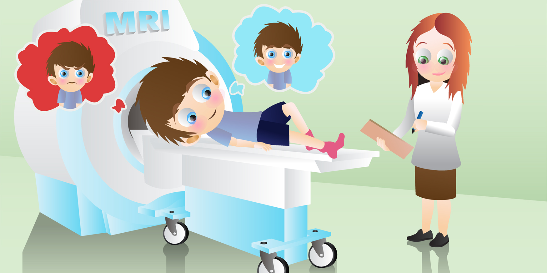
The way the brain processes and regulates emotions can be studied using a technique called magnetic resonance imaging (MRI). An MRI scanner looks like a big tunnel (see Figure 1A). Actually, it is just a very fancy camera that is able to take images of all the parts inside your body. For example, an MRI camera can take an image of the bones in your leg, of your beating heart, or of the organ we are interested in – the brain. We can use the MRI camera to look at the structure (shape and size) of the brain. When we want to see how the brain works, then we can use an MRI camera to look at brain function. Just as you need more food when you do sports, your brain also needs more energy when it becomes active, but instead of food it needs oxygen. Therefore, when a specific region in the brain is hard at work, it will get more oxygen transported to it by the bloodstream. We call this blood oxygen-rich. Oxygen-rich blood gives different signals to the MRI camera compared with blood that has less oxygen. Using this knowledge, researchers can create an image of both the brain’s structure and function. With special computer programs, we can make pictures like the ones in Figure 1B. One of the most amazing things is that the MRI camera can take pictures of your brain at work without even touching you! But there are some challenges for people who take part in research studies using an MRI. Two of the biggest challenges are that (1) you have to stay super still while the pictures are taken or they become blurry (for an explanation, see Figure 2) and (2) you have to protect your ears against the noise. Big cameras such as an MRI can be quite loud, which is why you need to wear special headphones. Staying still can be practiced with fun games, such as the freezing game, where you have to stay still like an ice statue. If you want to know more and see what MRI experiments involving young children look like, you can watch the following video (http://www.jove.com/video/1309/making-mr-imaging-child-s-play-pediatric-neuroimaging-protocol [2]).
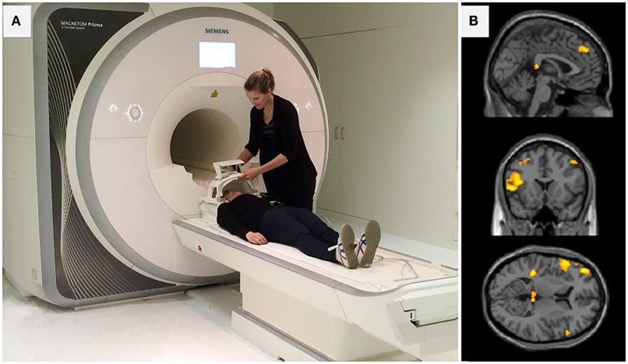
- Figure 1
- A. Two of our research team members showing you an MRI camera and how it is used. B. Different views of a child’s brain as taken by an MRI camera. The areas that are colored yellow are important for emotion processing and regulation.
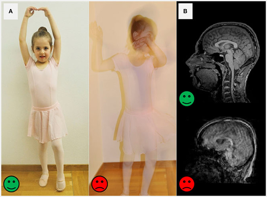
- Figure 2
- Why staying still during an MRI session is important: A. A picture taken by a regular camera can be very sharp when the person is standing super still (green happy face). But when the person is moving a lot, the picture becomes blurry (red sad face). B. The same is true when taking brain pictures. The pictures can turn out super sharp when the person stays still (green happy face) or blurry and hard for scientists to read for when the person wiggles around (red sad face).
What Does the Brain Look Like While Processing and Regulating Emotions?
Now, in the first section, you learned about feelings, which scientists call emotions. You heard that emotions can lead to a reaction in your body. You also know that sometimes we experience several emotions at once and that sometimes it is necessary to control a feeling and not to act on it. This process is called emotion regulation. In the second section, you learned how an MRI camera works and how it can be used to take images of the structure and function of the brain. In the next section, we want to combine these two things and talk about the parts of the brain that are responsible for processing and regulating emotion.
Using MRI cameras, scientists have shown that emotions are processed by many different areas of the brain. There is not just one place that is responsible for processing an emotion. Several brain regions work together as a team. This is why scientists say that emotions are processed by a network of brain regions. A network of brain regions that process emotions is called an emotion processing network (see Figure 3). Let us name some of those brain regions that are activated by emotions. They are the amygdala, the prefrontal cortex, the cingulate cortex, the hippocampus, and the basal ganglia [3]. Fancy names, but it is not these names you need to remember. What is important to understand is that there are many brain regions involved during emotion processing. All the different regions have their own job and they all work together to identify and control an emotion. The amygdala, for example, is a tiny part of the brain (it has the shape and size of an almond), and it is responsible for handling both positive and negative information. The amygdala is especially important when we experience the emotion of fear. Another region of the emotion processing network is the prefrontal cortex, which is named after its location: in the front of the brain. The prefrontal cortex is like a control center, helping to guide our actions, and therefore, this area is also involved during emotion regulation. Both the amygdala and the prefrontal cortex are part of the emotion network. Just like good friends, these different brain regions stay in touch and communicate frequently with each other. For example, the amygdala (the emotion center) can detect an important fearful event and transport that information to the prefrontal cortex (the control center). The prefrontal cortex gets the message that there is something scary happening. If necessary, this control center at the front of your head sends commands to other brain regions telling them to move your body and run away. To sum it up, many brain regions work together to process and react to an emotional situation (see Figure 3).
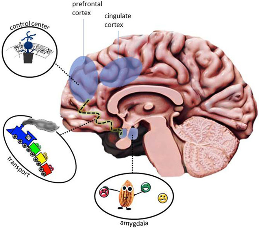
- Figure 3 - The emotion processing network includes several areas of the brain.
- Some of these areas are shown here shaded in blue and you can see their different jobs: the amygdala (almond) recognizes and sorts the emotions before transporting them to other areas. In the picture, this transportation is visualized by a train driving along the dotted track line to the most frontal part of the brain. Once the information arrives there, the prefrontal cortex and the cingulate cortex act as a control center (little man behind desk), deciding what has to be done next with the incoming emotions. Many areas work together to process an emotion! (illustration by Menks).
What Happens in the Brain When Emotion Processing Fails?
By now, you understand that feelings are complicated and that emotions are represented and processed by many regions in the brain. You also remember that successful emotion regulation is important for a persons’ well-being and central for the people around them. As mentioned before, it can be really difficult to be around people that are constantly cursing, hitting, or bullying the people around them because they cannot control their negative emotions. Unfortunately, some children struggle more than others with their emotions. Imagine you have a classmate named Jamie, who has problems with regulating emotions, especially anger and fear. Now picture that you make a silly joke with Jamie, but instead of laughing, Jamie gets very upset and maybe even starts fighting with you. This is an example of someone who has emotion regulation difficulties. Such difficulties in handling emotions can often be observed in very aggressive (frequently fighting and bullying) and antisocial (breaking rules) teenagers. Research studies have shown that these teenagers cannot always successfully identify their emotions. It can also be very hard for these children to control their emotions, like in the case of Jamie. This is not fun for you, if you become a victim of Jamie when he wants to fight you. But it is also not fun for Jamie, who might be expelled from school for his behavior. It is no fun either for his parents or the people around him. You can see that many individuals are affected by Jamie’s difficulties controlling his emotions.
Because we are interested in how the brain processes and regulates emotions, we do a lot of work with children who can successfully handle their emotions. We also invite children who struggle with emotion processing and regulation to see whether their brain structure and function looks any different from the children who do not have trouble with emotion processing. So far, there have been several small studies, suggesting that there are differences in brain function and structure in children with aggressive behavior [4]. But, as our MRI section describes, there are challenges when doing research studies with younger participants. For example, it is very hard for children to stay very still while the MRI takes pictures (Figure 2A). Because of this, most studies have a very small number of participants, and the results are not as clear. A method called “meta-analysis” helps to summarize the information from all of these very important small studies. Meta-analysis takes the results of many studies and combines them into one big finding. For example, we have combined all small studies done so far in children and teenagers with aggressive behavior [5]. While each study had a maximum size of about 40 participants, combining all of them into one meta-analysis allowed us to look at over 500 children at once. By doing so, we were able to show changes in both brain structure and brain activity (function) in the emotion processing network in aggressive teenagers (Figure 3).
May “The Force” be with You!
To summarize, emotions are feelings that are processed by a team of brain regions. Emotion processing is a complicated process, which sometimes does not work so well. Difficulties with emotion processing and regulation are found in children and teenagers with very aggressive and antisocial behavior. Using structural and functional neuroimaging techniques, we showed that areas of the emotion processing network of the brain are different in the youths with aggressive behavior. Luckily, the brain has the ability to change and adapt, especially when people are still young. The more we know about how our brain develops and how it processes and regulates emotions, the more we can help children with emotion processing problems. This knowledge also helps doctors to choose the most helpful treatment for these children. For example, if we know that a child struggles with recognizing an emotion, then that is what we teach them to practice. Or if we see that a child cannot control his emotions, we teach him ways to do so. In the end, we want to understand and teach others how to deal with feelings of anger, fear, and aggression in a good way. We hope that we can help those children struggling with their emotions and bring all of us a little closer to the “Jedi in us.”
Glossary
Emotions: ↑ Feelings, such as happiness, sadness, fear, anger, or joy.
Emotion Regulation: ↑ The process of adjusting, controlling, and adapting your own feelings depending on the background of a situation.
Magnetic Resonance Imaging (MRI) Camera: ↑ A machine that allows researchers and doctors to take pictures of the inside of someone’s body, such as bones, organs, or the brain.
Emotion Processing Network: ↑ All brain regions activated by emotions (feelings).
Meta-Analysis: ↑ This is a study that takes the results of several studies about a certain subject and calculates the results based on all these studies combined together.
Funding
CS has received funding through FemNAT-CD, a collaborative project by the European Union under the 7th Framework Program (grant agreement no. 602407). NR received funding through the Psychiatric University Clinics and the University of Basel.
Conflict of Interest Statement
The authors declare that the research was conducted in the absence of any commercial or financial relationships that could be construed as a potential conflict of interest.
References
[1] ↑ Gross, J. J., and Barrett, L. F. 2011. Emotion generation and emotion regulation: one or two depends on your point of view. Emot. Rev. 3:8–16. doi:10.1177/1754073910380974
[2] ↑ Raschle, N. M., Lee, M., Buechler, R., Christodoulou, J. A., Chang, M., Vakil, M., et al. 2009. Making MR imaging child’s play – pediatric neuroimaging protocol, guidelines and procedure. J. Vis. Exp. doi:10.3791/1309
[3] ↑ Phan, K. L., Wager, T., Taylor, S. F., and Liberzon, I. 2002. Functional neuroanatomy of emotion: a meta-analysis of emotion activation studies in PET and fMRI. Neuroimage 16:331–48. doi:10.1006/nimg.2002.1087
[4] ↑ Sterzer, P., Stadler, C., Poustka, F., and Kleinschmidt, A. 2007. A structural neural deficit in adolescents with conduct disorder and its association with lack of empathy. Neuroimage 37:335–42. doi:10.1016/j.neuroimage.2007.04.043
[5] ↑ Raschle, N. M., Menks, W. M., Fehlbaum, L. V., Tshomba, E., and Stadler, C. 2015. Structural and functional alterations in right dorsomedial prefrontal and left insular cortex co-localize in adolescents with aggressive behaviour: an ALE meta-analysis. PLoS ONE 10:e0136553. doi:10.1371/journal.pone.0136553