
Advances in Surgical Approaches for the Treatment of Glioma

67kviews
Editorial11 July 2023
Editorial: Advances in surgical approaches for the treatment of glioma

Intraoperative direct electrical stimulation (DES) can directly identify neural networks crucial for brain function and remains the gold standard for eloquent cortex localization (1,2). Although there remains some debate (3,4), awake brain mapping plus DES is generally associated with more extensive resection, better overall survival, and fewer severe persistent neurological deficits compared with resection under general anesthesia (5)(6)(7)(8). Wang et al. presented their experience in awake craniotomy with intraoperative DES mapping for gliomas invading eloquent areas. With a similar surgical strategy, Yao et al. reviewed their single-center experience of treating diffuse lower grade glioma (DLGG) in the central lobe (including precentral and postcentral gyri and paracentral lobule). Both studies reported favorable results. Moreover, Wang et al. established a high sensitivity of the Montreal Cognitive Assessment (MoCA), a relatively brief screening tool for assessment of this population's cognitive impairment.Compared with awake surgery, surgery under general anesthesia is less time-consuming, with lower intraoperative risk (9,10). Cui et al. reported their experience of removing glioma under general anesthesia assisted by intraoperative multimodal techniques (combined use of neuronavigation, iMRI, with/without DES/neuromonitoring). The large sample size (nearly 500 patients) was the strength of this study. The researchers confirmed that the application of multimodal techniques improved the extent of resection (EOR) and rate of gross total resection (GTR) while associated with a lower incidence of permanent language deficits (PLD). They also found clinical factors for predicting language deficit. It is worth noting that the researchers achieved a higher rate of GTR (72.7%) than all previous studies of awake craniotomy, with only a slightly higher incidence of PLD (13.4%). The established predictive model may provide surgeons and patients a reference in choosing an appropriate surgical strategy (e.g., awake craniotomy with DES for patients with the highest risk of language deficit, while multimodal techniques under general anesthesia for those with moderate risk). However, the model should be validated with more studies.Transcranial magnetic stimulation (TMS) can generate a magnetic field, inducing transient electric fields within the targeted brain cortex. High/low frequency stimulation excites/inhibits neuron activity (11). As another way to directly test the crucial neurocircuits of brain function besides DES, navigated TMS (nTMS) has a similar effect in predicting the location of eloquent areas (12)(13)(14)(15). Li et al. verified the reliability of individual-target TMS in preoperative mapping. They showed that compared to intraoperative DES in awake surgery, the combination of nTMS can lead to an improvement in language performance as well as in brain-structure preservation. Precise localization by preoperative methods may help reduce the time required for awake surgery.It has been reported that subtle neuropsychological disturbances are more frequent than traditional thoughts after glioma surgery (16)(17)(18). Therefore, some scholars even proposed performing awake surgery in all LGG patients regardless of tumor location and monitoring more subtle cognitive and behavioral functions intra-operatively (19). As studies showed mental disorders such as depression (20)(21)(22) and post-traumatic stress disorder (23,24) were associated with shorter survival periods of LGG patients, the underlying neurocircuitry of emotion may also be considered.Less traumatic procedures could reduce patients' pain, improve cosmetic appearance, mitigate neurophysiological reflex response, and reduce the risk of infection (25,26). Some previous studies have utilized an endoscope to resect deep-seated glioblastoma (27,28). Sakata et al. further expanded its use in resecting superficial glioblastomas. In their six-case series, all achieved gross total/near total resection. The author emphasized the importance of prior endovascular tumor embolization. Huge intraoperative blood loss (>1000 ml) only occurred in one case, for whom the preceding embolization could not be performed due to the non-localization of a proper feeder artery.

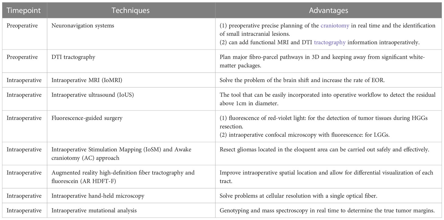
Jiang et al. reviewed four main intraoperative systems for delineating malignant tumors: intraoperative MRI (iMRI), fluorescence, Raman histology, and mass spectrometry. iMRI overcomes brain drift defects and has been shown to increase the complete resection rate (29,30). Besides, functional MRI (fMRI), diffusion tensor imaging (DTI), and MR spectroscopy (MRS) can be applied with patients asleep for the whole surgical procedure, giving a panoramic view of the functional brain cortex, fiber tracts, and metabolic change levels. To solve the problem that the DTI reconstruction process relies on the experience of operators and is time-consuming, Yuan et al. developed an inhouse software, "DiffusionGo." It can reconstruct DTI automatically and quickly. The researchers demonstrated its efficacy in language function preservation for three patients who underwent surgical resection of gliomas. DiffusionGo is especially suitable for intraoperative use, and its adaptation to iMRI may be a future research direction. Although clinical implementation should be further established with a larger sample size, this software has presented promising value.Yang et al. also mentioned in their review of surgical treatment options in gliomas, the augmented reality (AR) neuronavigation system, which integrates MR/CT images into the surgical field and presents three-dimensional virtual tissues to guide resection (31)(32)(33).Cui et al. did bibliometric research on artificial intelligence developments in nervous system diseases. They found "glioma" to be the leading research hotspot, and "machine learning," "brain metastasis," and "gene mutations" were at the research frontier. This result indicated a promising prospect for radiomics biomarkers and multi-omics studies. RS is a label-free imaging method that uses intrinsic biochemical markers to identify tumors (34,35) and has the advantages of being non-destructive, rapid and accurate. Presently, the above two techniques for histological tests are used for resected human tissue. Both methods can be equipped with miniaturized hand-held probes, so they can potentially test in vivo tumor tissue, offering guidance on the range of resection. However, their respective safety and accuracy should be further evaluated.

In summary, the 11 studies included in this Research Topic included advances in surgical approaches from various aspects, and future progress in this realm can be anticipated.

2,131 views

Minimally invasive treatment for glioblastoma through endoscopic surgery including tumor embolization when necessary: a technical note
Background: Although there have been some reports on endoscopic glioblastoma surgery, the indication has been limited to deep-seated lesions, and the difficulty of hemostasis has been a concern. In that light, we attempted to establish an endoscopic procedure for excision of glioblastoma which could be applied even to hypervascular or superficial lesions, in combination with pre-operative endovascular tumor embolization. Methods: Medical records of six consecutive glioblastoma patients who received exclusive endoscopic removal between September and November 2020 were analyzed. Preoperative tumor embolization was performed in cases with marked tumor stain and proper feeder arteries having an abnormal shape, for instance, tortuous or dilated, without passing through branches to the normal brain. Endoscopic tumor removal through a key-hole craniotomy was performed by using an inside-out excision for a deep-seated lesion, with the addition of an outside-in extirpation for a shallow portion when needed. Results: Endoscopic removal was successfully performed in all six cases. Before resection, endovascular tumor embolization was performed in four cases with no resulting complications, including ischemia or brain swelling. Gross total resection was achieved in three cases, and near total resection in the other three cases. Intraoperative blood loss exceeded 1000 ml in only one case, whose tumor showed a prominent tumor stain but no proper feeder artery for embolization. In all patients, a smooth transition to adjuvant therapy was possible with no surgical site infection. Conclusions: Endoscopic removal for glioblastoma was considered to be a promising procedure with minimal invasiveness and a favorable impact on prognosis.

4,597 views

12 citations

A review on surgical treatment options in gliomas
REVIEW
16 March 2023
A review on surgical treatment options in gliomas
Zhongxi Yang, 4 more and Xuan Chen

7,975 views

16 citations

ORIGINAL RESEARCH
24 March 2023
Automatic bundle-specific white matter fiber tracking tool using diffusion tensor imaging data: A pilot trial in the application of language-related glioma resection

3,708 views

1 citations

The clinical and neurocognitive functional changes with awake brain mapping for gliomas invading eloquent areas: Institutional experience and the utility of The Montreal Cognitive Assessment
ORIGINAL RESEARCH
22 February 2023
The clinical and neurocognitive functional changes with awake brain mapping for gliomas invading eloquent areas: Institutional experience and the utility of The Montreal Cognitive Assessment
Objective: Awake craniotomy with intraoperative brain functional mapping effectively reduces the potential risk of neurological deficits in patients with glioma invading the eloquent areas. However, glioma patients frequently present with impaired neurocognitive function. The present study aimed to investigate the neurocognitive and functional outcomes of glioma patients after awake brain mapping and assess the experience of a tertiary neurosurgical center in China over eight years. Methods: This retrospective study included 80 patients who underwent awake brain mapping for gliomas invading the eloquent cortex between January 2013 and December 2021. Clinical and surgical factors, such as the extent of resection (EOR), perioperative Karnofsky Performance Score (KPS), progression-free survival (PFS), and overall survival (OS), were evaluated. We also used the Montreal Cognitive Assessment (MoCA) to assess the neurocognitive status changes. Results: The most frequently observed location of glioma was the frontal lobe (33/80, 41.25%), whereas the tumor primarily invaded the language-related cortex (36/80, 45%). Most patients had supratotal resection (11/80, 13.75%) and total resection (45/80, 56.25%). The median PFS was 43.2 months, and the median OS was 48.9 months in our cohort. The transient (less than seven days) neurological deficit rate was 17.5%, whereas the rate of persistent deficit (lasting for three months) was 15%. At three months of follow-up, most patients (72/80, 90%) had KPS scores > 80. Meanwhile, compared to the preoperative baseline tests, the changes in MoCA scores presented significant improvements at discharge and three months follow-up tests. Conclusion: Awake brain mapping is a feasible and safe method for treating glioma invading the eloquent cortex, with the benefit of minimizing neurological deficits, increasing EOR, and extending survival time. The results of MoCA test indicated that brain mapping plays a critical role in preserving neurocognitive function during tumor resection.

4,180 views

9 citations

REVIEW
26 January 2023
Advances in the intraoperative delineation of malignant glioma margin
Shan Jiang, 1 more and Qisheng Tang
Advances in the intraoperative delineation of malignant glioma margin
Surgery plays a critical role in the treatment of malignant glioma. However, due to the infiltrative growth and brain shift, it is difficult for neurosurgeons to distinguish malignant glioma margins with the naked eye and the preoperative examinations. Therefore, several technologies were developed to portray precise tumor margins intraoperatively. Here we introduced four intraoperative technologies to delineate malignant glioma margin, including magnetic resonance imaging, fluorescence-guided surgery, Raman histology, and mass spectrometry. By tracing their detecting principles and developments, we reviewed their advantages and disadvantages respectively, and imagined future trends.

3,867 views

16 citations

ORIGINAL RESEARCH
3 February 2023
Preoperative individual-target transcranial magnetic stimulation demonstrates an effect comparable to intraoperative direct electrical stimulation in language-eloquent glioma mapping and improves postsurgical outcome: A retrospective fiber-tracking and electromagnetic simulation study
Sanzhong Li, 9 more and Zhou Fei

3,670 views

9 citations

Clinical feasibility of miniaturized Lissajous scanning confocal laser endomicroscopy for indocyanine green-enhanced brain tumor diagnosis
ORIGINAL RESEARCH
13 January 2023
Clinical feasibility of miniaturized Lissajous scanning confocal laser endomicroscopy for indocyanine green-enhanced brain tumor diagnosis
Duk Hyun Hong, 7 more and Shin-Hyuk Kang
Intraoperative real-time confocal laser endomicroscopy (CLE) can be an alternative modality for frozen tissue histology that enables visualizing the cytoarchitecture of living tissues with spatial resolution at the cellular level. We developed a new CLE with “Lissajous scanning pattern” and conducted a study to identify its feasibility for fluorescence-guided brain tumor diagnosis. Conventional hematoxylin and eosin (H&E) histological images were compared with indocyanine green (ICG)-enhanced CLE images in two settings: (1) in vitro and ex vivo glial tumors of mice and (2) surgically resected human brain tumors. The cultured U87 and GL261 tumor cells were implanted into the mouse brain, and the H&E stains were compared with CLE images at normal and tumor tissue ex vivo. To determine the invasion of the normal brain, two patient-derived glioma cells were used for orthotopic tumor formation and compared using two methods (CLE vs H&E staining). In human brain tumors, specimens from 69 patients were prospectively obtained after elective surgical resection and were also compared. The comparison was performed by an experienced neuropathologist. In the incubation of ICG in vitro, U87 and GL261 cell morphologies were well defined in the CLE images and depended on dimethyl sulfoxide (DMSO). In the ex vivo examination of xenograft model, dense and heterogeneous glioma cell cores and peri-lesional necrosis was well defined in both methods. In patient-derived glioma xenograft models, invasive tumor cell clusters were well identified in CLE images, which corresponded to H&E staining. In human brain tumors, CLE images well visualize the cytoarchitecture of the brain and tumor tissues and the pathognomonic microstructures according to the tumor subtypes were also clearly observed. Interestingly, in glioma, the cellularity of the tumor and the density of streak-like patterns were significantly associated with tumor grade in the CLE images. In addition, panoramic view reconstruction was successfully conducted for visualizing a gross tissue morphology. In conclusion, newly developed CLE with Lissajous laser scanning can be a helpful intraoperative device for real-time diagnosis, detection of tumor-free margins, and maximal safe resection of brain tumors.

5,311 views

12 citations

ORIGINAL RESEARCH
19 January 2023
Predictive model of language deficit after removing glioma involving language areas under general anesthesia
Meng Cui, 7 more and Xiaodong Ma

2,338 views

2 citations

REVIEW
5 January 2023
Raman spectroscopy: A prospective intraoperative visualization technique for gliomas
Yi Zhang, 7 more and Xuan Chen
The infiltrative growth and malignant biological behavior of glioma make it one of the most challenging malignant tumors in the brain, and how to maximize the extent of resection (EOR) while minimizing the impact on normal brain tissue is the pursuit of neurosurgeons. The current intraoperative visualization assistance techniques applied in clinical practice suffer from low specificity, slow detection speed and low accuracy, while Raman spectroscopy (RS) is a novel spectroscopy technique gradually developed and applied to clinical practice in recent years, which has the advantages of being non-destructive, rapid and accurate at the same time, allowing excellent intraoperative identification of gliomas. In the present work, the latest research on Raman spectroscopy in glioma is summarized to explore the prospect of Raman spectroscopy in glioma surgery.
Raman spectroscopy: A prospective intraoperative visualization technique for gliomas

5,292 views

28 citations