The theme of the research included in this special issue, which is driven by our experience within the EU MSCA European Training Network, BioTrib, is how best to facilitate improvements in pre-clinical in silico, in vitro and in vivo testing within an orthopaedic context to allow a reduction in the adverse events that occur in implants once marketed. The studies cover techniques such as artificial intelligence, image processing, laboratory and computer simulations, to achieve this end.Several of the studies are focused on addressing the limitations of using a "human in the loop" for e.g. diagnosis or implant selection. Cu et al. used a deep learning approach for localizing cruciate ligament rupture on knee MRI images. The over all aim of such work is to identify the location and tissue quality of injured ACLs to determine if the ACL repair surgery can be performed. The goal of the work of Burge et al. was to develop a computational tool for automatic selection of total knee replacement implant size using X-ray images. Both studies support the use of algorithms to achieve repeatable outcomes to reduce operator dependency. Some of the studies are comparing the biomechanical efficacy of two or more surgical procedures or devices, with the overall aim of ranking the procedures or improving device design. Here the benefit of preclinical in silico testing can be utilized to avoid subjecting patients to clinical trials of devices or procedures that may involve elevated risk. Fung et al., e.g., studied the efficacy of prophylactic ceramic-based cement augmentation of the proximal femur to prevent hip fracture. The authors provide insight into the potential efficacy of such a procedure, but prophylaxis for fracture prevention is a controversial topic among clinicians. Using an advanced experimental model, Techens et al. studied the biomechanical consequences of cement discoplasty. This surgical procedure is relatively novel, and experimental models that can provide insight into e.g. the changes in spine kinematics following the treatment, are needed.Another important aspect of pre-clinical testing of orthopaedic devices is addressed in the study by Kohli et al. Here, a bioreactor analyses of tissue ingrowth, ongrowth and remodelling around implants is used to reduce the need for animal testing. Some of the studies in this special issue, fall under the category of novel measurement techniques and protocols. In the study of Kandel et al., an automated system is introduced for polymer wear debris analysis in total disc arthroplasty using convolution neural network. Using dual-plane fluoroscopy to observe treadmill walking, Zhou et al. investigate whether multi-planar instability, laxity and reduced knee flexion during the support phase of walking are determinants of return to sports. On the other hand, D'Isidoro et al. use dual plane moving fluoroscopy-based analysis, to study total hip arthroplasty kinematics during unrestricted activities of daily living. These advanced measurement techniques are providing novel insights into aspects of total joint replacements that are difficult to achieve with other, more conventional methods.Overall, the papers on this research topic cover some recent advances in preclinical testing of musculoskeletal devices. The editors hope that this special issue will contribute to advancing the field of orthopaedics, by inspiring researchers, clinicians and industry alike, towards reduction in the need for animal testing, improving patient safety and lowering health care costs.
Editorial21 July 2023
Editorial: Advanced pre-clinical and pre-surgical assessment of musculo-skeletal medical devicesORIGINAL RESEARCH
7 March 2023
Yuzhao Lu, 5 more and Tianhao Xie
7 Authors
- YL
Yuzhao Lu
The First Affiliated Hospital of Nanchang University, China
- GH
Gai Hang
School of Medicine, China
- YF
Yu Feng
School of Medicine, China
- BC
Bo Chen
Ruijin Hospital, China
- SM
Shenghui Ma
School of Medicine, China
- YW
Yang Wang
The First Affiliated Hospital of Nanchang University, China
- TX
Tianhao Xie
General Hospital of Central Theater Command, China
2,498 views
4 citations

Purpose: Proximal tibial fractures are common, but the current available internal fixation strategies remain debatable, especially for comminuted fractures. This study aimed to compare the biomechanical stability of three internal fixation strategies for extra-articular comminuted proximal tibial fractures.
Methods: A total of 90 synthetic tibiae models of simulated proximal tibial fractures with segmental bone defects were randomly divided into three groups: single lateral plating (LP), double plating (DP) and intramedullary nailing (IN). Based on the different number of fixed screws, the above three groups were further divided into nine subgroups and subjected to axial compression, cyclic loading and static torsional testing.
Results: The subgroup of intramedullary nailing with five proximal interlocking screws showed the highest axial stiffness of 384.36 ± 35.00 N/mm. The LP group obtained the lowest axial stiffness performance with a value of 96.59 ± 16.14 N/mm. As expected, the DP group offered significantly greater biomechanical stability than the LP group, with mean static axial stiffness and mean torque increasing by approximately 200% and 50%, respectively. According to static torsional experiments, the maximum torque of the DP subgroup was 3308.32 ± 286.21 N mm, which outperformed all other groups in terms of torsional characteristics.
Conclusion: Utilizing more than four distal screws did not provide improved biomechanical stability in the LP or DP groups, while a substantial increase in the biomechanical stability of DP was obtained when an additional medial plate was used. For the intramedullary nailing group, increasing the number of proximal interlocking screws could significantly improve biomechanical stability, and the intramedullary nailing with three proximal interlocking screws had similar static and cyclic stiffness as the DP group. The intramedullary nailing with five proximal screws had better axial stability, whereas DP had better torsional stability.
ORIGINAL RESEARCH
20 February 2023
Nupur Kohli, 4 more and Richard J. van Arkel
6 Authors
- NK
Nupur Kohli
Biomechanics Group, United Kingdom
- KT
Konstantinos Theodoridis
Biomechanics Group, United Kingdom
- TA
Thomas A. G. Hall
Biomechanics Group, United Kingdom
- IS
Inigo Sanz-Pena
Biomechanics Group, United Kingdom
- DC
David C. A. Gaboriau
FILM, United Kingdom
- RJ
Richard J. van Arkel
Biomechanics Group, United Kingdom
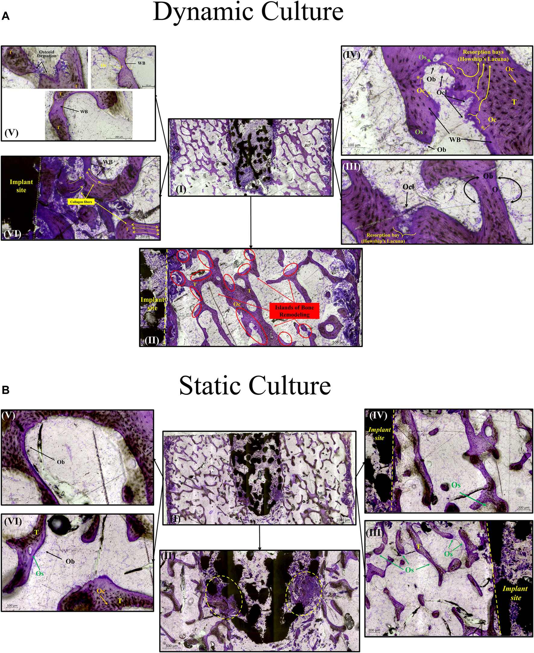
Preclinical assessment of bone remodelling onto, into or around novel implant technologies is underpinned by a large live animal testing burden. The aim of this study was to explore whether a lab-based bioreactor model could provide similar insight. Twelve ex vivo trabecular bone cylinders were extracted from porcine femora and were implanted with additively manufactured stochastic porous titanium implants. Half were cultured dynamically, in a bioreactor with continuous fluid flow and daily cyclic loading, and half in static well plates. Tissue ongrowth, ingrowth and remodelling around the implants were evaluated with imaging and mechanical testing. For both culture conditions, scanning electron microscopy (SEM) revealed bone ongrowth; widefield, backscatter SEM, micro computed tomography scanning, and histology revealed mineralisation inside the implant pores; and histology revealed woven bone formation and bone resorption around the implant. The imaging evidence of this tissue ongrowth, ingrowth and remodelling around the implant was greater for the dynamically cultured samples, and the mechanical testing revealed that the dynamically cultured samples had approximately three times greater interfacial shear strength (p<0.05). Ex vivo bone models enable the analysis of tissue remodelling onto, into and around porous implants in the lab. While static culture conditions exhibited some characteristics of bony adaptation to implantation, simulating physiological conditions with a bioreactor led to an accelerated response.

ORIGINAL RESEARCH
10 November 2022
Kateryna Kardash, 1 more and Miguel A. Otaduy
This work describes a computational methodology for the design of braces for adolescent idiopathic scoliosis. The proposed methodology relies on a personalized simulation model of the patient’s trunk, and automatically searches for the brace geometry that optimizes the trade-off between clinical improvement and patient comfort. To do this, we introduce a formulation of differentiable biomechanics of the patient’s trunk, the brace, and their interaction. We design a simulation model that is differentiable with respect to both the deformation state and the brace design parameters, and we show how this differentiable model is used for the efficient update of brace design parameters within a numerical optimization algorithm. To evaluate the proposed methodology, we have obtained trunk models with personalized geometry for five patients of adolescent idiopathic scoliosis, and we have designed Boston-type braces. In a simulation setting, the designed braces improve clinical metrics by 45% on average, under acceptable comfort conditions. In the future, the methodology can be extended beyond synthetic validation, and tested with physical braces on the actual patients.