Molecular Diagnostics of Pediatric Cancer

138kviews
Editorial11 October 2021
Editorial: Molecular Diagnostics of Pediatric Cancer

Pediatric tumors are defined as tumors arising from the complex physiological growth process of embryonic stem cells (1). They differ from malignant adult tumors in cellular origin, epidemiology, genetic complexity, driver mutations, and potential mutational processes, and they are generally considered to be rare events (2). This research topic collects research related to molecular markers, signaling pathways, drug development and treatment, and emerging molecular technologies of pediatric tumors.

Chen et al. reviewed the progress of molecular epidemiology of hepatoblastoma (HB), focusing on the studies of single nucleotide polymorphisms (SNPs) related to the risk of HB. As treatment regimens for medulloblastoma (MB) are becoming subgroup-specific, methods are needed to discriminate its subgroups. Gershanov et al. used the SARC algorithm that reduces the set of 22 genes to only 6 genes, which could distinguish four MB subgroups reliably. The gene set identified is small enough to allow clinicians to easily obtain the qPCR-based classification of MB subtypes to better determine treatment options. Wang et al. found that the sensitivity of the NB5 method to detect neuroblastoma (NB) with micrometastases in bone marrow (BM) and peripheral blood (PB) was significantly higher than that of bone marrow biopsy (BMB). Liver and bone metastases are factors that affect the sensitivity of NB5 detection in the bone marrow and peripheral blood. Zhanghuang et al. illustrated that targeting the PI3K-AKT signaling pathway and microRNA-related proteins had high potential values for treating malignant rhabdoid tumors of the kidney (MRTK). Poot et al. described recent advances in the therapeutical development of pediatric cancer and illustrates how this methodology affects diagnosis and provides additional treatment options for these patients. These studies contribute to a better understanding, diagnosis, and treatment of pediatric cancer.

Pediatric cancers are characterized by high molecular heterogeneity. For instance, CTNNB1, NFE2L2, AXIN1, APC, MYCN1, and IGF2 may be potential biomarkers for the diagnosis of HB. Hu et al. demonstrated that pediatric HB patients with causal genetic alterations had significantly lower complete remission (CR) rates than patients with wide-type gene counterparts (P<0.05). Moreover, regarding acute lymphoblastic leukemia (ALL), Liu et al. found that METTL3 gene polymorphism was associated with an increased risk of ALL in children and suggested that METTL3 gene polymorphism may be a potential biomarker for the selection of chemotherapy agents for pediatric ALL. Cai et al. proved that Prp19 regulates the expression of YAP through YAP pre-mRNA splice, thus affecting the invasion, migration, and EMT of NB cells. It was the first report to demonstrate that Prp19 is a potential therapeutic target and prognostic biomarker in patients with NB. Shi et al. showed that the high expression of CDC20 was involved in the tumorigenesis of Wilms tumor (WT), and inhibition of CDC20 could suppress the proliferation and migration of WT cells and arrest the cell cycle in the G2/M phase, suggesting that CDC20 could be a potential biomarker of WT. Liu et al. established a multinomial predictive survival model and a survival-associated ceRNA network, which provides a new potential biomarker for improving prognosis and treatment of WT patients. Taken together, these biomarkers may be able to predict clinical outcomes and hold great promise in clinical application of pediatric cancer.

The immune system is closely related to the occurrence and development of pediatric cancer, and understanding the immune microenvironment is helpful to the treatment of pediatric cancer (3). Li et al. used single-cell RNA sequences to reveal the characteristics of malignant cells and the immune microenvironment in subcutaneous panniculitis-like T-cell lymphoma (SPTCL), providing a better understanding of the transcriptional characteristics and immune microenvironment of this rare tumor. Feng et al. explored the immune microenvironment of langerhans cell histiocytosis (LCH). They found that serum levels of immune indicators are somewhat representative of disease severity, and associated laboratory tests can be used to improve risk stratification and guide immunotherapy.

The rapid rise of gene sequencing and bioinformatics and the opening of relevant tumor databases provide opportunities to elucidate the molecular mechanisms of pediatric cancer and precise drug target therapy of pediatric cancer. Feng et al. applied artificial intelligence methods to improve the accuracy of gene express-based survival prediction for neuroblastoma. Ruan et al. showed that monitoring circulating tumor DNA (ctDNA) with next-generation sequencing-based analysis could provide more information about genetic mutations to guide the precise treatment of acute myeloid leukemia (AML) in children. Sun et al. established a random forest classifier and identified 10 HB core genes. These findings may help in the diagnosis, prediction, and targeted treatment of HB. Li et al. provided an overview of the techniques currently available in vitro and in vivo models of pediatric brain tumors and discussed the opportunities presented by new techniques such as 3D culture and organic-like compounds that can overcome the limitations of the simplicity of single-layer culture and the complexity of living models to accommodate greater precision in drug development for pediatric brain tumors. Wang et al. reported the first case of acute promyelocytic with FIP1L1/RARA identified by next-generation sequencing (NGS). NGS analysis is recommended as a routine test for patients with variant acute promyelocytic leukemia (APL). Cimmino et al. found that 9 out of 11 patients carried at least one pathogenic variant and developed a targeted NGS approach to identify tumor-specific alterations in ctDNA in NB patients. This information can be combined with clinical and pathological data at NB diagnosis. The goal of these molecular diagnostic studies for pediatric cancer is to translate them into the clinic to achieve more accurate diagnosis, more accurate risk stratification, and more effective and less toxic treatments.

In conclusion, the “Molecular Diagnostics of Pediatric Cancer” research topic highlights the most recent advance of diagnostic molecular biomarkers and novel therapeutic targets for pediatric cancer.

2,821 views

3 citations

ORIGINAL RESEARCH
9 September 2021
Novel Associations Between METTL3 Gene Polymorphisms and Pediatric Acute Lymphoblastic Leukemia: A Five-Center Case-Control Study
Xiaoping Liu, 12 more and Hua Jiang

3,289 views

16 citations

ORIGINAL RESEARCH
26 August 2021
Identification of CDC20 as a Novel Biomarker in Diagnosis and Treatment of Wilms Tumor
Qinlin Shi, 5 more and Guanghui Wei

5,718 views

6 citations

ORIGINAL RESEARCH
6 August 2021
Genotypic Characteristics of Hepatoblastoma as Detected by Next Generation Sequencing and Their Correlation With Clinical Efficacy
Huimin Hu, 6 more and Dongsheng Huang

4,332 views

7 citations

ORIGINAL RESEARCH
29 July 2021
Targeted Next-Generation Sequencing of Circulating Tumor DNA, Bone Marrow, and Peripheral Blood Mononuclear Cells in Pediatric AML
Min Ruan, 18 more and Xiaofan Zhu

6,133 views

15 citations

ORIGINAL RESEARCH
24 June 2021
Myeloid Sarcoma Type of Acute Promyelocytic Leukemia With a Cryptic Insertion of RARA Into FIP1L1: The Clinical Utility of NGS and Bioinformatic Analyses
Yongren Wang, 5 more and Yongjun Fang

3,254 views

9 citations

Classifying Medulloblastoma Subgroups Based on Small, Clinically Achievable Gene Sets
ORIGINAL RESEARCH
10 June 2021
Classifying Medulloblastoma Subgroups Based on Small, Clinically Achievable Gene Sets
Sivan Gershanov, 8 more and Mali Salmon-Divon

6,805 views

13 citations

ORIGINAL RESEARCH
14 May 2021
The Application of and Factors Influencing, the NB5 Assay in Neuroblastomas
Zuopeng Wang, 6 more and Kai Li

3,161 views

2 citations

Identification of Ten Core Hub Genes as Potential Biomarkers and Treatment Target for Hepatoblastoma
ORIGINAL RESEARCH
1 April 2021
Identification of Ten Core Hub Genes as Potential Biomarkers and Treatment Target for Hepatoblastoma
Rui Sun, 3 more and Long Li
Background: This study aimed to systematically investigate gene signatures for hepatoblastoma (HB) and identify potential biomarkers for its diagnosis and treatment. Materials and Methods: GSE131329 and GSE81928 were obtained from the Gene Expression Omnibus (GEO) database. Differentially expressed genes (DEGs) between hepatoblastoma and normal samples were identified using the Limma package in R. Then, the similarity of network traits between two sets of genes was analyzed by weighted gene correlation network analysis (WGCNA). Cytoscape was used to visualize and select hub genes. GO enrichment and KEGG pathway analyses of hub genes were carried out using ClueGO. The random forest classifier was constructed based on the hub genes using the GSE131329 dataset as the training set, and its reliability was validated using the GSE81928 dataset. The resulting core hub genes were combined with the InnateDB database to identify the innate core genes. Results: A total of 4244 DEGs in HB were identified. WGCNA identified four modules that were significantly correlated with the disease status. A total of 114 hub genes were obtained within the top 20 genes of each node rank. GO enrichment and KEGG pathway analyses of hub genes were focused on MAPK, cell cycle, p53, and other crucial pathways involved in HB. A random forest classifier was constructed using the 114 hub genes as feature genes, resulting in a 95.5% true positive rate when classifying HB and normal samples. A total of 35 core hub genes were obtained through the mean decrease in accuracy and mean decrease Gini of the random forest model. The classification efficiency of the random forest model was 81.4%. Finally, CDK1, TOP2A, ADRA1A, FANCI, XRCC1, TPX2, CCNB2, CDK4, GLYATL1, and CFHR3 were identified by cross-comparison with the InnateDB database. Conclusion: Our study established a random forest classifier that identified 10 core genes in HB. These findings may be beneficial for the diagnosis, prediction, and targeted therapy of HB.

7,004 views

18 citations

ORIGINAL RESEARCH
7 May 2021
Immune Microenvironment in Langerhans Cell Histiocytosis: Potential Prognostic Indicators
Chuchu Feng, 8 more and Jianpei Fang

3,194 views

13 citations

REVIEW
1 April 2021
In Vivo and Ex Vivo Pediatric Brain Tumor Models: An Overview
Zhiqin Li and Sigrid A. Langhans
In Vivo and Ex Vivo Pediatric Brain Tumor Models: An Overview
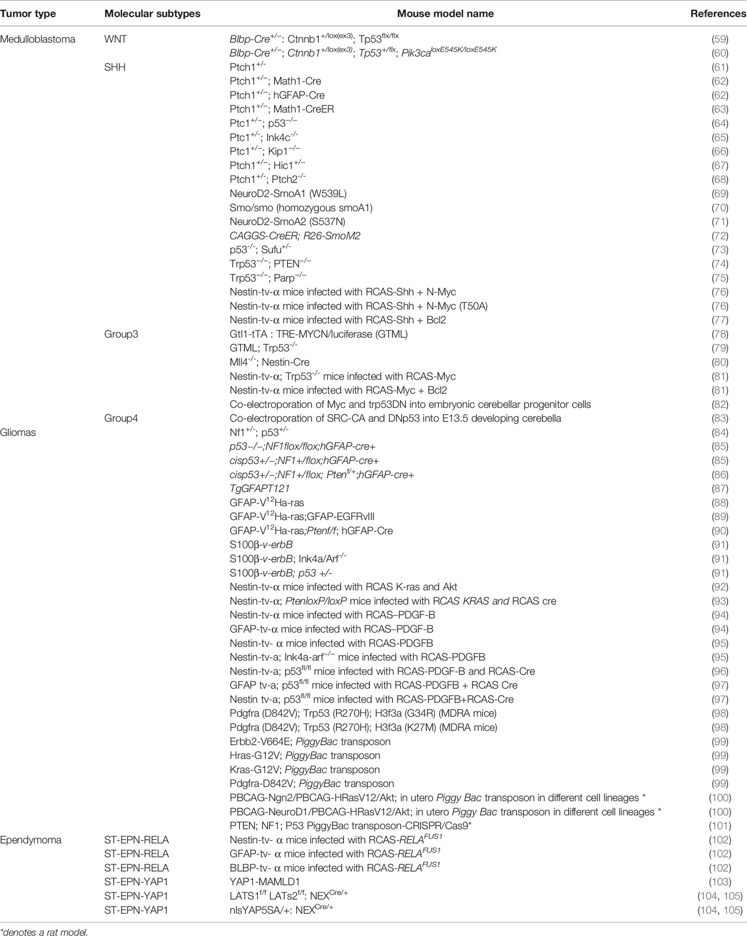
After leukemia, tumors of the brain and spine are the second most common form of cancer in children. Despite advances in treatment, brain tumors remain a leading cause of death in pediatric cancer patients and survivors often suffer from life-long consequences of side effects of therapy. The 5-year survival rates, however, vary widely by tumor type, ranging from over 90% in more benign tumors to as low as 20% in the most aggressive forms such as glioblastoma. Even within historically defined tumor types such as medulloblastoma, molecular analysis identified biologically heterogeneous subgroups each with different genetic alterations, age of onset and prognosis. Besides molecularly driven patient stratification to tailor disease risk to therapy intensity, such a diversity demonstrates the need for more precise and disease-relevant pediatric brain cancer models for research and drug development. Here we give an overview of currently available in vitro and in vivo pediatric brain tumor models and discuss the opportunities that new technologies such as 3D cultures and organoids that can bridge limitations posed by the simplicity of monolayer cultures and the complexity of in vivo models, bring to accommodate better precision in drug development for pediatric brain tumors.

11,475 views

38 citations

Single-Cell RNA-seq Reveals Characteristics of Malignant Cells and Immune Microenvironment in Subcutaneous Panniculitis-Like T-Cell Lymphoma
ORIGINAL RESEARCH
18 March 2021
Single-Cell RNA-seq Reveals Characteristics of Malignant Cells and Immune Microenvironment in Subcutaneous Panniculitis-Like T-Cell Lymphoma
Zifeng Li, 19 more and Xiaowen Zhai
Background: Subcutaneous panniculitis-like T-cell lymphoma (SPTCL) is a malignant primary T-cell lymphoma that is challenging to distinguish from autoimmune disorders and reactive panniculitides. Delay in diagnosis and high misdiagnosis rate affect the prognosis and survival of patients. The difficulty of diagnosis is mainly due to an incomplete understanding of disease pathogenesis. Methods: We performed single-cell RNA sequencing of matched subcutaneous lesion tissue, peripheral blood, and bone marrow from a patient with SPTCL, as well as peripheral blood, bone marrow, lymph node, and lung tissue samples from healthy donors as normal controls. We conducted cell clustering, gene expression program identification, gene differential expression analysis, and cell-cell interaction analysis to investigate the ecosystem of SPTCL. Results: Based on gene expression profiles in a single-cell resolution, we identified and characterized the malignant cells and immune subsets from a patient with SPTCL. Our analysis showed that SPTCL malignant cells expressed a distinct gene signature, including chemokines families, cytotoxic proteins, T cells immune checkpoint molecules, and the immunoglobulin family. By comparing with normal T cells, we identified potential novel markers for SPTCL (e.g., CYTOR, CXCL13, VCAM1, and TIMD4) specifically differentially expressed in the malignant cells. We also found that macrophages and fibroblasts played the primary role in tumor-immune interactions of SPTCL. Conclusions: This work offers insight into the heterogeneity of subcutaneous panniculitis-like T-cell lymphoma, providing a better understanding of the transcription characteristics and immune microenvironment of this rare tumor.

7,237 views

12 citations

ORIGINAL RESEARCH
30 March 2021
Clinical and Molecular Differentiation Between Malignant Rhabdoid Tumor of the Kidney and Normal Tissue: A Two-Case Report
Chenghao Zhanghuang, 8 more and Bing Yan

3,155 views

12 citations

ORIGINAL RESEARCH
26 February 2021
A Survival-Related Competitive Endogenous RNA Network of Prognostic lncRNAs, miRNAs, and mRNAs in Wilms Tumor
HengChen Liu, 6 more and ZhaoZhu Li

3,302 views

12 citations

The Current Status and Future Potential of Theranostics to Diagnose and Treat Childhood Cancer
MINI REVIEW
19 November 2020
The Current Status and Future Potential of Theranostics to Diagnose and Treat Childhood Cancer
Alex J. Poot, 1 more and Max M. van Noesel

12,037 views

24 citations

ORIGINAL RESEARCH
2 November 2020
Prp19 Is an Independent Prognostic Marker and Promotes Neuroblastoma Metastasis by Regulating the Hippo-YAP Signaling Pathway
Yuanxia Cai, 6 more and Zhixiang Wu
Prp19 Is an Independent Prognostic Marker and Promotes Neuroblastoma Metastasis by Regulating the Hippo-YAP Signaling Pathway
Pre-mRNA processing factor 19(Prp19) was previously reported to involve in tumor progression. However, the Prp19 expression and functions remain elusive in neuroblastoma. Here we aim to identify the functions and mechanisms of Prp19 in neuroblastoma. Neuroblastic tumor tissue microarrays and two independent validation datasets indicated that Prp19 was associated with high-risk markers, bone marrow metastasis and served as a prognostic marker for worse clinical outcome in neuroblastoma. Gain- and loss-of-expression assays revealed that Prp19 promoted invasion, migration and epithelial–mesenchymal transition (EMT) of neuroblastoma cells in vitro. Bioinformatics analysis of RNA-seq data showed that the expressions of YAP and its downstream genes were significantly inhibited after downregulation of Prp19. Prp19 and YAP expression in metastatic lymph nodes was higher than in situ neuroblastoma tissue. Further experiments showed that Prp19 regulated YAP expression and consequently infect cell invasion, migration and EMT in neuroblastoma by pre-mRNA splicing of YAP. In conclusion, our findings provide the first evidence that Prp19 is a potential therapeutic target and prognostic biomarker for patients with neuroblastoma.

4,566 views

19 citations