 José Miguel Palacios-Jaraquemada1
 Álbaro Jose Nieto-Calvache2,3*Rozy Aditya Aryananda4Nicolás Basanta5
- 1Department of Gynaecology, Otamendi Hospital, Buenos Aires, Argentina
- 2Clínica de Espectro de Acretismo Placentario, Fundación Valle del Lili, Cali, Colombia
- 3Latin American Group for the Study of Placenta Accreta Spectrum, Cali, Columbia
- 4Dr. Soetomo Academic General Hospital, Universitas Airlangga, Surabaya, Indonesia
- 5Department of Obstetrics an Gynaecology, Hospital General de Agudos Juan A Fernández, Buenos Aires, Argentina
Placenta accreta spectrum (PAS) is an entity with a wide range of clinical presentations. From cases with “mild” lesions requiring moderate surgical effort and are associated with few complications (1), to severe ones with a challenging management and life-threatening risk (2).
Individualizing the management of PAS patients is essential, especially when a significant percentage of women undergoing PAS surgery ultimately do not have this diagnosis or present superficial or focal lesions (3). In addition, let's not forget that some women wish to preserve their fertility (4) or that vascular interventions or the hysterectomy itself can cause additional morbidity (5).
However, customizing the management of PAS seems complicated. Choosing the ideal management for each patient is a task for personal and institutional reasons.
Although multiple treatments have been described (6, 7), frequently, each PAS team adopts one strategy for all cases and specializes in its performance. Applying this type of intervention becomes the norm for all patients with PAS presenting at a specific hospital. Additionally, historic international consensus focuses on hysterectomy as the standard treatment, mentioning other management alternatives as secondary options “only” for exceptional cases (6, 8). There is practically no doubt that supervised training modify this previous concept.
On the other hand, the preoperative classification based on the severity of the placenta invasion needs a subsequent histological analysis (9). Therefore, pathologic tissue analysis does not help make decisions on the table, and histological study is subject to multiple biases.
The placenta invasion topography is closely related to surgical complexity and maternal morbidity (10). Placenta invasion below the peritoneal reflection implies more significant risks due to reduced space, extrauterine arterial pedicles’ multiplicity, and requirement for adequate vascular control strategies (11, 12).
It is essential to validate an applicable classification before non-reversible maneuvres (that is, before incising the uterus and causing bleeding) that also suggests a specific type of treatment according to the characteristics of each case. The accurate dissection of coalescence pelvic fascia's spaces allows using avascular spaces with minimal tissue manipulation to have a precise diagnosis and avoid possible complications, this is the principle of intraoperative PAS staging. Additionally, the surgical staging allows getting objective PAS information that could be missing after prenatal ultrasound (13).
Although it is possible to make a diagnostic approach based on the results of prenatal images (ultrasonography and MRI), it is during the laparotomy for the cesarean section when the surgeon can use safe and straightforward dissection techniques to establish the affected area of the uterus, and consequently, the risk of massive bleeding or organ injury.
Topographic classification seeks to define which uterine wall is affected (anterior, lateral, or posterior), the presence of lesions above the level of peritoneal reflection (high lesions) or below that level (low or “subperitoneal” lesions), and the nature of the lesion (characterized by neovascularization or with the presence of fibrosis between the uterus and neighboring organs). The main objective of the intraoperative topographic classification of PAS (14, 15) is to use the most suitable PAS treatment according to objective surgical findings.
Each of the possible affected uterine areas is related to well-defined anatomical arterial pedicles and neighboring structures (urinary and vascular) that determine the surgical difficulty and the recommended dissection and management strategies (16) (Figure 1).
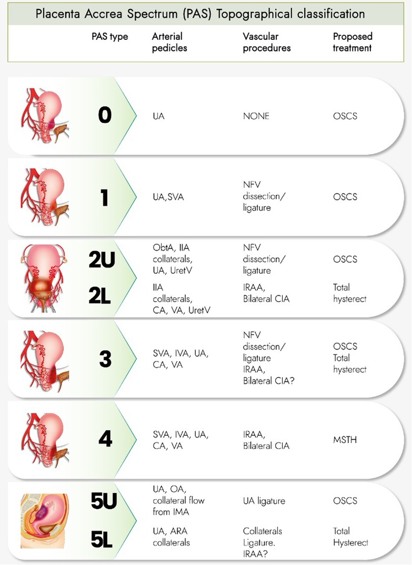
Figure 1. Intraoperative staging makes it possible to determine which uterine wall is affected and the relationship of the lesion to the vesicouterine peritoneal fold (above or below that level), as well as the predominance of neovascularization or the presence of vesicouterine fibrosis.
The topographic classification establishes a “conceptual 3D map” and different management options for each case. For example, what the PAS teams call “A, B, and C plans,” necessary when facing the wide variety of PAS clinical presentations, does not necessarily require the same management route in all cases.
Some publications described the advantages of topographic classification in retrospective studies (14–16). Hopefully, the comparison between individualized arterial pedicles control and the mandatory use of interventional radiology in all the cases could be promissory in large prospective multicentric studies. Likewise, the topographic classification, followed by a protocolized and individualized management, can enhance the postoperative histological analysis.
In each topography, some arterial pedicles are identified that provide most of the irrigation to the PAS area and that determine the recommended vascular procedures and the type of treatment necessary (One Step Conservative Surgery [OSCS], Total hysterectomy or Modified SubTotal Hysterectomy [MSTH]).
Type 0 PAS: uterine “window” or dehiscense. Type 1 PAS: uterine segment upper part involvement. Type 2 PAS: parametrial involvement (2U: upper parametrial involvement, 2 L: lower parametrial involvement). Type 3 PAS: cervix or uterine segment lower part involvement (below the peritoneal reflection). Type 4 PAS: type 3 PAS plus vesicouterine fibrosis. Type 5 PAS: uterine posterior wall involvement (5U: involvement of the upper part of that wall. 5 L: Lesions below the level of the peritoneal reflection).
Author contributions
JP, AN, RA and NB contributed to the design, planning, data analysis and manuscript writing. All authors contributed to the article and approved the submitted version.
Conflict of interest
The authors declare that the research was conducted in the absence of any commercial or financial relationships that could be construed as a potential conflict of interest.
Publisher's note
All claims expressed in this article are solely those of the authors and do not necessarily represent those of their affiliated organizations, or those of the publisher, the editors and the reviewers. Any product that may be evaluated in this article, or claim that may be made by its manufacturer, is not guaranteed or endorsed by the publisher.
Abbreviations
UA, uterine artery; SVA, superior vesical artery; NFV, Newly formed vessels; ObtA, obturator artery; IIA, internal iliac artery; UretV, Ureteral vessels; CA, cervical artery; VA, vaginal arteries; IVA, inferior vesical artery; OA, ovarian artery; IMA, inferior mesenteric artery; ARA, anterior rectal artery; IRAA, infrarenal aortic artery; CIA, common iliac artery.
References
1. Kingdom JC, Hobson SR, Murji A, Allen L, Windrim RC, Lockhart E, et al. Minimizing surgical blood loss at cesarean hysterectomy for placenta previa with evidence of placenta increta or placenta percreta: the state of play in 2020. Am J Obstet Gynecol. (2020) 223(3):322–9. doi: 10.1016/j.ajog.2020.01.044.32007492
2. Nieto-Calvache AJ, Palacios-Jaraquemada JM, Osanan G, Cortes-Charry R, Aryananda RA, Bangal VB, et al. Lack of experience is a main cause of maternal death in placenta accreta spectrum patients. Acta Obstet Gynecol Scand. (2021) 100(8):1445–53. doi: 10.1111/aogs.14163.33896009
3. Hussein AM, Elbarmelgy RA, Elbarmelgy RM, Thabet MM, Jauniaux E. Prospective evaluation of the impact of post-cesarean section uterine scarification in the perinatal diagnosis of placenta accreta spectrum [published online ahead of print, 2021 Jul 5]. Ultrasound Obstet Gynecol. (2021) 59(4):474–82. doi: 10.1002/uog.23732.
4. Einerson BD, Watt MH, Sartori B, Silver R, Rothwell E. Lived experiences of patients with placenta accreta spectrum in Utah: a qualitative study of semi-structured interviews. BMJ Open. (2021) 11(11):e052766. doi: 10.1136/bmjopen-2021-052766
5. Whittington JR, Pagan ME, Nevil BD, Kalkwarf KJ, Sharawi N el, Hughes DS, et al. Risk of vascular complications in prophylactic compared to emergent resuscitative endovascular balloon occlusion of the aorta (REBOA) in the management of placenta accreta spectrum [published online ahead of print, 2020 Aug 11]. J Matern Fetal Neonatal Med. (2020) 35(16):1–4. doi: 10.1080/14767058.2020.1802717.29804488
6. Allen L, Jauniaux E, Hobson S, Papillon-Smith J, Belfort MA. FIGO Placenta Accreta Diagnosis and Management Expert Consensus Panel. FIGO consensus guidelines on placenta accreta spectrum disorders: nonconservative surgical management. Int J Gynaecol Obstet. (2018) 140(3):281–90. doi: 10.1002/ijgo.12409.29405317
7. Sentilhes L, Kayem G, Chandraharan E, Palacios-Jaraquemada J, Jauniaux E. FIGO placenta accreta diagnosis and management expert consensus panel. FIGO consensus guidelines on placenta accreta spectrum disorders: conservative management. Int J Gynaecol Obstet. (2018) 140(3):291–8. doi: 10.1002/ijgo.12410.29405320
8. Collins SL, Alemdar B, van Beekhuizen HJ, Bertholdt C, Braun T, Calda P, et al. Evidence-based guidelines for the management of abnormally invasive placenta: recommendations from the International Society for Abnormally Invasive Placenta. Am J Obstet Gynecol. (2019) 220(6):511–26. doi: 10.1016/j.ajog.2019.02.054.30849356
9. Jauniaux E, Ayres-de-Campos D, Langhoff-Roos J, Fox KA, Collins S. FIGO placenta accreta diagnosis and management expert consensus panel. FIGO classification for the clinical diagnosis of placenta accreta spectrum disorders. Int J Gynaecol Obstet. (2019) 146(1):20–4. doi: 10.1002/ijgo.12761.31173360
10. Cali G, Forlani F, Lees C, Timor-Tritsch I, Palacios-Jaraquemada J, Dall’Asta A, et al. Prenatal ultrasound staging system for placenta accreta spectrum disorders. Ultrasound Obstet Gynecol. (2019) 53(6):752–60. doi: 10.1002/uog.20246.30834661
11. Palacios-Jaraquemada JM, D’Antonio F, Buca D, Fiorillo A, Larraza P. Systematic review on near miss cases of placenta accreta spectrum disorders: correlation with invasion topography, prenatal imaging, and surgical outcome. J Matern Fetal Neonatal Med. (2020) 33(19):3377–84. doi: 10.1080/14767058.2019.1570494.30700221
12. Palacios-Jaraquemada JM, D’Antonio F, Buca D, Fiorillo A, Larraza P, et al. Comparing three-dimensional models of placenta accreta spectrum with surgical findings [published online ahead of print, 2021 May 17]. Int J Gynaecol Obstet. (2021) 33(19):3377–84. doi: 10.1002/ijgo.13743
13. Silveira C, Kirby A, Melov SJ, Nayyar R. Placenta accreta spectrum: we can do better [published online ahead of print, 2022 Jan 4]. Aust N Z J Obstet Gynaecol. (2022) 62(3):376–82. doi: 10.1111/ajo.13471
14. Palacios-Jaraquemada JM, Fiorillo A, Hamer J, Martínez M, Bruno C. Placenta accreta spectrum: a hysterectomy can be prevented in almost 80% of cases using a resective-reconstructive technique. J Matern Fetal Neonatal Med. (2022) 35(2):275–82. doi: 10.1080/14767058.2020.1716715.31984808
15. Nieto-Calvache AJ, Palacios-Jaraquemada JM, Aryananda RA, Rodriguez F, Ordoñez CA, Messa Bryon A, et al. How to identify patients who require aortic vascular control in placenta accreta spectrum disorders? [published online ahead of print, 2021 Oct 2]. Am J Obstet Gynecol MFM. (2021) 4(1):100498. doi: 10.1016/j.ajogmf.2021.100498.34610485
Keywords: placenta accerta, individualize, PAS management, hysterectomie, topographic classification
Citation: Palacios-Jaraquemada JM, Nieto-Calvache ÁJ, Aryananda RA and Basanta N (2023) Advantages of individualizing the placenta accreta spectrum management. Front. Reprod. Health 4:1096175. doi: 10.3389/frph.2022.1096175
Received: 11 November 2022; Accepted: 28 November 2022;
Published: 6 January 2023.
Edited by:
Gabriele Saccone, Federico II University Hospital, ItalyReviewed by:
Michal Silber, University of Pennsylvania, United States© 2023 Palacios-Jaraquemada, Nieto-Calvache, Aryananda and Basanta. This is an open-access article distributed under the terms of the Creative Commons Attribution License (CC BY). The use, distribution or reproduction in other forums is permitted, provided the original author(s) and the copyright owner(s) are credited and that the original publication in this journal is cited, in accordance with accepted academic practice. No use, distribution or reproduction is permitted which does not comply with these terms.
*Correspondence: Álbaro Jose Nieto-Calvache YWxiYXJvLm5pZXRvQGZ2bC5vcmcuY28=
Specialty Section: This article was submitted to Gynecology, a section of the journal Frontiers in Reproductive Health