 Flor Daniela Alday-Montañez1*
 Brandon Daniel Lariz-Nevárez1
 Víctor Josué Carrasco-Urrutia2
 Daniel Dickens-Terrazas3Adali Barragán-Farías2
 Gloria Erika Mejía-Carmona1
 Robert Arthur Kirken4
 Alfonso Enrique Bencomo-Alvarez5
 Naún Lobo-Galo1
 Alejandra Vargas-Caraveo1
 Ángel Gabriel Díaz-Sánchez1
 Elisa Robles-Escajeda4*
 Alejandro Martínez-Martínez1*
- 1Department of Chemical-Biological Sciences, Autonomous University of Ciudad Juarez, Ciudad Juarez, Chihuahua, Mexico
- 2Department of Advanced Gynaecology and Robotic Surgery, Endofem, Ciudad Juarez, Chihuahua, Mexico
- 3Department of Pathology, IMSS, Zone Hospital Number 6, Ciudad Juarez, Chihuahua, Mexico
- 4Border Biomedical Research Center, College of Science, University of Texas at El Paso, El Paso, TX, United States
- 5Department of Host-Microbe Interactions, St. Jude Children’s Research Hospital, Memphis, TN, United States
Background: Endometriosis affects approximately 10% of women of reproductive age; this prevalence may be underestimated, mostly in developing countries, including Mexican and Hispanic populations, due to socioeconomic barriers and limited access to specialized diagnosis. Although laparoscopy remains the gold standard for diagnosis, highlighting the need for non-invasive biomarkers. Haplotype expression of specific miRNAs acts as a circulating signature in both healthy and disease states, including endometriosis. However, their applicability in Hispanic populations has been unexplored.
Method: This study evaluated the discriminatory capacity of a miRNA expression haplotype in the blood plasma of a Hispanic cohort with laparoscopic confirmed diagnosis (15 patients with endometriosis and 7 from a reference group). The expression levels of miR-451a, miR-3613, miR-125b, let-7b, miR-150, and miR-342 were quantified using qRT-PCR, and their diagnostic performance was assessed through individual ROC curves and multivariate classification models: Logistic regression, CRT, and stacking-based ensemble model.
Results: The miRNA expression haplotype demonstrated high diagnostic accuracy with logistic regression (AUC = 0.914), CRT (AUC = 0.990), and an ensemble model using stacking (AUC = 0.990). Individually, miR-451a (AUC = 0.79), miR-3613 (AUC = 0.714), and let-7b (AUC = 0.667) were the most relevant markers and demonstrated more relevance in the expression haplotype.
Conclusion: These findings suggest that a miRNA-based diagnostic panel could provide a highly sensitive and specific alternative for diagnosing endometriosis in Hispanic populations. However, validation in larger cohorts is essential to confirm reproducibility and assess its clinical utility in different healthcare settings.
1 Introduction
Endometriosis is characterized by the ectopic presence of endometrial tissue, associated to chronic inflammation influenced by estrogens and the menstrual cycle (1). It affects 10% of women of reproductive age, accounting for approximately 190 million women worldwide, 30% to 50% of whom experience chronic pelvic pain and infertility (2). Its incidence is underestimated due to the normalization of menstrual pain and the complexity of diagnosis (3). Laparoscopy is both at once: the diagnosis gold standard, as well as treatment, even though it is an invasive technique with multiple operational limitations, including high costs, the need for highly specialized personnel, and its late indication, underscoring the need for a non-invasive, specific, low-cost, and sensitive biomarker (4).
MicroRNAs (miRNAs) have been studied as potential biomarkers for endometriosis, their diagnostic value lies in their stability in systemic circulation and their association with specific pathological metabolic states (5). Unlike other RNAs, miRNAs are protected from degradation through their release in exosomes or their association with proteins such as Argonaute 2 (Ago2), Nucleophosmin 1 (NPM1), and high-density lipoproteins (6). The release of miRNAs into the systemic circulation has been related to pathophysiological processes, such as apoptosis and necrosis in passive secretion, and intercellular communication mediated by exosomes and microvesicles in active secretion (7). The different expression patterns of miRNAs have been associated with various pathologies, including cancers, metabolic disorders, and endometriosis (8).
The diagnostic potential of miRNAs has been found in non-Hispanic populations. A previous study identified a diagnostic signature composed of 109 miRNAs in saliva by next-generation RNA sequencing and a machine learning algorithm, achieving a sensitivity of 96.7% and a specificity of 100% (9). Likewise, a blood-based signature of miRNAs was developed using NGS and artificial intelligence, which showed a sensitivity of 96.8%, a specificity of 100%, and an AUC of 0.984 (10). In addition, a panel of serum miRNAs made it possible to distinguish between different stages of the disease with an AUC curve of 0.940 (8).
Although miRNA expression is intrinsically related to metabolic status, it is a fact that epigenetic factors can cause variations in their expression levels among different ethnic groups, which can affect their performance as “universal” biomarkers and their usefulness in diagnosis in specific groups (11). Therefore, validation of miRNA-specific expression haplotypes for specific populations to diagnose endometriosis is critical (3). Although studies such as those conducted by the National Health and Nutrition Examination Survey (NHANES) report a low prevalence of endometriosis in the Hispanic or Mexican-American population (12, 13) factors such as socioeconomic, cultural, access to health care, and the normalization of menstrual pain, may contribute to an underestimation of its prevalence and incidence in this and other diseases (14, 15). The lack of specific studies on endometriosis in the Hispanic population, with the only relevant work in USA being that of Moustafa et al. (8), highlights the urgent need for specialized research in this population.
The objective of this research was to evaluate a set of six miRNAs in the detection of endometriosis in a Hispanic population in the city of Juarez, México.
2 Materials and methods
This research was approved by the Ethics Committee of the Autonomous University of Ciudad Juarez (permit CEI-2023-1-870) and is part of the project entitled “Molecular aspects associated with the recruitment theory of transformed endometrial stem cells in the development of breast cancer”. Various institutions on the border between Ciudad Juarez, Mexico, and El Paso, USA, collaborated, including Hospital Angeles, Star-Medica Hospital, Specialty Medical Center, Lourdes Surgical Center, Autonomous University of Ciudad Juarez, University of Texas at El Paso, Quiescente Molecular, and Endofem.
The miRNAs included in the expression haplotype associated with endometriosis in Hispanic patients were identified through a systematic review of previous studies on differential expression of miRNAs in this disease, with emphasis on their diagnostic potential. The search was carried out in PubMed, Google Scholar, and Scielo databases, using keywords such as “miRNAs”, “microRNAs”, “endometriosis”, “circulating”, “microRNome”, “detection”, and “diagnosis”.
2.1 Patients and samples
Based on the methodology proposed by Moustafa et al. (8), an analytical cross-sectional prospective study was conducted, using a double-blinded, non-probabilistic convenience sampling focusing specifically on Hispanic women.
All samples were collected between November 2022 and July 2023 from 8 AM to 10 AM in patients that were fasting for at least 8 h. Prior to venipuncture, each patient signed an informed consent and answered a survey of demographics features. Subsequently, 4 ml of blood was extracted by venipuncture, using tubes with EDTA-K2 (Vacutainer®). The inclusion criteria for the participants were Hispanic women with suspected endometriosis or gynecological pathologies related to chronic pelvic pain, who after blood collection would undergo laparoscopy. The exclusion criteria were patients diagnosed with different forms of gynecologic cancer. Blood was immediately centrifuged at 2,500 rpm for 10 min at room temperature, samples with visible hemolysis were discarded. The plasma was aliquoted and stored at −20°C until analysis.
2.2 qRT-PCR
Total RNA, including miRNAs, was extracted from plasma using the miRNeasy Serum/Plasma Advance kit (Cat. No. 217184) following the manufacturer's instructions. The relative quantification of miRNAs was carried out by RT-qPCR using the miRCury LNA RT kit (Cat. No. 339340) for cDNA synthesis. The amplification of the miRNAs was performed with the Open qPCR equipment of CHAI BIO, using the CHAI Green qPCR Master Mix buffer (Cat. No. R02201S). The specific primers for each miRNA were acquired through the GeneGlobeID platform, belonging to the miRCury LNA miRNA PCR Assays kit (Cat. No. 339306, Table 1). The qPCR reactions were prepared according to the proportions described in the manufacturer's protocol. The samples were subjected to a specific thermocycling program for each miRNA, with the small nuclear RNA U6 (YP02119464) used as the reference gene. UniSP6 external quality control (YP00203954) was included in all reactions to ensure the validity of the results. The data obtained were analyzed according to threshold cycles (Ct) and normalized against the reference gene.
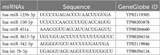
Table 1. Sequences of miRNAs included in this study.
2.3 Data analysis
To evaluate the usefulness of the expression profile of miRNAs in the detection of endometriosis, different statistical analyses were applied, such as ROC curves (Receiver Operating Characteristic), confidence intervals (95% CI) computed via bootstrap resampling (n = 1,000), logistic regression, decision algorithms using the CRT (Classification and Regression Trees) method, and the ensemble model using the Stacking technique using the statistical tools of jamovi (16), IBM SPSS (17), and R Studio (18, 19).
3 Results
Based on the systematic review conducted following the PRISMA methodology, a total of 518 abstracts were retrieved, of which 333 were excluded because they were review articles, duplicates, or were not focused on endometriosis. Subsequently, of the remaining 185 articles, 172 were discarded because they focused on elucidating the pathophysiological role of miRNAs (n = 23) or used alternative samples, e.g., FFPE or saliva, n = 3 (Figure 1).

Figure 1. PRISMA diagram of the systematic review carried out for the design of the expression haplotype of miRNAs diagnostic of endometriosis in Hispanic patients.
From among the relevant studies, miRNAs that had shown the greatest diagnostic potential and that had been previously evaluated in Hispanic populations were selected. Thus, the haplotype of this study included hsa-miR-125b-5p (MIMAT0000423), hsa-miR-150-5p (MIMAT0000451), hsa-miR-451a (MIMAT0001631), hsa-miR-3613-5p (MIMAT0017990), hsa-miR-342-5p (MIMAT0004694), and hsa-let-7b-5p (MIMAT0000063) (Table 1).
Subsequently, a total of 84 blood samples were collected from patients with clinical suspicion of endometriosis, all of whom underwent laparoscopic surgery following sample collection. Sample quality was assessed based on the absence of hemolysis, successful amplification of exogenous RNA (UniSP6), detection of the endogenous control gene (snRNA U6), and availability of a confirmed diagnosis. During plasma isolation and initial quality screening, visibly hemolyzed samples were excluded, yielding 60 suitable samples. These were processed for miRNA purification and reverse transcription, with UniSP6 (Ct = 20 ± 2) serving as an exogenous quality control. Three additional samples were excluded due to failure in U6 amplification, resulting in 57 high-quality samples. Finally, after double-blind diagnostic confirmation via laparoscopy, a subset of samples was further excluded due to unresolved quality issues, leaving 22 samples for analysis: 15 from patients with confirmed endometriosis (stages I, II, and IV according to the rASRM classification), and 7 from the reference group (Table 2).
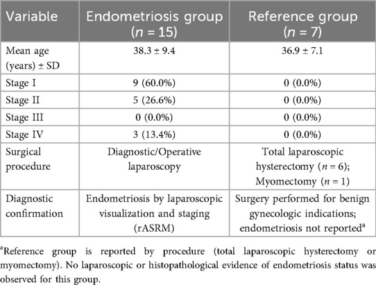
Table 2. Available demographic and clinical characteristics of study participants.
The demographic characteristics revealed that the mean age of patients with endometriosis was 38.3 ± 9.4 years, while the reference group presented a mean age of 36.9 ± 7.1 years. In the endometriosis group, 60% were diagnosed at stage I, 26.6% at stage II, and 13.4% at stage IV; no stage III cases were identified. In the reference group, all participants underwent total laparoscopic hysterectomy for benign gynecological conditions. Data regarding body mass index, menstrual cycle phase, and hormonal medication use were not available for all participants. Menstrual cycle information was not accurately recorded due to irregularities and hormonal treatments; however, previous studies have reported that the expression of the miRNAs included in this study is independent of the menstrual cycle phase, BMI, and age (8).
The quantification of the relative expression of the haplotype miRNAs was performed double-blinded by RT-qPCR, using the U6 gene as a constitutive control. Normality tests (Shapiro–Wilk, P ≤ 0.05) indicated that the data did not follow a normal distribution, so the nonparametric Mann–Whitney U-test was used to compare the differences between groups (Figure 2). This analysis showed that, of the miRNAs evaluated, only miR-451a was significantly downregulated in patients with endometriosis (P = 0.032). Although the rest of the miRNAs did not reach statistical significance, a trend toward decreased expression was observed in the endometriosis group.
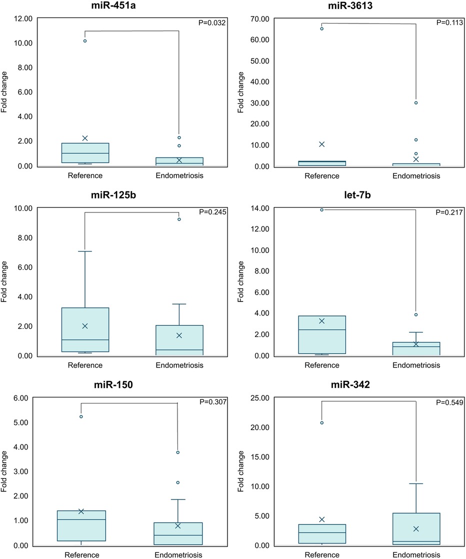
Figure 2. miR-451a has the potential to be a biomarker for endometriosis by itself; is the only miRNA with a statistically significant difference (P < 0.05) as compared to the reference group. Means comparison between the reference and endometriosis groups is shown (Mann–Whitney U-test).
Given that the comparison of sample means is limited in non-parametric data and with a small sample size, it was considered of greater relevance to evaluate the discriminative power of each miRNA using individual ROC curves (Figure 3). In this analysis, the AUC ranged from 0.581 (miR-342) to 0.790 (miR-451a). Individual sensitivities ranged from 40% (miR-125b) to 86.6% (miR-451a and let-7b), while specificity ranged from 57.1% (let-7b and miR-150) to 100% (miR-125b). Considering all metrics (Table 3), miR-451a had the highest individual diagnostic power, followed by miR-3613 and let-7b.
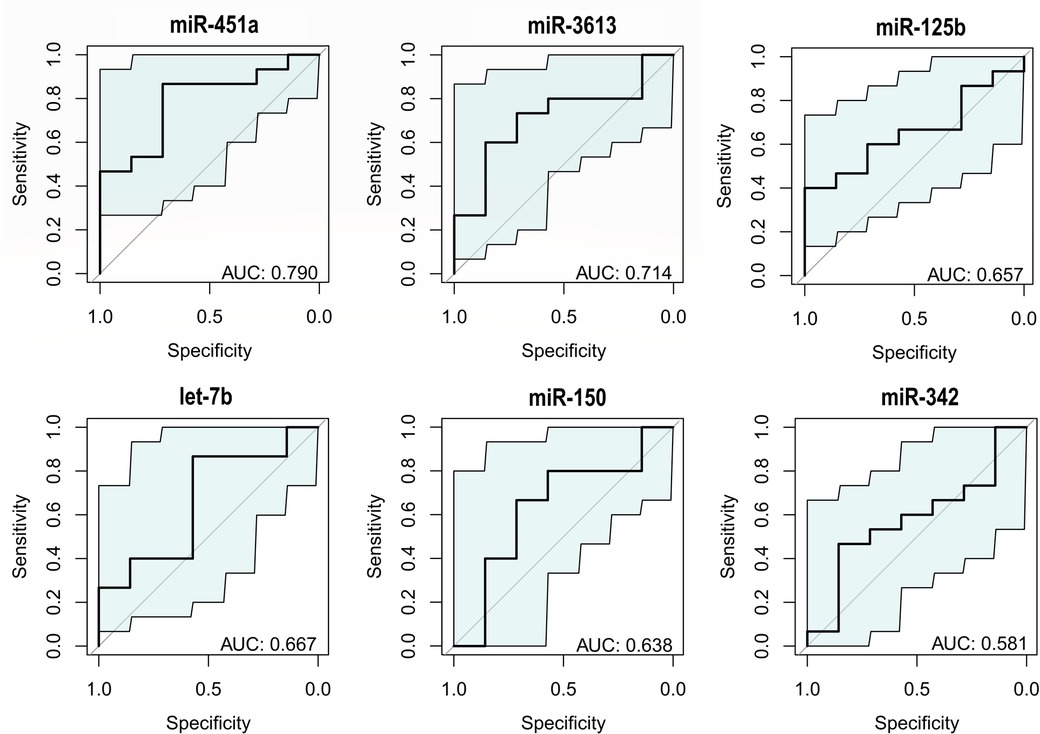
Figure 3. miR-451a exhibits the highest discriminative power (AUC = 0.790). Individual ROC curves of miRNAs performed by pROC package in R Studio (18, 19).
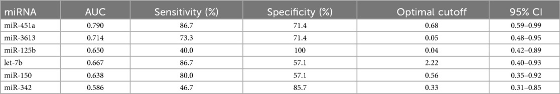
Table 3. Metrics obtained by individual ROC analysis of miRNAs performed by pROC package in R Studio (18, 19).
The integration of classification algorithms, using logistic regression and CRT models, allowed the evaluation of the diagnostic capacity of the set of 6 miRNAs. In the logistic regression model, the incorporation of miRNAs as predictor variables improved the classification of the sample, reaching an overall percentage of 81.8% and a high discrimination power (AUC = 0.914; sensitivity 80%; specificity 100%) (Table 4), being the most significant variables in the equation miR-451a (P = 0.176), let-7b (P = 0.175), and miR-3613 (P = 0.219). In parallel, the CRT analysis generated a decision algorithm at three levels, in which miR-451a (improvement = 0.146), miR-3613 (improvement = 0.069), and miR-let-7b (improvement = 0.121) stood out, achieving a correct classification of 95.5% (cross-validation was performed; AUC = 0.990; sensitivity 100%; specificity 85.7%). In addition, a stacking ensemble model was implemented that combined both algorithms, obtaining a discriminative capacity of AUC = 0.990, with a sensitivity of 93.3%, and a specificity of 100% (Figure 4).
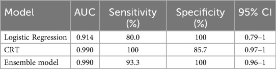
Table 4. Accuracy of binary classification models to assess the diagnostic capacity of miRNAs in Hispanic patients with and without endometriosis.
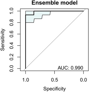
Figure 4. Expression haplotype of miRNAs (miR-451a, miR-3613, let-7b, miR-342, miR-125b, and miR-155) showed a high discriminative capacity (AUC = 0.990) between Hispanic individuals with and without endometriosis. ROC curve corresponds to a stacking-based ensemble model combining Logistic Regression and Classification and Regression Tree (CRT) analyses.
4 Discussion
In this study, the discriminative capacity of a miRNAs expression haplotype to differentiate between Hispanic women with and without endometriosis was evaluated. Up to our knowledge, this is the first study specifically targeting this population with miRNAs approach. The results show that the expression haplotype, composed of six miRNAs and analyzed using a stacking-based ensemble model, which integrates logistic regression and classification trees (CRT), reached an AUC of 0.990, with a specificity of 100% and a sensitivity of 93.3%. These findings indicate a high robustness and reproducibility of the model, suggesting its potential as a complementary tool in the diagnosis of endometriosis before laparoscopy in Hispanic patients. This could represent a non-invasive alternative with relevant clinical implications, allowing an earlier diagnosis, optimizing the management of the disease and reducing its socioeconomic impact.
So far, most studies on miRNA expression in endometriosis have been conducted in North American, European, and Asian populations, limiting the generalizability of their findings to other ethnic groups. For example, a study conducted in an Iranian population in 2025 demonstrated moderate to acceptable diagnostic accuracy for endometriosis based on differential expression of eleven miRNAs, with miR-340 showing a particularly high diagnostic value (AUC = 0.846) (20). Another study, conducted in a Chinese population, reported that a panel comprising miR-199a, miR-122, miR-145, and miR-141, combined with serum CA125 levels, achieved an AUC of 0.939 for diagnosing endometriosis (21). To date, the only study that has included Hispanic patients—though in a limited subset—was conducted by Moustafa et al. (8). Therefore, the present study represents a significant advancement in biomarker research for endometriosis and helps fill a critical gap in the literature by providing population-specific data for Hispanic women, a group historically underrepresented in miRNA expression studies. This is particularly important, as genetic and epigenetic differences between populations can influence biomarker expression, underscoring the need to develop or validate diagnostic tools tailored to specific population groups (11).
Currently, there are several strategies for the diagnosis of endometriosis in patients with suspected disease, with diagnostic powers ranging from 82% to 98.4%. These include the implementation of questionnaires and symptomatology classification (22), imaging techniques such as transvaginal ultrasound (23), and magnetic resonance imaging (24), as well as biomarkers such as CA125, IL-6, glycodelin, IGFBP-3, and VEGF (25). In recent years, additional circulating biomarkers, including specific miRNA and piRNA signatures (21, 26, 27), extracellular vesicle profiles (28, 29), and metabolomic patterns (30) have been investigated, showing promising but variable performance across different populations. Moreover, bioinformatic approaches, such as machine learning and artificial intelligence, have been explored (31), as well as genomic and transcriptomic analyses (10). However, many of these strategies may not be suitable for the Hispanic population due to limitations in accuracy, sensitivity, or specificity, as well as economic and operational barriers. Therefore, the implementation of a diagnostic method with high specificity and sensitivity, based on a defined miRNA expression haplotype validated in the Hispanic population, represents a viable and potentially scalable alternative, particularly since it relies on precise and accessible molecular biology techniques, such as RT-qPCR.
An advantage of the expression haplotype including miR-451a, miR-3613, miR-125b, let-7b, miR-150, and miR-342, is that previous studies have shown that their expression does not vary significantly with menstrual cycle progression, contraceptive use, or body mass index (8). In this study, a general trend towards the downregulation of miRNAs was observed in patients with endometriosis; however, only miR-451a showed a statistically significant decrease (P = 0.032). Regarding individual discriminative capacity, the miRNAs analyzed presented AUC values between 0.581 and 0.790, with miR-451a (AUC = 0.790), miR-3613 (AUC = 0.714) and let-7b (AUC = 0.667) standing out as the most relevant.
miR-451a has previously been reported with up to 10-fold overexpression as fold change, in patients with endometriosis (8, 32). However, other studies with a similar approach discarded miR-451a as a potential biomarker, since, despite its high discriminating power (AUC = 0.828), it was downregulated in patients with endometriosis, contrary to what was previously reported in other populations (33), but that data is consistent with the results obtained in this study. This discrepancy highlights the need for further exploration of this specific miRNA, as factors unrelated to the disease could influence its expression levels. A relevant example is hemolysis in samples, not our case because we discarded those, since miR-451a is predominantly expressed in erythrocytes, which could alter its detection in plasma or serum in the presence of hemolysis (28).
In contrast, miR-3613 and let-7b showed a behavior consistent with previous studies, presenting significant downregulation in patients with endometriosis (8, 28, 33). In addition, the discriminative capacity of these three miRNAs (miR-451a, miR-3613 and let-7b) is consistent with previous studies, and their greatest contribution was observed in our analysis of the complete expression haplotype by logistic regression and CRT. In both models, these three miRNAs showed a great influence, which reinforces their importance in the development of a diagnostic panel for endometriosis.
Bioinformatics miRNA target prediction tools, such as miRPathDB, TargetMiner and KEGG, have demonstrated the involvement of these miRNAs in the regulation of key physiological pathways, including cell proliferation, cell cycle regulation, metastasis, angiogenesis, and hormone regulation, among others. Although endometriosis is not considered a disease with malignant behavior, it shares biological characteristics with neoplasms, which suggests a similar pathogenic mechanism in certain aspects, which is why some of the pathophysiological routes of cancer were taken as a reference (Figure 5).
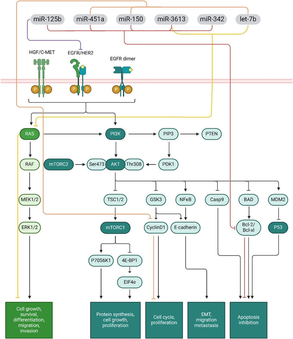
Figure 5. Signaling pathways and functions of the miRNAs included in the expression haplotype evaluated for endometriosis diagnosis, where their participation in processes such as cell cycle regulation, proliferation, survival, and apoptosis stand out. Diagram modified from Lei et al. (34) using Biorender, miRNAs targets verified with miRPathDB, KEGG and TargetMiner.
When analyzing the potential targets of miRNAs, their participation in the negative regulation of genes such as Bcl-2, cyclin D, RAS, and EGFR were identified, which act as proto-oncogenes by promoting cell proliferation (34). Under physiological conditions, the proper expression of these miRNAs maintains control of the cell cycle, preventing uncontrolled growth. However, the downregulation of the miRNAs observed in this, and other studies avoids the negative regulation of these key genes and could be related to atypical cell proliferation, ectopic implantation of endometrial tissue, and metastasis-like features in endometriosis (Figure 5).
The results of this study are promising in the development of a non-invasive diagnostic method for endometriosis in the Hispanic population, based on molecular biology techniques combined with advanced classification models (logistic regression, CRT, and stacking). These methodologies have been optimized for the analysis of a short sample, which represents an advance in the research of biomarkers for this disease. In a potential clinical pathway, this haplotype-based assay could be implemented as an initial screening tool in primary or gynecological care settings for women with suggestive symptoms, thereby guiding referrals for imaging or surgical confirmation only in high-probability cases. Such integration could shorten diagnostic delays, reduce the need for invasive procedures, and optimize healthcare resources. Further large-scale validation will be essential before formal incorporation into diagnostic guidelines.
However, the study has certain limitations, with the sample size being the most relevant. In addition, there is a possible sample selection bias, given that all patients come from the same region. An additional limitation is the partial availability of demographic and clinical data (e.g., BMI, menstrual cycle phase, hormonal medication use), inherent to translational medicine studies. Nevertheless, previous reports indicate that these variables are unlikely to influence the analyzed miRNAs (35, 36). Another consideration is the inclusion of women with other gynecological pathologies in the reference group could generate variability in the results. However, laparoscopy is the only confirmatory method for both the positive and negative diagnosis of endometriosis and clinicians must differentiate patients with and without the disease based on clinical criteria. Thus, including a reference group with other gynecological conditions was considered an appropriate approach. It is also possible that interlaboratory variability in miRNA quantification represents an additional source of bias. It is important to note that validation of these findings in diverse hispanic subpopulations would require a multicenter, collaborative effort, integrating patients from different regions to ensure representativeness. While such a study would be logistically demanding, it remains a crucial step to confirm the generalizability of the diagnostic model. These aspects highlight the need to validate findings in larger cohorts to assess the reproducibility of the diagnostic model. In addition, to better understand the expression levels of the miRNAs analyzed and their biological relevance, it is essential to perform functional studies that confirm their participation in the pathophysiology of endometriosis.
In summary, the miRNAs expression haplotype identified in this study, particularly miR-451a, miR-3613, and let-7b; showed a high discriminating power between patients with endometriosis vs. other pelvic pain but without endometriosis in the studied population (AUC = 0.990). These results reinforce the idea that a diagnostic signature based on multiple miRNAs is more effective than the analysis of single markers. However, its validation in other cohorts is essential before considering its clinical implementation.
5 Conclusions
The miRNA expression haplotype composed of miR-451a, miR-3613, miR-125b, let-7b, miR-150, and miR-342: showed a high capacity for discrimination between patients with and without endometriosis in the Hispanic population. The advanced classification models used in this study confirmed their performance: logistic regression (AUC = 0.914), CRT (AUC = 0.990), and stacking-based ensemble model (AUC = 0.990). These findings support its potential use as a diagnostic tool, future validation in a larger cohort must be performed before clinical trials.
Data availability statement
The raw data supporting the conclusions of this article will be made available by the authors, without undue reservation.
Ethics statement
The studies involving humans were approved Research Ethics Committee of the Autonomous University of Ciudad Juárez (permit CEI-2023-1-870). The studies were conducted in accordance with the local legislation and institutional requirements. The participants provided their written informed consent to participate in this study.
Author contributions
FA-M: Conceptualization, Data curation, Formal analysis, Funding acquisition, Investigation, Methodology, Project administration, Resources, Software, Supervision, Validation, Visualization, Writing – original draft, Writing – review & editing. BL-N: Methodology, Writing – review & editing. VC-U: Methodology, Writing – review & editing. DD-T: Methodology, Writing – review & editing. AB-F: Methodology, Writing – review & editing. GM-C: Methodology, Writing – review & editing. RK: Writing – review & editing, Supervision. AB-A: Supervision, Writing – review & editing, Formal analysis, Investigation. NL-G: Investigation, Supervision, Writing – review & editing, Conceptualization, Methodology, Validation. AV-C: Conceptualization, Methodology, Validation, Formal analysis, Data curation, Investigation, Supervision, Writing – review & editing. AD-S: Conceptualization, Investigation, Methodology, Supervision, Validation, Writing – review & editing. ER-E: Conceptualization, Investigation, Methodology, Supervision, Validation, Writing – review & editing, Data curation, Formal analysis, Funding acquisition, Project administration, Resources, Software, Visualization, Writing – original draft. AM-M: Conceptualization, Data curation, Formal analysis, Funding acquisition, Investigation, Methodology, Project administration, Resources, Software, Supervision, Validation, Visualization, Writing – original draft, Writing – review & editing.
Funding
The author(s) declare that financial support was received for the research and/or publication of this article. Secretary of Science, Humanities, Technology and Innovation (SECIHTI CBF2023-2024-4026), University of Texas at El Paso by US Mexico Faculty Collaboration Fellowship 2024–2025, and Autonomous University of Ciudad Juárez.
Acknowledgments
This study was sponsored by the Secretary of Science, Humanities, Technology and Innovation (SECIHTI CBF2023-2024-4026), the University of Texas at El Paso through the US-Mexico Faculty Collaboration Fellowship 2024–2025, and the Autonomous University of Ciudad Juárez. The authors are grateful to Dr- Angélica María Escárcega-Ávila for her statistical guidance and all participants, their relatives, and caregivers. The authors would like to thank the administrators and staff of the selected hospitals for their support. FDAM, and GEMC thank SECITHI for their scholarships.
Conflict of interest
The authors declare that the research was conducted in the absence of any commercial or financial relationships that could be construed as a potential conflict of interest.
Generative AI statement
The author(s) declare that Generative AI was used in the creation of this manuscript. Generative AI was used only as word editor and grammar correction for a better understanding of the ideas.
Any alternative text (alt text) provided alongside figures in this article has been generated by Frontiers with the support of artificial intelligence and reasonable efforts have been made to ensure accuracy, including review by the authors wherever possible. If you identify any issues, please contact us.
Publisher's note
All claims expressed in this article are solely those of the authors and do not necessarily represent those of their affiliated organizations, or those of the publisher, the editors and the reviewers. Any product that may be evaluated in this article, or claim that may be made by its manufacturer, is not guaranteed or endorsed by the publisher.
References
1. Bulun SE, Yilmaz BD, Sison C, Miyazaki K, Bernardi L, Liu S, et al. Endometriosis. Endocr Rev. (2019) 40(4):1048–79. doi: 10.1210/er.2018-00242
2. Kvaskoff M, Mahamat-Saleh Y, Farland LV, Shigesi N, Terry KL, Harris HR, et al. Endometriosis and cancer: a systematic review and meta-analysis. Hum Reprod Update. Oxford University Press. (2021) 27:393–420. doi: 10.1093/humupd/dmaa045
3. Matías-González Y, Sánchez-Galarza A, Rosario-Hernández E, Flores-Caldera I, Rivera-Segarra E. Stigma and social support and their impact on quality of life and self-esteem among women with endometriosis in Latin-America and the Caribbean. PLOS Glob Public Heal. (2022) 2(12):1–14. doi: 10.1371/journal.pgph.0001329
4. Koninckx PR, Fernandes R, Ussia A, Schindler L, Wattiez A, Al-Suwaidi S, et al. Pathogenesis based diagnosis and treatment of endometriosis. Front Endocrinol. (2021) 12:1–13. doi: 10.3389/fendo.2021.745548
5. Garate X, La Greca A, Neiman G, Blüguermann C, Santín Velazque NL, Moro LN, et al. Identification of the miRNAome of early mesoderm progenitor cells and cardiomyocytes derived from human pluripotent stem cells. Sci Rep. (2018) 8(1):1–14. doi: 10.1038/s41598-018-26156-3
6. Condrat CE, Thompson DC, Barbu MG, Bugnar OL, Boboc A, Cretoiu D, et al. miRNAs as biomarkers in disease: latest findings regarding their role in diagnosis and prognosis. Cells. (2020) 9(2):276. doi: 10.3390/cells9020276
7. Cui M, Wang H, Yao X, Zhang D, Xie Y, Cui R, et al. Circulating MicroRNAs in cancer: potential and challenge. Front Genet. (2019) 10:626. doi: 10.3389/fgene.2019.00626
8. Moustafa S, Burn M, Mamillapalli R, Nematian S, Flores V, Taylor HS. Accurate diagnosis of endometriosis using serum microRNAs. Am J Obstet Gynecol. (2020) 223(4):557.e1–557.e11. doi: 10.1016/j.ajog.2020.02.050
9. Bendifallah S, Suisse S, Puchar A, Delbos L, Poilblanc M, Descamps P, et al. Salivary MicroRNA signature for diagnosis of endometriosis. J Clin Med. (2022) 11(3):612. doi: 10.3390/jcm11030612
10. Bendifallah S, Dabi Y, Suisse S, Jornea L, Bouteiller D, Touboul C, et al. MicroRNome analysis generates a blood-based signature for endometriosis. Sci Rep. (2022) 12(1):1–13. doi: 10.1038/s41598-022-07771-7
11. Rotival M, Siddle KJ, Silvert M, Pothlichet J, Quach H, Quintana-Murci L. Population variation in miRNAs and isomiRs and their impact on human immunity to infection. Genome Biol. (2020) 21(1):1–31. doi: 10.1101/2020.01.31.928580
12. Hall MS, Talge NM, Upson K. Urinary cadmium and endometriosis prevalence in a US nationally representative sample: results from NHANES 1999–2006. Hum Reprod. (2023) 38(9):1835–42. doi: 10.1093/humrep/dead117
13. Hu PW, Zhang XL, Yan XT, Qi C, Jiang GJ. Association between depression and endometriosis using data from NHANES 2005–2006. Sci Rep. (2023) 13(1):1–8. doi: 10.1038/s41598-023-46005-2
14. Alday-Montañez FD, Dickens-Terrazas D, Mejia-Carmona GE, Robles-Escajeda E, Kirken RA, Bencomo-Alvarez AE, et al. Pathogenic variants in BRCA1 and BRCA2 genes associated with female breast and ovarian cancer in the Mexican population. J Med Life. (2025) 18(1):38–47. doi: 10.25122/jml-2024-0213
15. Bencomo-Alvarez AE, Gonzalez MA, Rubio AJ, Olivas IM, Lara JJ, Padilla O, et al. Ethnic and border differences on blood cancer presentation and outcomes: a Texas population-based study. Cancer. (2021) 127(7):1068–79. doi: 10.1002/cncr.33347
16. The jamovi project. jamovi (Version 2.5) [Computer Software]. (2024). Available online at: https://www.jamovi.org
18. Robin X, Turck N, Hainard A, Tiberti N, Lisacek F, Sanchez J-C, et al. pROC: an open-source package for R and S+ to analyze and compare ROC curves. BMC Bioinformatics. (2011) 8:12–77. doi: 10.1186/1471-2105-12-77
19. RStudio Team. RStudio: Integrated Development for R. Boston, MA: RStudio, PBC (2020). Available online at: http://www.rstudio.com/
20. Mohammadi SD, Moeini A, Rastegar T, Amidi F, Saffari M, Zhaeentan S, et al. Diagnostic accuracy of plasma microRNA as a potential biomarker for detection of endometriosis. Syst Biol Reprod Med. (2025) 71(1):61–75. doi: 10.1080/19396368.2025.2465268
21. Chen G, Guo J, Li W, Zheng R, Shang H, Wang Y. Diagnostic value of the combination of circulating serum miRNAs and CA125 in endometriosis. Medicine. (2023) 102(48):e36339. doi: 10.1097/md.0000000000036339
22. Dantkale KS, Agrawal M. A comprehensive review of the diagnostic landscape of endometriosis: assessing tools, uncovering strengths, and acknowledging limitations. Cureus. (2024) 16(3):e56978. doi: 10.7759/cureus.56978
23. Guerriero S, Ajossa S, Pagliuca M, Borzacchelli A, Deiala F, Springer S, et al. Advances in imaging for assessing pelvic endometriosis. Diagnostics. (2022) 12(12):1–22. doi: 10.3390/diagnostics12122960
24. Lorusso F, Scioscia M, Rubini D, Stabile Ianora AA, Scardigno D, Leuci C, et al. Magnetic resonance imaging for deep infiltrating endometriosis: current concepts, imaging technique and key findings. Insights Imaging. (2021) 12(1):105. doi: 10.1186/s13244-021-01054-x
25. Nisenblat V, Bossuyt PM, Shaikh R, Farquhar C, Jordan V, Scheffers CS, et al. Blood biomarkers for the non-invasive diagnosis of endometriosis. Cochrane Database Syst Rev. (2016) 2016(5):CD012179. doi: 10.1002/14651858.cd012179
26. Zafari N, Tarafdari AM, Izadi P, Noruzinia M, Yekaninejad MS, Bahramy A, et al. A panel of plasma miRNAs 199b-3p, 224-5p and Let-7d-3p as non-invasive diagnostic biomarkers for endometriosis. Reprod Sci. (2021) 28(4):991–9. doi: 10.1007/s43032-020-00415-z
27. Dabi Y, Suisse S, Marie Y, Delbos L, Poilblanc M, Descamps P, et al. New class of RNA biomarker for endometriosis diagnosis: the potential of salivary piRNA expression. Eur J Obstet Gynecol Reprod Biol. (2023) 291:88–95. doi: 10.1016/j.ejogrb.2023.10.015
28. Papari E, Noruzinia M, Kashani L, Foster WG. Identification of candidate microRNA markers of endometriosis with the use of next-generation sequencing and quantitative real-time polymerase chain reaction. Fertil Steril. (2020) 113(6):1232–41. doi: 10.1016/j.fertnstert.2020.01.026
29. Wu Y, Yuan W, Ding H, Wu X. Serum exosomal miRNA from endometriosis patients correlates with disease severity. Arch Gynecol Obstet. (2022) 305(1):117–27. doi: 10.1007/s00404-021-06227-z
30. Li Q, Xu L, Lin Y, Yuan M, Jiao X, Ren Q, et al. Serum metabolites as diagnostic biomarkers in patients with endometriosis. Reprod Sci. (2024) 31(12):3719–28. doi: 10.1007/s43032-024-01536-5
31. Bendifallah S, Puchar A, Suisse S, Delbos L, Poilblanc M, Descamps P, et al. Machine learning algorithms as new screening approach for patients with endometriosis. Sci Rep. (2022) 12(1):1–12. doi: 10.1038/s41598-021-04637-2
32. Cosar E, Mamillapalli R, Ersoy GS, Cho SY, Seifer B, Taylor HS. Serum microRNAs as diagnostic markers of endometriosis: a comprehensive array-based analysis. Fertil Steril. (2016) 106(2):402–9. doi: 10.1016/j.fertnstert.2016.04.013
33. Walasik I, Klicka K, Grzywa TM, Szymusik I, Włodarski P, Wielgoś M, et al. Circulating miR-3613-5p but not miR-125b-5p, miR-199a-3p, and miR-451a are biomarkers of endometriosis. Reprod Biol. (2023) 23(4):1–8. doi: 10.1016/j.repbio.2023.100796
34. Lei Z-N, Teng Q-X, Tian Q, Chen W, Xie Y, Wu K, et al. Signaling pathways and therapeutic interventions in gastric cancer. Signal Transduct Target Ther. (2022) 7(1):358. doi: 10.1038/s41392-022-01190-w
35. Rekker K, Saare M, Roost AM, Salumets A, Peters M. Circulating microRNA profile throughout the menstrual cycle. PLoS One. (2013) 8(11):1–6. doi: 10.1371/journal.pone.0081166
Keywords: translational medicine, pelvic pain, menstruation, dysmenorrhea, dyspareunia, dyschezia, dysuria
Citation: Alday-Montañez FD, Lariz-Nevárez BD, Carrasco-Urrutia VJ, Dickens-Terrazas D, Barragán-Farías A, Mejía-Carmona GE, Kirken RA, Bencomo-Alvarez AE, Lobo-Galo N, Vargas-Caraveo A, Díaz-Sánchez ÁG, Robles-Escajeda E and Martínez-Martínez A (2025) miRNA expression haplotype in Hispanics with endometriosis. Front. Reprod. Health 7:1663755. doi: 10.3389/frph.2025.1663755
Received: 11 July 2025; Accepted: 8 September 2025;
Published: 25 September 2025.
Edited by:
Mohd Helmy Mokhtar, National University of Malaysia, MalaysiaReviewed by:
Mohammad Abbaszadeh, Tehran University of Medical Sciences, IranRamiro Cabrera Carranco, Instituto Doyenne, Mexico
Copyright: © 2025 Alday-Montañez, Lariz-Nevárez, Carrasco-Urrutia, Dickens-Terrazas, Barragán-Farías, Mejía-Carmona, Kirken, Bencomo-Alvarez, Lobo-Galo, Vargas-Caraveo, Díaz-Sánchez, Robles-Escajeda and Martínez-Martínez. This is an open-access article distributed under the terms of the Creative Commons Attribution License (CC BY). The use, distribution or reproduction in other forums is permitted, provided the original author(s) and the copyright owner(s) are credited and that the original publication in this journal is cited, in accordance with accepted academic practice. No use, distribution or reproduction is permitted which does not comply with these terms.
*Correspondence: Alejandro Martínez–Martínez YWxlamFuZHJvLm1hcnRpbmV6QHVhY2oubXg=; Elisa Robles–Escajeda ZXJvYmxlczNAdXRlcC5lZHU=; Flor Daniela Alday–Montañez Zmxvci5hbGRheUB1YWNqLm14