 Qian Li1,2,3†
 Abigail Weiland1†
 Xuemei Chen4
 Xi Lan1
 Xiaoning Han1
 Frederick Durham1Xi Liu1
 Jieru Wan1
 Wendy C. Ziai1,5
 Daniel F. Hanley5
 Jian Wang1*
- 1Department of Anesthesiology and Critical Care Medicine, Johns Hopkins University School of Medicine, Baltimore, MD, United States
- 2Department of Biochemistry and Molecular Biology, School of Basic Medical Sciences, Capital Medical University, Beijing, China
- 3Advanced Innovation Center for Human Brain Protection, Beijing, China
- 4Department of Human Anatomy, College of Basic Medical Sciences, Zhengzhou University, Zhengzhou, China
- 5Department of Neurology, Johns Hopkins University School of Medicine, Baltimore, MD, United States
by Li, Q., Weiland, A., Chen, X., Lan, X., Han, X., Durham, F., Liu, X., Wan, J., Ziai, W. C., Hanley, D. F., and Wang, J. (2018). Front. Neurol. 9:581. doi: 10.3389/fneur.2018.00581
In the published article, there was an error in Figure 2Ci as published. The wrong image was inadvertently used. The corrected Figure 2 appears below.
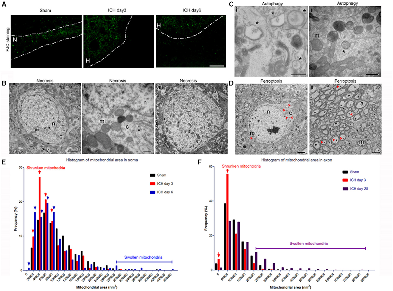
Figure 2. Mixed forms of neuronal death after intracerebral hemorrhage (ICH). (A) Fluoro-Jade C histology shows degenerating neurons in the perihematoma region at 3 and 6 days post-ICH or along the needle track in the striatum of the sham-operated mice. Dashed lines indicate the margin of hematoma. N, needle track; H, hematoma. (B) Necrotic neuronal soma (i–iii) show loss of a distinct nuclear membrane, enlarged mitochondria, and changes in chromatin structure at 3 days post-ICH. (C) Autophagy in neuronal soma at 3 days post-ICH; autophagosomes are labeled with asterisks. Inset shows a high-power image of an autophagosome. (D) Neuronal soma and axons show signs of ferroptosis with evidence of shrunken mitochondria (red arrowheads) at 3 days post-ICH. (E) Quantification of mitochondrial area frequency in neuronal somas at various time points after ICH. Arrows indicate increased frequency of shrunken mitochondria on days 3 and 6. Number of mitochondria: sham, n = 429; ICH day 3, n = 306; ICH day 6, n = 296. (F) Quantification of mitochondrial area frequency in axons at various time points after ICH. Arrows indicate increased frequency of shrunken mitochondria on day 3. Number of mitochondria: sham, n = 433; ICH day 3, n = 314; ICH day 28, n = 603. Scale bars: (A) 100 μm; (Bi,iii, D) 2 μm; (Bii, Cii) 500 nm. n, nucleus; c, cytoplasm; m, mitochondria; a, axon. n = 6 animals per group.
In the published article, there was an error in the Funding statement. The NSFC (U1704166), the Henan Province Science and Technology Cooperation Project (No. 182106000061) or the NIH grants (R01NS078026, R01AT007317, R56NS096549, R21NS101614, R21NS102899 and UG3NS106937) did not support this work. The correct Funding statement appears below.
Funding
This research was supported by the American Heart Association (Grant-in-Aid, 17GRNT33660766 to JWang; Scientist Development Grant, 16SDG30980031 to XH; Postdoctoral Fellowship Awards, 16POST29640010 to QL, 17POST33660191 to XLa, and 18POST33970007 to JWan), and a Stimulating and Advancing ACCM Research (StAAR) grant from the Department of Anesthesiology and Critical Care Medicine, Johns Hopkins University.
The authors apologize for this error and state that this does not change the scientific conclusions of the article in any way. The original article has been updated.
Publisher's note
All claims expressed in this article are solely those of the authors and do not necessarily represent those of their affiliated organizations, or those of the publisher, the editors and the reviewers. Any product that may be evaluated in this article, or claim that may be made by its manufacturer, is not guaranteed or endorsed by the publisher.
Keywords: cell death, intracerebral hemorrhage, synapse, transmission electron microscopy, white matter injury
Citation: Li Q, Weiland A, Chen X, Lan X, Han X, Durham F, Liu X, Wan J, Ziai WC, Hanley DF and Wang J (2024) Corrigendum: Ultrastructural characteristics of neuronal death and white matter injury in mouse brain tissues after intracerebral hemorrhage: coexistence of ferroptosis, autophagy, and necrosis. Front. Neurol. 15:1385719. doi: 10.3389/fneur.2024.1385719
Received: 13 February 2024; Accepted: 22 February 2024;
Published: 01 March 2024.
Edited and reviewed by: Bruce Miller, University of California, San Francisco, United States
Copyright © 2024 Li, Weiland, Chen, Lan, Han, Durham, Liu, Wan, Ziai, Hanley and Wang. This is an open-access article distributed under the terms of the Creative Commons Attribution License (CC BY). The use, distribution or reproduction in other forums is permitted, provided the original author(s) and the copyright owner(s) are credited and that the original publication in this journal is cited, in accordance with accepted academic practice. No use, distribution or reproduction is permitted which does not comply with these terms.
*Correspondence: Jian Wang, jwang79@jhmi.edu
†These authors have contributed equally to this work