 Natasha M. Drissi1,2
 Attila Szakács3†
 Suzanne T. Witt2†
 Anna Wretman4
 Martin Ulander5Henriettae Ståhlbrandt6
 Niklas Darin3
 Tove Hallböök3
 Anne-Marie Landtblom5,7
 Maria Engström1,2*
- 1Department of Medical and Health Sciences (IMH), Linköping University, Linköping, Sweden
- 2Center for Medical Image Science and Visualization (CMIV), Linköping University, Linköping, Sweden
- 3Department of Paediatrics, Institute of Clinical Sciences, Sahlgrenska Academy, University of Gothenburg, Gothenburg, Sweden
- 4Department of Behavioral Science and Learning, Linköping University, Linköping, Sweden
- 5Department of Clinical and Experimental Medicine, Linköping University, Linköping, Sweden
- 6Department of Radiology, Medical Diagnostics, Highland Hospital, Eksjö, Sweden
- 7Department of Neurology, Uppsala University, Uppsala, Sweden
A Corrigendum on
Altered Brain Microstate Dynamics in Adolescents with Narcolepsy
by Drissi, N. M., Szakács, A., Witt, S. T., Wretman, A., Ulander, M., Ståhlbrandt, H., et al. (2016). Front. Hum. Neurosci. 10:369. doi: 10.3389/fnhum.2016.00369
In the original article, there was a mistake in the legend for Figure 2 as published. The legend states that the error bars represent the standard deviation, this is incorrect. The error bars in Figure 2 represent the standard error. The correct legend appears below.
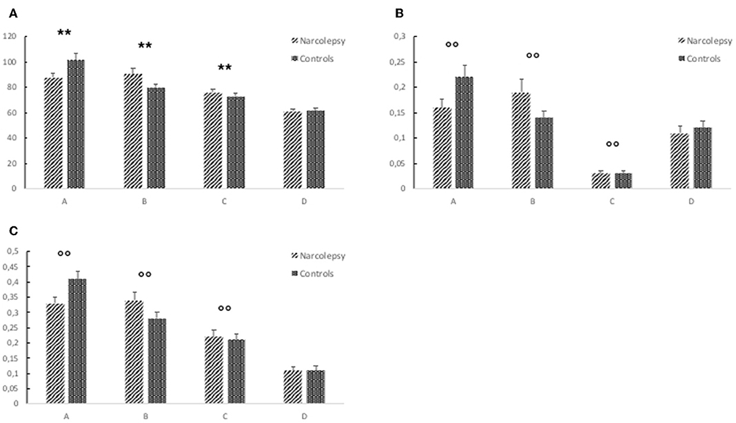
Figure 2. Results from the electroencephalography (EEG) microstates analysis. The figure shows (A) mean duration of each microstate (in ms), (B) mean global explained variance (GEV), and (C) ratio of total time covered for each microstate. The error bars represent the standard error. Descriptive data can be found in Table 3. “**” Indicates a significant post hoc difference. “°°” Indicates a trend-level post hoc difference.”
Additionally, in the original article, there was a mistake in Figure 2 as published. The figure legend indicating which bars represent narcolepsy and control have been reversed, so that the diagonal stripes are incorrectly shown to represent the narcolepsy group while the dots represent the control group. This should be reversed to be in line with the data in Table 3 as well as in the Results, where the group differences are described correctly. The corrected Figure 2 appears below.
The authors apologize for these errors and state that they do not change the scientific conclusions of the article in any way. The original article has been updated.
Keywords: narcolepsy, default mode network, functional magnetic resonance imaging (fMRI), electroencephalography (EEG), microstates, resting state networks, orexin, sleep
Citation: Drissi NM, Szakács A, Witt ST, Wretman A, Ulander M, Ståhlbrandt H, Darin N, Hallböök T, Landtblom A-M and Engström M (2019) Corrigendum: Altered Brain Microstate Dynamics in Adolescents With Narcolepsy. Front. Hum. Neurosci. 13:385. doi: 10.3389/fnhum.2019.00385
Received: 23 September 2019; Accepted: 15 October 2019;
Published: 06 November 2019.
Edited and reviewed by: Mingzhou Ding, University of Florida, United States
Copyright © 2019 Drissi, Szakács, Witt, Wretman, Ulander, Ståhlbrandt, Darin, Hallböök, Landtblom and Engström. This is an open-access article distributed under the terms of the Creative Commons Attribution License (CC BY). The use, distribution or reproduction in other forums is permitted, provided the original author(s) and the copyright owner(s) are credited and that the original publication in this journal is cited, in accordance with accepted academic practice. No use, distribution or reproduction is permitted which does not comply with these terms.
*Correspondence: Maria Engström, bWFyaWEuZW5nc3Ryb21AbGl1LnNl
†These authors have contributed equally to this work