 Cyril Bertheaux
 Rosario Toscano1
 Céline Borg
- 1Université de Lyon, ENISE, LTDS, UMR 5513 CNRS, Saint-Étienne, France
- 2ISAE-ENSMA, Chasseneuil-du-Poitou, France
- 3University Hospital of Saint-Étienne, SNA-EPI Laboratory, EA 4607, CHU, Université de Lyon, Saint-Priest-en-Jarez, France
- 4University Hospital of Saint-Étienne, CMRR Neuropsychology, Department of Neurology, Université de Lyon, Saint-Priest-en-Jarez, France
The emotion generated by the touch of materials is studied via a protocol based on blind assessment of various stimuli. The human emotional reaction felt toward a material is estimated through (i) explicit measurements, using a questionnaire collecting valence and intensity, and (ii) implicit measurements of the activity of the autonomic nervous system, via a pupillometry equipment. A panel of 25 university students (13 women, 12 men), aged from 18 to 27, tested blind twelve materials such as polymers, sandpapers, wood, velvet and fur, randomly ordered. After measuring the initial pupil diameter, taken as a reference, its variation during the tactile exploration was recorded. After each touch, the participants were asked to quantify the emotional value of the material. The results show that the pupil size variation follows the emotional intensity. It is significantly larger during the touch of materials considered as pleasant or unpleasant, than with the touch of neutral materials. Moreover, after a time period of about 0.5 s following the stimulus, the results reveal significant differences between pleasant and unpleasant stimuli, as well as differences according to gender, i.e., higher pupil dilatation of women than men. These results suggest (i) that the autonomic nervous system is initially sensitive to high arousing stimulation, and (ii) that, after a certain period, the pupil size changes according to the cognitive interest induced and the emotional regulation adopted. This research shows the interest of the emotional characterization of materials for product design.
Introduction
The touch is an essential sense to the human because it allows the contact with our environment, the perception of wind, humidity, temperature changes, relief, roughness, softness, grip (Barnett, 1972). The touch is the most developed sense at birth (Field, 2001). Various studies have shown the influence of massage on brain development and improvement of visual function in premature rats, primates and humans (Stack and Muir, 1990; Field, 1998; Kaffman and Meaney, 2007; Guzzetta et al., 2009), with effects after the first week of stimulation in puppies (Sale et al., 2004). The lack of caresses is known to be a cause of death in orphaned babies (Spitz, 1945) and in baby monkeys (Harlow and Harlow, 1962). Study on maternal care of baby rats established that adult rats descended from a mother who gave numerous licks/groomings showed lower levels of fear reactivity than rats descended from a mother who dispensed very little (Menard et al., 2004; Dunbar, 2010). The pleasant properties of caresses on cats, dogs, humans have often been demonstrated (Harlow, 1959; De Waal, 1989; Lindgren et al., 2010). The therapeutic properties of touch are well known, and it seems that the touch has a significant impact on the neuro-hormonal system (Bush, 2001). Different hormonal changes have been observed in tactile stimulations: change in the rate of cortizol in children after a simple contact with the skin of their mother, change of insulin levels with significance effects on type 2 diabetes, secretion of oxytocin during a pleasant touch (Feldman et al., 2010; Baldini et al., 2013; Clermont, 2018). Human skin is considered as a social organ (Löken and Olausson, 2010). Recent studies suggest (i) that the degree of allowed touch can predict the closeness of a relationship (Suvilehto et al., 2015), and (ii) that the degree of actual touch can predict functional connectivity of the social brain (Brauer et al., 2016). Other studies on social relation showed that a caress activates different emotional cerebral areas, depending on whether participants believe it to come from a person of the same or of the opposite sex (Dolinski, 2010; Gazzola et al., 2012).
The psychophysical dimensions of touch have also been studied (Taylor and Lederman, 1975; Jones and Lederman, 2006; McGlone and Reilly, 2010; Zahouani et al., 2013; Abdouni et al., 2017, 2018). The “epicritical” sensitivity corresponds to the fine and discriminative touch (shape, contours, texture, and relief) (Mountcastle, 2005), while the “protopathic” sensitivity corresponds to the cutaneous sensitivity triggered by strong stimulation, like temperature. The “protopathic” sensitivity generates a defense reaction of the body (Melzack and Wall, 1965).
The “epicritical touch” is achieved by Meissner’s corpuscles and Merkel’s receptors. Meissner’s corpuscles are speed detectors that encode motion. Merkel’s receptors are sensitive to intensity, pressure and temporal information on stimuli. The “protopathic touch” is achieved by Pacini’s corpuscles and Ruffini’s receptors. Pacini’s corpuscles are sensitive to vibrations and record temporal information on stimuli. Ruffini’s receptors are sensitive to the stretching of the skin. The skin also includes thermoreceptors, nociceptors and itch receptors that are sensitive to thermal sensations, pain and skin irritation and itching (Nordin, 1990; Misery and Ständer, 2010; Misery, 2014). Touch also involves proprioceptors, which are sensitive to changes of position and angular velocity of an articulation, tendon. There are three main types of proprioceptors: free nerve endings, Golgi receptors and Pacini corpuscles (Hasan, 1992; Edin, 2001).
Okamoto et al. (2012) have identified five dimensions of the “epicritical” touch that affect our tactile perceptions: hardness (hard, soft), temperature (hot, cold), friction properties (wet, dry, sticky, slippery), fine roughness (rough, smooth) and macro roughness (uneven, relief). Lederman and Klatzky (1987) have defined the gesture adapted to the tactile characterization of objects and surfaces. Gibson (1962) and Wall (2000) propose to distinguish the “Active” or voluntary touch, and the “Passive” or involuntary touch. During an “Active” touch, the mechanoreceptors of human skin treat tactile, proprioceptive and kinesthetic information which are relayed to the central nervous system to produce the emotional and sensory quality of the material. Some studies have focused on the study of perceptive sensory or emotional lexicons (Melzack, 1975; Essick et al., 1999, 2010; Cardello et al., 2003; Guest et al., 2011). Other studies have investigated the links between physical properties of materials and associated hedonic sensations (Chen et al., 2009: Datta, 2016). These different studies offer a better understanding of the emotional aspects of materials in order to master their emotional impact in a sensory design process.
The aim of this paper is to investigate pupil size variation in response to touch of different materials. It is the first study relating emotional touch with pupillometry. After describing the link between touch and emotional development, the experimental procedure is detailed, and the results are discussed.
Touch and Emotional Development
Studies on emotional touch have advanced through the study of a patient (GL) with Guillain-Barré syndrome (Forget and Lamarre, 1987). This patient had lost almost all large myelinated afferents, resulting in a total deficit of discriminative touch, over the whole of the body. Despite this deficit, she was able to describe the movement of a soft brush on her arm, as a light, pleasant and gentle touch. A little before, Zotterman (1939) had identified non-myelinated C-Tactile fibers (CT) in cats. These fibers have also been found in the hairy skin of mammals (Leem et al., 1993; Li and Ginty, 2014; Liljencrantz and Olausson, 2014; Rutlin et al., 2014; Walsh et al., 2015; Moayedi et al., 2016). The qualitative feelings of affective stimuli are comparable in hairy and glabrous skin. Recent studies suggest that different CT fibers have been found in the glabrous skin and have different biochemical and structural characteristics (Nagi and Mahns, 2010, 2013; Djouhri, 2016). The hedonic feelings felt with the fingertips still remains to be explained. Recent studies have identified MrgprB4 neurons in mouse as being involved in the pleasure felt during massage. In the experience realized, the MrgprB4 neuron was only activated the mouse was stroked with a brush. Results suggest that CT fibers with MRGPRB4 have distinct sensitivity to other anatomically similar CT neurons with receptors (MRGPRD) sensitive to unpleasant stimulation (Vrontou et al., 2013). This new knowledge could partly explain the gentle touch felt by the extremity of the finger.
A study realized on healthy subjects by Functional magnetic resonance images (fMRI) show that soft brush stroking activates the somatosensory areas S1 and S2, as well as insular cortex, notably the posterior part of the contralateral insular cortex (Olausson et al., 2002). S1 and S2 receive fibers Aβ projections and are known to play crucial roles in discriminative touch. When similar brushing stimuli are applied to GL, no activation is found in the somatosensory areas when the posterior insular region is activated (Olausson et al., 2010). The unmyelinated CT afferents, therefore, probably have excitatory projections mainly to emotion-related cortical systems such as the insular cortex (Vallbo et al., 1993; Vallbo et al., 1999). Other studies (Rolls et al., 2003) showed that the orbitofrontal cortex is involved in representing both positive and negative affect produced by touch, as it is the case with a stimulation through other sensory modalities (Rolls, 2000).
The emotional response triggered by a stimulus can be studied according to its three components: the expressive component (or behavioral response) measured by electromyogram of facial muscles, the cognitive component (feeling experienced) evaluated by self-assessment questionnaires, and the physiological component (or autonomic activity). Recent technologies allow now scientists to measure the emotions more accurately. For instance, in an array of pictures, those inducing fear (e.g., snakes) are located faster than the others (Öhman et al., 2001). Moreover, between 60 and 100 milliseconds after displaying an unpleasant stimulus, those inducing fear produce larger amplitude of visual evoked potentials than the others (Stolarova et al., 2006).
The first data showing a clear correlation between pupil diameter change and emotion were published in the sixties (Hess and Polt, 1960). Later, Bradley et al. (2008) have used emotional images to study their effect on the pupillary response. They divided pictures of the International Affective Picture System (IAPS; Lang et al., 2005) into three equal parts being considered, respectively, as pleasant, neutral and unpleasant. The results showed that the diameter of the pupil is significantly affected by the emotional pictures: the pupil becomes more dilated with emotional pictures, whatever its valence (i.e., pleasant or unpleasant). Additionally, Bradley et al. (2008) reported that the pupil diameter changes are correlated with changes in the conductivity of the skin, but not with the heart rate. This is consistent with the idea that the pupil diameter reflects the activity of the sympathetic nervous system. Recently, other studies suggest that non-visual emotions can also increase the diameter of pupils. This has been observed with participants listening to emotional sounds (Partala and Surakka, 2003; Babiker et al., 2013) or testing emotional products (Lemercier et al., 2014). In neuropsychology domain, by studying the effect of emotional stimuli on pupil diameter of people with Parkinson’s disease, Dietz et al. (2011) have found a normal sympathetic excitation to affective stimuli (indexed by pupil diameter), but no oculomotor differences.
The pupillary response reflects the activity of the autonomic nervous response, especially the parasympathetic and sympathetic systems (Charier et al., 2017). The parasympathetic system innervates the sphincter pupillae and controls the pupil constriction, whereas the sympathetic nervous system causes the excitation of the dilator pupillae (Beatty and Lucero-Wagoner, 2000). Initially, the major role of these muscles is to adjust the amount of light allowed to enter the eye (Ellis, 1981). Recently, a review of 134 studies (Kreibig, 2010) has shown that many theories agree on the relationship between emotion and the organization of autonomic nervous system activity. In this way, many authors propose to adopt an observation of the results according to the type of emotion examined. This is consistent with the results of Bradley et al. (2008). An increase of pupil diameter and sweating is observed in pleasant and unpleasant conditions, whereas the heart rate slows down for negative stimuli and increases with neutral and pleasurable condition. Other researchers have studied heart rate variability in different emotional states (Onorati et al., 2013; Park and Thayer, 2014).
It is now well-known that touch involves a sensory and emotional integration (Chaplan et al., 1994; Armaied et al., 2005; Rolls and Kesner, 2006; Spapé et al., 2015; Schirmer and Adolphs, 2017). Nevertheless, few studies have examined the activity of the autonomic nervous system during an exploration by touching a surface. Thus, the present study proposes to measure the emotional touch of different surfaces with explicit (verbal rating scale) and implicit (pupillometry) measures. We expect that the tactile exploration of an emotional material will increase the activity of the dilator muscle. Therefore, pupil diameter is supposed to increase with material surfaces with high emotional Intensity, regardless their hedonic valence.
Experimental Procedure
Participants and Materials
A preliminary study was conducted to select test materials, within a library containing 50 different samples (wood, fabrics, furs, skins, metals, plastics, slimes, sandpaper). This library was created according to the recommendations on the representation of the pleasant touch specified in the literature (Greenspan and McGillis, 1991; Francis et al., 1999; Rolls et al., 2003; Thieulin, 2017). Six participants were asked to estimate the emotional valence and intensity of the surfaces, and two set of six materials were selected. The set 1 contains six surfaces obtained by 3D printing of six different neutral materials. Three of them are made of 100% polylactic material (green, pink, turquoise), and the others contain 50% of wood, copper and titanium powder, respectively. The measured roughness coefficients of these samples range from 0.255 to 0.417. The set 2 is made of six emotional surfaces. Two of them are considered as pleasant (very soft velvet and artificial fur). The three others are different grades of sandpaper, with an increasing roughness, i.e., N1 (slightly rough), N2 (moderate rough) and N3 (very rough). They are considered as unpleasant.
A panel of twenty-five students, 12 men and 13 women aged from 18 to 27, was selected. According to the “Edinburg” test which allows calculating the coefficient of laterality (Q.L.) on the basis of the examination of 12 gestural tasks (Oldfield, 1971), the panel was found to be right-handed, except for a female subject. According to the “Von Frey” test, which measures the changes in tactile sensitivity by using five calibrated filaments (Von Frey, 1896), all participants have a normal tactile perception. Finally, the emotional state of the participants before the test was estimated by using the questionnaire of the “HAD – Hospital Anxiety and Depression scale” (Zigmond and Snaith, 1983). Four subjects presented a score above the pathological limit. Therefore, the HAD scores were introduced as covariate in our analyses. Since these data were found to induce no measurements bias, the twenty-five students were kept for the test.
Procedure
Emotion outcome was assessed (i) via a self-report judgment (explicit measure), to collect information on intensity and valence (Russell, 1980), and (ii) via measurements of the autonomous nervous system using a pupillometer. Since the works of Osgood (1969) and Russell (1980), stimuli can be characterized by two dimensions, valence and intensity. Valence refers to the hedonic nature of information on a continuum ranging from “positive, pleasant” to “negative, unpleasant” while the intensity corresponds to the level of physiological activation provoked by emotional information on a continuum ranging from “calm” to “excited”. Many authors (Bradley et al., 2008) have shown that pupillary responses might serve as a reliable index of emotional intensity and other works have shown the interest of collecting the emotional value of a material (Bertheaux et al., 2018). This physiological response is considered as more automatic and implicit than explicit, verbalized self-report measures. Pupil diameter is modulated by emotional intensity (linked to somaesthetic cues). Studies comparing and correlating emotional responses about their peripheral physiological mechanisms and self-report judgment, are limited in number.
The participants were sitting on a comfortable chair, the arms resting on a table to limit the movement of the body, in an artificially lighted room with a small opening on the outside. Luminance measurements gave values ranging from 240 to 250 Lux. A 850 × 850 mm white wooden tray with a target point in its center was placed at 40 cm in front of the participants. This experimental equipment was used to focus the gaze toward a target point to facilitate the measurement of the pupil and to avoid any distractions. To perform a blind experiment, the materials were placed in an experiment box with 6 materials in 6 housing spaced 25 mm. The box dimensions were 700 mm long, 140 mm high and 200 mm deep. The samples were 70 × 70 × 5 mm to allow exploration of the surface with 3 fingers per tangential and circular touch. Randomly ordered set 1 and 2 were presented successively to the participants.
During the test, an experimenter was placed behind the subject so that the participant was not tempted to look at him during the answers. He described each step to the participant who executed the experiment. A second experimenter, located beside the participant, registered markers in order to sequence each measurement step. Figure 1 gives the different steps of the procedure. The first step consisted in a blind touch during 15 s of a randomly chosen material of the set. This step erased the “surprise” effect, and the associated data are not processed. It was followed by a recorded “Rest phase” of 30 s, which was used to define the initial pupil diameter of the participant d0. Then, participants silently touched each material during 15 s with the dominant hand. After this touching period, the experimenter asked them about his feelings and noted their answers during 30 s. Valence was assessed using a scale from –4 (very unpleasant) to +4 (very pleasant), and intensity is ranged from 0 (very calm) to 5 (very excited).
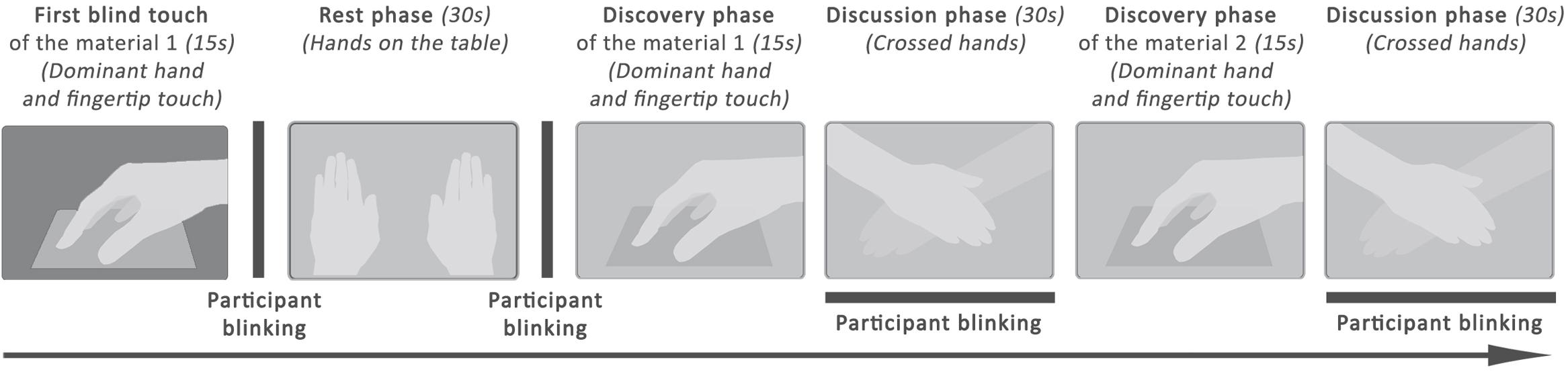
Figure 1. Experimental procedure applied on each set.
The scales used for valence and intensity are adapted from the SAM scale (Self Assesment Mankini), which proposes to measure the emotion on three dimensions: pleasure (valence), excitation (intensity) and dominance (Bradley and Lang, 1994). During the “Discussion Phase”, the participant said whether the sample was pleasant (yes or no) and indicated his feeling on the valence scale (−4 to 4). Then, the participant said whether he has felt an emotion (yes or no), and quantified it with the intensity scale (0 to 5). A written example is shown in Table 1.
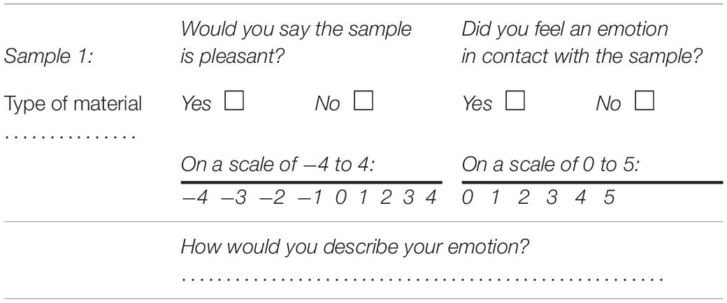
Table 1. Sample of survey used in the subject assessment.
Pupil diameter measurements were performed at 60 Hz with an ISCAN-ETL-100H pupilometer device connected to an infrared camera. The size of the pupil in pixels is detected in the vertical and horizontal directions, and the mean value at time t gives de pupil diameter d(t).
Results and Discussion
Questionnaire
Figure 2 gives the histograms of emotional values collected by the questionnaire for two samples, “Green polylactic (PLA)” (Figure 2A) and “synthetic fur” (Figure 2B). In Figure 2A the values are clearly distributed around a mean value of 0.6 (intensity) and 1.76 (valence). The emotional value of this sample is considered as “neutral”. In Figure 2B, the intensity values are highly positive, between 3 and 4, with a mean value of 3.12, and the valence is mainly positive, with a relatively high mean value of 3.84. The emotional value of this sample is therefore considered as “pleasant”. However, it can be observed in Figure 2B that two participants have considered the synthetic fur as very unpleasant, with two ratings at −3. This can probably be explained by the cultural background of these two participants who raised in an environment with wild animals. Actually, the furry animals are often considered as pleasant. Often during our childhood, we experiment the soothing and relaxing effect of touch with a cat, a dog, a teddy bear … (Servais, 2007). It does not represent a danger. Nevertheless, the two students lived in a country where animals are dangerous, so that they did not consider the fur as pleasant as the other students.
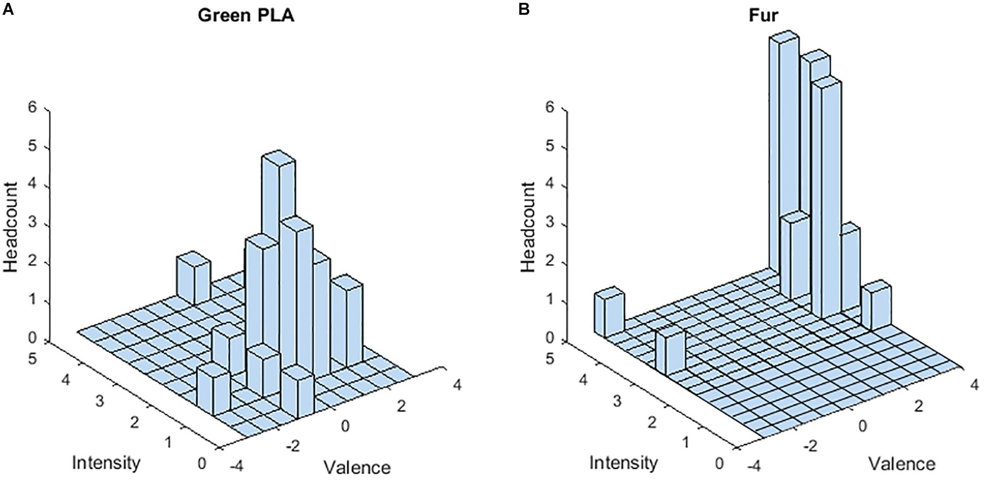
Figure 2. Emotional values (valence and Intensity) of two samples. Histograms of Green PLA (A) and Fur (B) as a function of Intensity and Valence.
Considering all the participants, the mean value of the verbalized valence was found to be 2.74 ± 1 for the two pleasant materials (very soft velvet and artificial fur), −0.41 ± 0.55 for the three unpleasant materials (sandpapers), and 0.77 ± 0.07 for the seven remaining neutral materials. A Friedman test on the valence ratings showed that these three conditions of emotion were significantly different (χ2 = 50, p ≤ 0.0001), and a posteriori Wilcoxon analysis showed significant differences between the three dimensions (p < 0.0001). Similarly, the mean intensity ratings for the pleasant, unpleasant and neutral touch stimulus across all subjects were, respectively, 3 ± 0.16, 2 ± 0.10, and 1.9 ± 0.17. A Friedman test on these intensity ratings showed that the three conditions of emotion were significantly different (χ2 = 48, p ≤ 0.0001). A posteriori Wilcoxon analysis showed significantly higher score for pleasant stimuli compared to neutral and unpleasant stimuli (z = 4.37, p ≤ 0.0001 and z = 4.37, p = 0.0001, respectively), and the pattern of results indicated higher mean intensity score for negative stimuli compared to neutral touch (z = 4.13, p = 0.0001). No specific gender differences appeared and the HAD score had no influence on the scales scoring.
Pupil Diameter Change
A cleaning algorithm has been applied to all the recorded diameters to detect blinks, absence of measurements, and measures considered aberrant (pupil size too high). After detection, the algorithm uses a numeric interpolation and replaces this data with a corrected data bridge that covers the mapped interval (Schlomer et al., 2010). For each material and for the rest phase, the recorded pupil diameters were exploited for 2 s. This duration was defined according to the work of Bradley et al. (2008), which shows that the constriction of the pupil intervenes from 0.6 to 1.6 s after the presentation of an image (emotional stimuli).
The data processing was finally performed by calculating the relative evolution of the pupil diameter of each subject, for each material, during the discovery phase, as [d(t)-d0]/d0. The d0 value is obtained by averaging the pupil diameter values during the last 2 s of the rest phase for each subject. Participants observed two periods of Rest phase, one taken during the measurement of set 1 and the other during set 2. The d(t) value is the pupil diameter of the participant measured at time t during the first 2 s of the discovery phase.
Figure 3 shows the time and participant averaged pupil dilatations for all materials, classified from unpleasant to pleasant, together with the mean verbalized valence and arousal. According to the Bienaymé-Tchebychev Inequality Test, in the case where the data do not follow a normal law, the probability that the random variable will be realized in interval ±2 σ is 75%, and becomes 89% in the range of ±3 σ. It can be observed in this figure that high emotional materials present larger pupil dilatations than neutral materials, but that pupil dilations for unpleasant and pleasant materials are very similar.
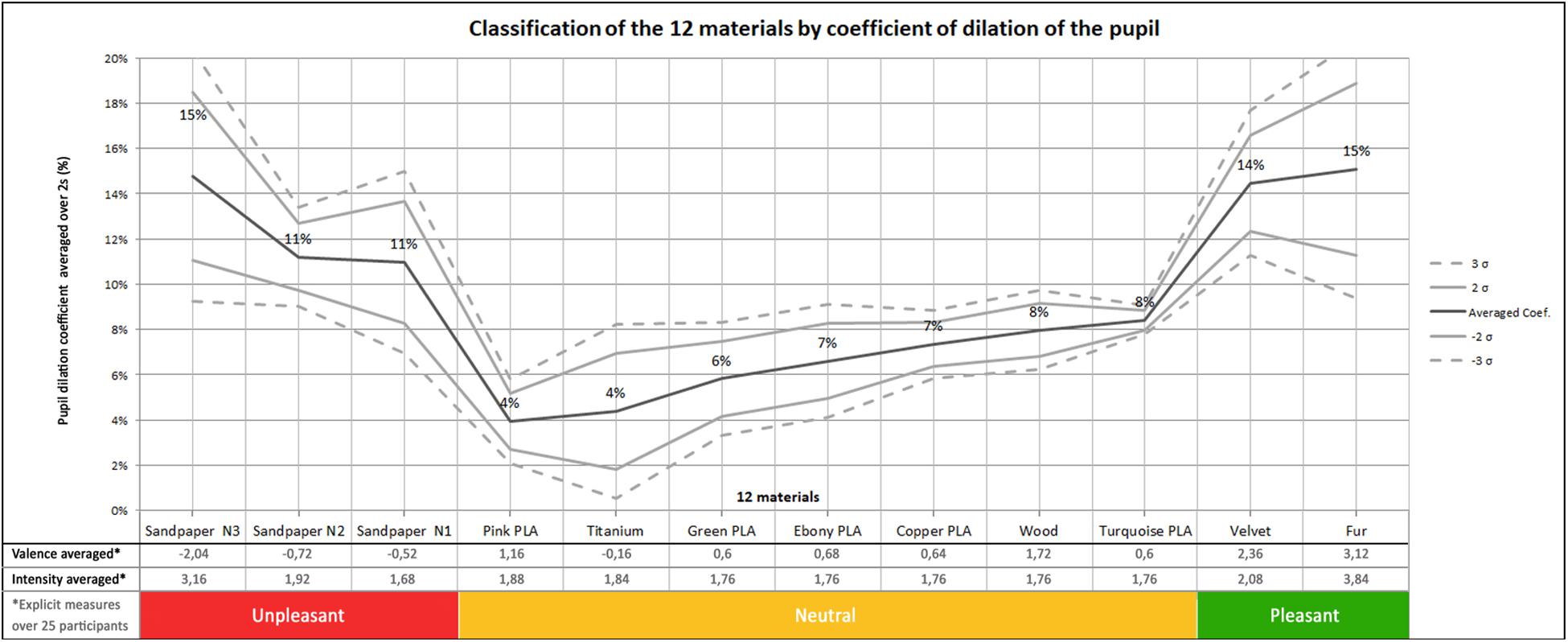
Figure 3. Classification of materials by coefficient of dilation of the pupil on 3 conditions: unpleasant, neutral, pleasant.
Figure 3 suggests that our statistical analyses can be performed with the following three classes of materials:
– Three Unpleasant: sandpapers from N1 to N3;
– Seven Neutral: all PLA, Titanium and Wood;
– Two Pleasant: Velvet and Fur.
We performed the Shapiro–Wilk test on this dataset. At 5% risk, our results showed that our data don’t follow a normal distribution. Therefore, all data were rank transformed (Conover and Iman, 1981) and submitted to an ANCOVA. The HAD score was used as covariate since previous studies showed that the level of mood can influence the processing of emotional materials (Mogg and Bradley, 1999; Mogg et al., 2000). We included in the present analyses the inter-subject factor “group” (male and female) and the within-subject factor “emotion” (pleasant, unpleasant and neutral). In addition, pairwise post hoc comparisons with the Bonferroni test and planned comparisons were performed when appropriate. The ANCOVA showed no effect of the HAD score on the overall results, but the “group” and emotion” factors were found to be significant.
The effect of emotion is significant [F(2,24) = 10.87, p = 0.0004]. Figure 4 shows the pupil dilation coefficient of the participants during the discovery phase. These data are averaged over the three classes of materials: neutral, unpleasant, pleasant. The following observations can be made on this graph:
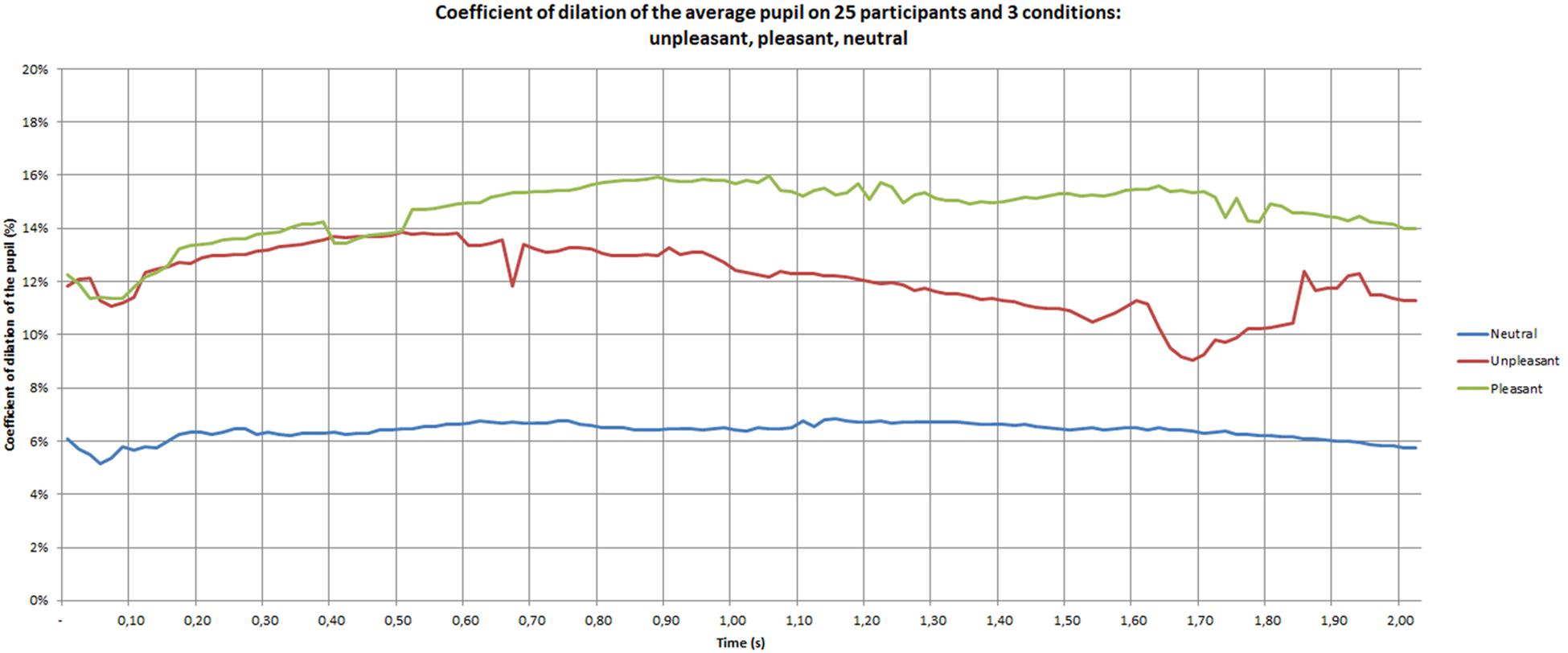
Figure 4. Pupil dilation coefficient of the participants during the discovery phase.
– The neutral materials lead to a lower pupil dilation than the pleasant [F(1,12) = 146.07, p = 0.0001] and unpleasant materials [F(1,12) = 11.99, p = 0.004];
– A constriction of the pupil is located around 0.05 s;
– After the constriction zone, the neutral curve is fairly constant in time, the pleasant curve increases during 0.5 s, and then stay roughly constant and the unpleasant curve stay constant during 0.5 s, before decreasing with greater variability on the end.
An observation conducted on the temporal data showed that the emotional conditions of pleasant and unpleasant materials become different from 0.50 to 2 s (p = 0.03).
The group factor (male and female) was found to also significant [F(2,24) = 3.42, p = 0.04]. The pupil is more dilated in women than in men under both pleasant and unpleasant conditions (p = 0.05 and 0.0001, respectively).
As shown in Figure 5, the pupil dilation coefficients of women are situated between 13 and 19% for the unpleasant and pleasant conditions, while they range between 4 and 6% for the neutral materials. The post hoc analysis showed a difference between unpleasant and pleasant (p = 0.01), unpleasant and neutral (p = 0.001) and pleasant and neutral (p < 0.0001) materials. The positive stimuli seemed to be the most emotional for women. In women, the curves showed that after 0.50 s the positive stimulus increased and the neutral and negative settled down.
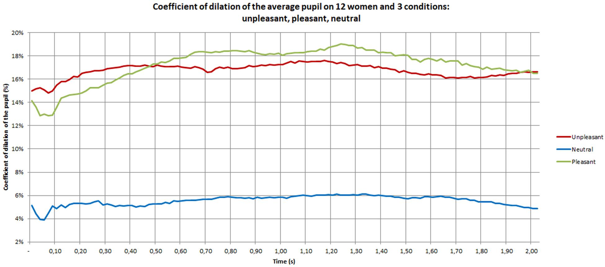
Figure 5. Pupil dilation coefficient of women during the discovery phase.
The pupil dilation coefficients of men are depicted in Figure 6. They range between 2 and 12% for the unpleasant and pleasant conditions, and between 6 and 8% for the neutral condition. Furthermore, the curve presents much more variation than for women. The post hoc analysis showed a difference between unpleasant and pleasant (p = 0.004), unpleasant and neutral (p = 0.009) and pleasant and neutral (p < 0.0001) materials. During the 2 s observed, the neutral curve is nearly constant. The pleasant stimulus shows large oscillation with a first peak at 1 s and a second peak at 1.70 s. The negative stimulus is located above the neutral and below the positive during the first second, and then a sharp decreased occurs that places the negative stimulus below neutral and pleasant conditions. Pleasant stimuli seem to be the most emotional for men.
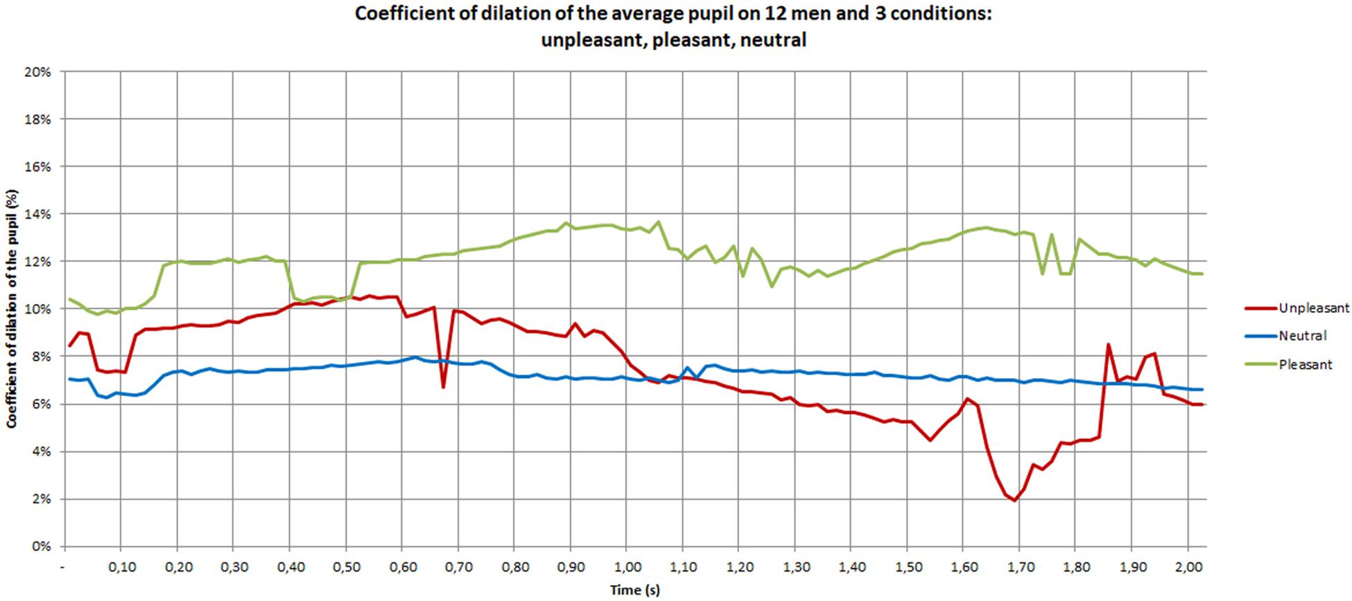
Figure 6. Pupil dilation coefficient of men during the discovery phase.
Figures 7–9 give the pupil dilation of men, women, and averaged, in the case of pleasant, unpleasant, and neutral materials. It can be observed in these figures that:
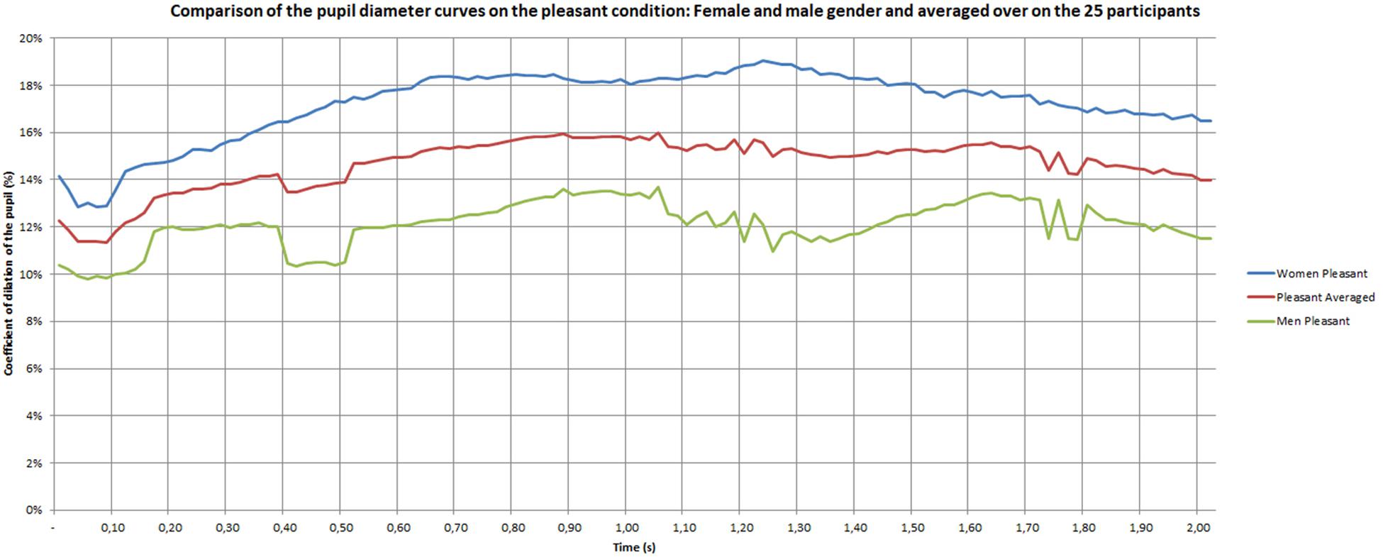
Figure 7. Pupil dilatation coefficient for pleasant materials.
– for the pleasant materials: the pupil dilation is larger for women than for men, with similar time evolutions, but with similar curves slightly noisy in the men case (Figure 7);
– for unpleasant materials: the pupil dilation is greater for women than for men with different time evolutions: nearly constant for women, and decreasing after 0.70 s for men (Figure 8);
– for neutral materials: the pupil dilations are roughly similar for men and women (Figure 9).
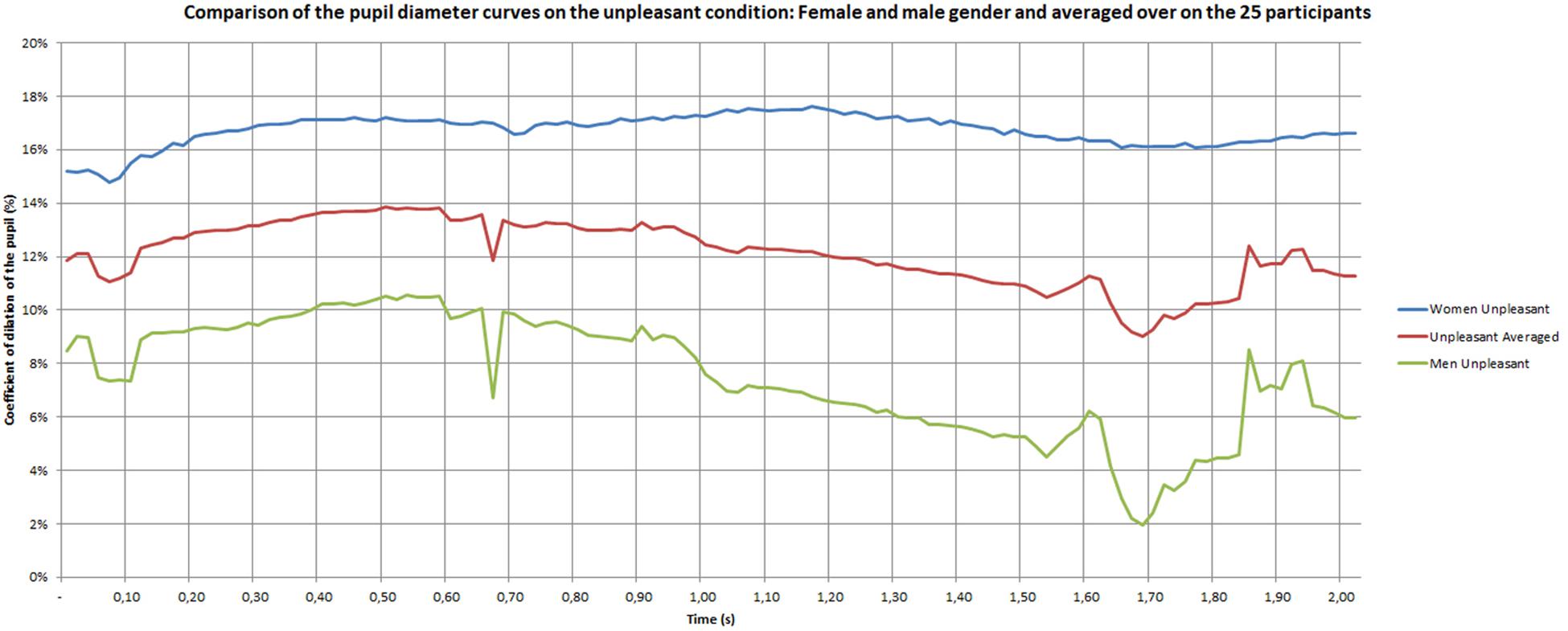
Figure 8. Pupil dilatation coefficient for unpleasant materials.
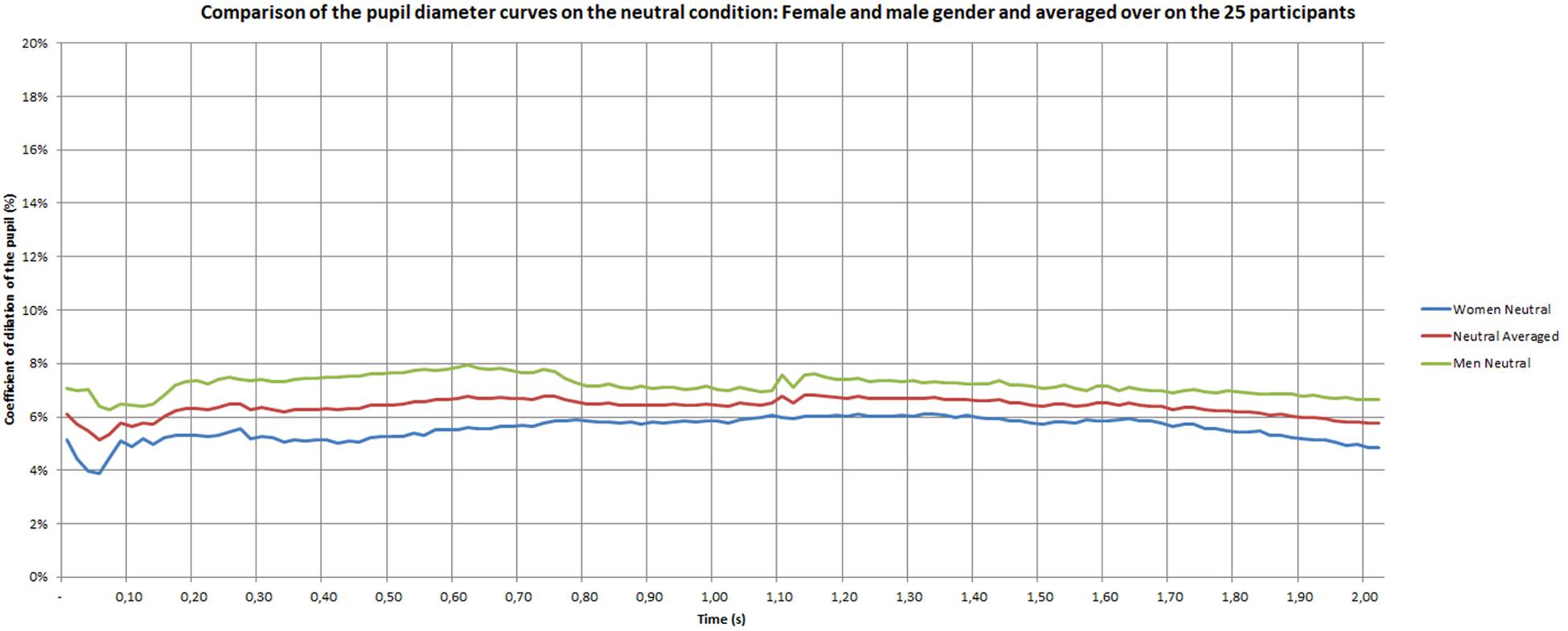
Figure 9. Pupil dilatation coefficient for neutral materials.
In Figure 10, a Principal Component Analysis (PCA) classifies the materials according to the mean value of the pupil dilation. Principal component analysis is a method of eliminating bias. On the horizontal principal axis, which represents 93.10% of the values, the materials are clearly ordered according to their Intensity, from neutral to emotional. This ranking is close to the classification made by the participants using subjective measures. On the vertical axis, which represents 2.56% of the values, the ordering of the materials is more difficult to analyze.
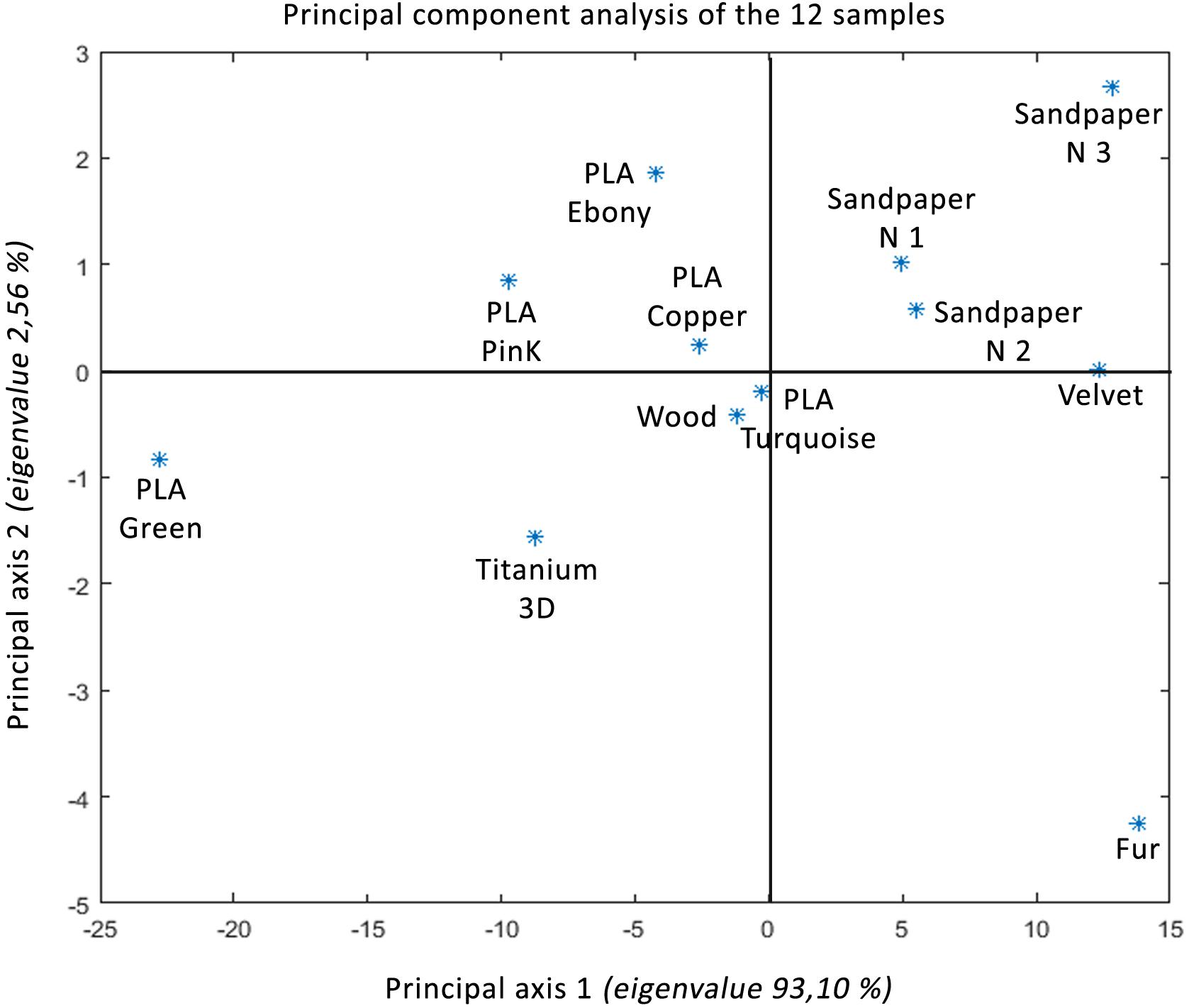
Figure 10. Principal component analyzis of the twelve samples.
Discussion
The aim of this study was to evaluate pupillometry as a measurement technique for emotion. For this purpose, an experimental procedure has been developed, and the touch of different surfaces has been used as a stimulus. The results show clearly an increase in pupil diameter within the first 2 s of the touch. As observed by Bradley et al. (2008), the initial increase is followed at about 0.04 until to 0.10 s by a slow decrease. After, the averaged pupil dilation follows a similar type of curve whatever the emotional materials until a period of about 0.5 s. The general pattern of results indicate a larger pupil size dilation in response to both negative (unpleasant) and positive (pleasant) stimuli, when compared with the dilation induced by neutral stimuli. After this time period of 0.50 s, the emotional response takes different ways. The pupil diameter tends to decrease for negative stimuli, whereas it stays constant for positive stimuli.
Pupil diameter can be considered as a physiological marker of the autonomic nervous system. The size of the pupil is fixed by the relative activity of the two iris muscles, the sphincter and the dilator (Beatty and Lucero-Wagoner, 2000). While pupil constriction is maintained by parasympathetic activity, pupil dilation is essentially induced by the sympathetic pathway (Andreassi, 2000). Because the size of the pupil is modulated by the autonomic nervous system, our results suggest that this system reacts differently to emotional and to neutral stimuli. This pattern of results confirms many studies suggesting that emotional intensity during picture viewing, sound auditioning, or tasting, is associated with high pupil dilation (Partala and Surakka, 2003; Bradley et al., 2008; Kret et al., 2013; Henderson et al., 2014; Lemercier et al., 2014). Bradley et al. (2008) indicated that the dilation of pupil diameter covaried with other autonomic measures of intensity, such as the skin conductance, confirming that pupillary responses can be considered as a reliable index of emotional intensity. More recently, Laeng et al. (2016) showed that emotional music may reveal changes in the diameter pupil and that a neuromodulator role of the central norepinephrine system is involved in this phenomenon. To our best knowledge, nobody has undertaken any studies on the response of the autonomic nervous system to touch.
Some studies indicated that changes in pupil diameter specifically occur within the first few hundred milliseconds after stimulus onset, with responses peaking after 1 to 2 s (Loewenstein and Loewenfeld, 1962; Andreassi, 2000; Beatty and Lucero-Wagoner, 2000; Nieuwenhuis et al., 2011). Beyond 2 s of regulation, we can also suppose larger pupil diameter. For example, Bradley et al. (2008) showed differences when participants viewed emotional or neutral pictures beyond 2 s. Partala and Surakka (2003) observed this effect with auditory stimuli. Our results show a dynamic pattern during the 2 s time period. Initially, unpleasant touch does not induce any difference compared to pleasant touch, but a difference can be observed after 0.50 s. In the same way, Partala and Surakka (2003) results showed that no pupil dilatation was observed before 0.40 s and that the maximum pupil dilatation was observed after 2 to 3 s. In addition, they showed that the time duration of the pupil diameter were somewhat different for female and male subjects. The positive stimuli provoke the strongest pupil dilations for female subjects, whereas the negative stimuli provoked the strongest dilations for male subjects.
As Partala and Surakka (2003), the present study showed an effect of gender but in a different way. The strongest pupil dilations appeared for both emotional conditions with women. This is constant with the duration time. For men, the emotional conditions induced a higher pupil dilations compared to neutral stimuli at the beginning. Nevertheless, a strongest decrease for negative emotion appeared with the time period. The explanation of a higher sensitivity in female participants could be explained by the difference of the skin thickness. It exceeds 60 microns for men (between 20 and 30 years old) and is often below 50 microns for women (Kawabe et al., 1985). For example, Peters et al. (2009) indicate that women can perceive finer surface details than men, and that tactile perception is improved with decreasing finger size (Van Boven et al., 2000; Goldreich and Kanics, 2003, 2006). An explanation of this phenomenon of better perception in women comes from the high density of Merkel’s receptors and Meissner’s corpuscles due to the small size of their fingers (Dillon et al., 2001; Peters et al., 2009). Other differences were found between men and women, like variability of vibrotactile detection thresholds, the volume of the area of contact with the material and a higher density of ridges in the fingerprint (Gutiérrez-Redomero et al., 2011; Venkatesan et al., 2015).
In the pain studies, many authors showed that the threshold of nociception by pressure decreases with age, especially in men. In contrast, age seems to have no effect on thermal sensations (Pickering et al., 2002). Another study showed that the temporal sum of mechanically evoked pain is seen with higher scores in women than in men (Sarlani and Greenspan, 2002). Studies of pupil changes due to painful pressure stimuli showed that increasing pupil diameter was a highly significant indicator of pain intensity. In this domain, female subjects show greater dilation than men at high pressure levels (Ellermeier and Westphal, 1995). These data lead us to the conclusion that the pupil dilation seems differently influenced by pain or emotion following the gender.
From a cognitive approach, Bayer et al. (2011) indicated that pupillary responses are sensitive to both task load and emotional content. Kuchinke et al. (2007) showed that word frequency significantly affected pupil dilation. The pupil diameter appeared to increase for low frequency words certainly because this type of words induced a more important cognitive load. In addition, cultural aspects are important in the touch of materials. This was demonstrated by Fisher (2004) in a study which suggests that consumer perceptions of plastics are physical and emotional. This researcher considers that the properties of plastics are “humanized” by different industrial processes making this material harmless, sensual and familiar unlike the animal or natural properties of some materials who remind us to our natural environment that can arouse more excitement. The appreciation of these materials varies according to the context, the emotional charge produced by its visual and tactile properties (Gibson, 1977) or cultural aspects due to habits. For example, sandpaper is mainly used by men in construction; manufacturing and its surface appearance may seem more familiar to them. Therefore, pupil dilation may reflect the time course of cognitive and attentional mechanisms inherent in emotion processing. Therefore, we can imagine that the pupillometry informs us as an emotional intensity (Rolls et al., 2003; Bradley et al., 2008) and can allow to show a pattern of results, with increase and decrease regulation (Urry et al., 2009), as it is the case in our study.
There is some evidence that self-report emotional answers vary with certain physiological changes associated with emotion (Levenson, 1992). In our study, we show that the materials are subjectively classified in the same order as the classification of the coefficient of dilation observed for the 12 materials. Thus, the main axis 1 seems to correspond to the bodily excitation of the tactile stimulus (Intensity). Nevertheless, the ratings of intensity showed that negative and positive stimuli were experienced as differently arousing. Our pattern of results justifies the use of verbal response in complement with physiological data. On one side, unpleasant touch was assessed as significantly less arousing than pleasant touch. On the other side, the positive intensity effect manifested specifically later during the pupillary response compared to the negative intensity effect. In accordance with subjective ratings, increasing positive emotions led to the most prominent pupil size enlargements during 2 s. These results confirmed that pupil diameter was modulated by the level of emotional intensity. Results suggested that pupil responses reflected the time course of the intensity perceived. According to Gross (1998), emotion regulation refers to both implicit, explicit physiological, behavioral and cognitive processes. The inclusion of multiple measures of autonomic intensity with subjective and behavioral emotional ratings might help to understand effects reflecting changes in emotional Intensity and cognitive demand.
In sum, the present study opens new research avenues. Our data evidence that the pupillary response in accordance with subjective data might include distinct temporal components reflecting emotional and cognitive regulation. It would be interesting to develop pupil diameter variation as a computer signal input. For example, it has been possible to develop signal analysis methods to detect successfully emotion from electrical activity of facial muscles.
Data Availability Statement
The datasets generated for this study are available on request to the corresponding author CBe, Y3lyaWwuYmVydGhlYXV4QGVuaXNlLmZy.
Ethics Statement
Ethical review and approval was not required for the study on human participants in accordance with the local legislation and institutional requirements. The patients/participants provided their written informed consent to participate in this study.
Author Contributions
CBe is doing his thesis at Université de Lyon, ENISE, LTDS, UMR 5513 CNRS on the emotional impact of touch and more particularly in the tactile exploration of materials. This research work aims to determine the emotional value of materials. For this multidisciplinary thesis, a partnership with the laboratories SNA-EPI Laboratory, EA 4607 (DC, pupil and ANS specialist) and CMRR, neuropsychology of the CHU Saint-Étienne (CBo, Thesis guide, specialist in emotion and cognition) was established to provide knowledge in metrology of the autonomic nervous system and in cognition. The rest of the management is made up of engineers researchers (RF, Thesis Director and RT, Thesis Co-director) and statisticians (J-CR, Thesis guide) who have contributed their skills to the various protocols and results collected. All authors contributed to all the steps until the writing of this article. Since 2016, several test protocols, data organization, interpretation, correction, mathematical and statistical treatments have made it possible to propose these results and manuscript revision, read, and approved the submitted version.
Conflict of Interest
The authors declare that the research was conducted in the absence of any commercial or financial relationships that could be construed as a potential conflict of interest.
References
Abdouni, A., Djaghloul, M., Thieulin, C., Vargiolu, R., Pailler-Mattei, C., and Zahouani, H. (2017). Biophysical properties of the human finger for touch comprehension: influences of ageing and gender. J. R. Soc. 4:170321. doi: 10.1098/rsos.170321
Abdouni, A., Vargiolu, R., and Zahouani, H. (2018). Impact of finger biophysical properties on touch gestures and tactile perception: aging and gender effects. Sci. Rep. 8:12605. doi: 10.1038/s41598-018-30677-2
Andreassi, L. J. (2000). “Pupillary response and behavior,” in Psychophysiology: Human Behavior & Physiological Response (New York, NY: Psychology Press), 218–233.
Armaied, E., Vargiolu, R., Bergheau, J. M., and Zahouani, H. (2005). Aging Effect on Tactile Perception: Experimental and Modelling Studies. Saint-Étienne:: Université de Lyon.
Babiker, A., Faye, I., and Malik, A. (2013). “Pupillary behavior in positive and negative emotions,” in Proceedings of the IEEE International Conference on Signal and Image Processing Applications, Melaka, 2013, Melaka.
Baldini, S., Restani, L., Baroncelli, L., Coltelli, M., Franco, R., Cenni, M. C., et al. (2013). Enriched early life experiences reduce adult anxiety-like behavior in rats: a role for insulin-like growth factor 1. J. Neurosci. 33, 11715–11723. doi: 10.1523/JNEUROSCI.3541-12.2013
Barnett, K. (1972). A theoretical construct of the concepts of touch as they relate to nursing. Nurs. Res. 21, 102–110.
Bayer, M., Sommer, W., and Schacht, A. (2011). Emotional words impact the mind but not the body: evidence from pupillary responses. Psychophysiology 48, 1554–1562. doi: 10.1111/j.1469-8986.2011.01219.x
Beatty, J., and Lucero-Wagoner, B. (2000). “The pupillary system,” in Handbook of Psychophysiology, eds J. T. Cacioppo, L. G. Tassinary, and G. G. Berntson, (New York, NY: Cambridge University Press), 142–162.
Bertheaux, C., Rosario, T., Fortunier, R., and Borg, C. (2018). Integration of perception and emotions in a new sensory design process. Inderscience. Int. J. Design Eng. 8:2018.
Bradley, M. M., and Lang, P. J. (1994). Measuring emotion: the self-assessment manikin and the semantic differential. J. Behav. Ther. Exp. Psychiatry 25, 49–59. doi: 10.1016/0005-7916(94)90063-9
Bradley, M. M., Miccoli, L., Escrig, M. A., and Lang, P. J. (2008). The pupil as a measure of emotional Intensity and autonomic activation. J. Psychophysiol. 45, 602–607. doi: 10.1111/j.1469-8986.2008.00654.x
Brauer, J., Xiao, Y., Poulain, T., Friederici, A. D., and Schirmer, A. (2016). Frequency of maternal touch predicts resting activity and connectivity of the developing social brain. Cereb. Cortex 26, 3544–3552. doi: 10.1093/cercor/bhw137
Bush, E. (2001). The use of human touch to improve the well-being of older adults: a holistic nursing intervention. J. Holistic Nurs. 19, 256–270. doi: 10.1177/089801010101900306
Cardello, A. V., Winterhalter, C., and Schutz, H. G. (2003). Predicting the handle and comfort of military clothing fabrics from sensory and instrumental data: development and application of new psychophysical methods. Textile Res. J. 73, 221–237. doi: 10.1177/004051750307300306
Chaplan, S. R., Bach, F. W., Pogrel, J. W., Chung, J. M., and Yaksh, T. L. (1994). Quantitative assessment of tactile allodynia in the rat paw. J. Neurosci. Methods 53, 55–63. doi: 10.1016/0165-0270(94)90144-9
Charier, D., Bertheaux, C., Zantour, D., Roche, F., and Molliex, S. (2017). Intérêt de la pupillométrie pour l’évaluation de la douleur. Douleurs 18, 213–222.
Chen, X., Barnes, C. J., Childs, T. H. C., Henson, B., and Shao, F. (2009). Materials’ tactile testing and characterisation for consumer products’ affective packaging design. Materials Design 30, 4299–4310. doi: 10.1016/j.matdes.2009.04.021
Clermont, A. D. (2018). Le toucher est-il le media par lequel l’ostéopathie a une action hormonale?.
Conover, W. J., and Iman, R. L. (1981). Rank transformations as a bridge between parametric and nonparametric statistics. Am. Statist. 35, 124–129. doi: 10.1080/00031305.1981.10479327
Datta, B. C. (2016). Emotive Materials: Towards a Shared Language of the Meaning of Materials. Doctoral dissertation, Cambridge, MA: Massachusetts Institute of Technology.
Dietz, J., Bradley, M. M., Okun, M. S., and Bowers, D. (2011). Emotion and ocular responses in Parkinson’s disease. Neuropsychologia 49, 3247–3253. doi: 10.1016/j.neuropsychologia.2011.07.029
Dillon, Y. K., Haynes, J., and Henneberg, M. (2001). The relationship of the number of Meissner’s corpuscles to dermatoglyphic characters and finger size. J. Anat. 199, 577–584. doi: 10.1046/j.1469-7580.2001.19950577.x
Djouhri, L. (2016). Electrophysiological evidence for the existence of a rare population of C-fiber low threshold mechanoreceptive (C-LTM) neurons in glabrous skin of the rat hindpaw. Neurosci. Lett. 613, 25–29. doi: 10.1016/j.neulet.2015.12.040
Dolinski, D. (2010). Touch, compliance, and homophobia. J. Nonverb. Behav. 34, 179–192. doi: 10.1007/s10919-010-0090-1
Dunbar, R. I. (2010). The social role of touch in humans and primates: behavioural function and neurobiological mechanisms. Neurosci. Biobehav. Rev. 34, 260–268. doi: 10.1016/j.neubiorev.2008.07.001
Edin, B. B. (2001). Cutaneous afferents provide information about knee joint movements in humans. J. Physiol. 531, 289–297. doi: 10.1111/j.1469-7793.2001.0289j.x
Ellermeier, W., and Westphal, W. (1995). Gender differences in pain ratings and pupil reactions to painful pressure stimuli. Pain 61, 435–439. doi: 10.1016/0304-3959(94)00203-q
Ellis, C. J. (1981). The pupillary light reflex in normal subjects. Br. J. Ophthalmol. 65, 754–759. doi: 10.1136/bjo.65.11.754
Essick, G. K., James, A., and McGlone, F. P. (1999). Psychophysical assessment of the affective components of non-painful touch. Neuroreport 10, 2083–2087. doi: 10.1097/00001756-199907130-00017
Essick, G. K., McGlone, F., Dancer, C., Fabricant, D., Ragin, Y., Phillips, N., et al. (2010). Quantitative assessment of pleasant touch. Neurosci. Biobehav. Rev. 34, 192–203. doi: 10.1002/ejp.1391
Feldman, R., Gordon, I., and Zagoory-Sharon, O. (2010). The cross-generation transmission of oxytocin in humans. Horm. Behav. 58, 669–676. doi: 10.1016/j.yhbeh.2010.06.005
Field, T. M. (1998). Touch therapy effects on development. Int. J. Behav. Dev. 22, 779–797. doi: 10.1080/016502598384162
Fisher, T. H. (2004). What we touch, touches us: materials, affects, and affordances. Design Issues 20, 20–31. doi: 10.1162/0747936042312066
Forget, R., and Lamarre, Y. (1987). Rapid elbow flexion in the absence of proprioceptive and cutaneous feedback. Hum. Neurobiol. 6, 27–37.
Francis, S., Rolls, E. T., Bowtell, R., McGlone, F., O’Doherty, J., Browning, A., et al. (1999). The representation of pleasant touch in thebrain and its relationship with taste and olfactory areas. Neuroreport 10, 453–459. doi: 10.1097/00001756-199902250-00003
Gazzola, V., Spezio, M. L., Etzel, J. A., Castelli, F., Adolphs, R., and Keysers, C. (2012). Primary somatosensory cortex discriminates affective signicance in social touch. Proc. Natl. Acad. Sci. U.S.A. 109, E1657–E1666. doi: 10.1073/pnas.1113211109
Goldreich, D., and Kanics, I. M. (2003). Tactile acuity is enhanced in blindness. J. Neurosci. 23, 3439–3445. doi: 10.1523/jneurosci.23-08-03439.2003
Goldreich, D., and Kanics, I. M. (2006). Performance of blind and sighted humans on a tactile grating detection task. Percept. Psychophys. 68, 1363–1371. doi: 10.3758/bf03193735
Greenspan, J. D., and McGillis, S. L. (1991). Stimulus features relevant to the perception of sharpness and mechanically evoked cutaneous pain. Somatosensory Motor Res. 8, 137–147. doi: 10.3109/08990229109144738
Gross, J. J. (1998). The emerging field of emotion regulation: an integrative review. Rev. Gen. Psychol. 2, 271–299. doi: 10.1037//1089-2680.2.3.271
Guest, S., Dessirier, J. M., Mehrabyan, A., McGlone, F., Essick, G., Gescheider, G., et al. (2011). The development and validation of sensory and emotional scales of touch perception. Attent. Percept. Psychophys. 73, 531–550. doi: 10.3758/s13414-010-0037-y
Gutiérrez-Redomero, E., Alonso, M. C., and Dipierri, J. E. (2011). Sex differences in fingerprint ridge density in the Mataco-Mataguayo population. Homo 62, 487–499. doi: 10.1016/j.jchb.2011.05.001
Guzzetta, A., Baldini, S., Bancale, A., Baroncelli, L., Ciucci, F., Ghirri, P., et al. (2009). Massage accelerates brain development and the maturation of visual function. J. Neurosci. 29, 6042–6051. doi: 10.1523/JNEUROSCI.5548-08.2009
Hasan, Z. (1992). Role of proprioceptors in neural control. Curr. Opin. Neurobiol. 2, 824–829. doi: 10.1016/0959-4388(92)90140-g
Henderson, R. R., Bradley, M. M., and Lang, P. (2014). Modulation of the initial light reflex during affective pictureviewing. Psychophysiology 5, 815–818. doi: 10.1111/psyp.12236
Hess, E. H., and Polt, J. M. (1960). James. pupil size as related to interest value of visual stimuli. Science 132, 349–350. doi: 10.1126/science.132.3423.349
Jones, L. A., and Lederman, S. J. (2006). “Tactile sensing,” in Human Hand Function (Cambridge: Oxford University Press), 44–74.
Kaffman, A., and Meaney, M. J. (2007). Neurodevelopmental sequelae of postnatal maternal care in rodents: clinical and research implications of molecular insights. J. Child Psychol. Psychiatr. 48, 224–244. doi: 10.1111/j.1469-7610.2007.01730.x
Kawabe, T. T., Maccallum, D. K., and Lillie, J. H. (1985). Variation in basement membrane topography in human thick skin. Anat. Rec. 211, 142–148. doi: 10.1002/ar.1092110205
Kreibig, S. D. (2010). Autonomic nervous system activity in emotion: a review. Biol. Psychol. 84, 394–421. doi: 10.1016/j.biopsycho.2010.03.010
Kret, M. E., Roelobs, E., and Stekelenburg, J. J. (2013). Emotional signals from faces, bodies and scenes influence observers’ face expressions, fixations and pupil-size. Front. Hum. Neurosci. 7:810. doi: 10.3389/fnhum.2013.00810
Kuchinke, L., Võ, M. L. H., Hofmann, M., and Jacobs, A. M. (2007). Pupillary responses during lexical decisions vary with word frequency but not emotional valence. Int. J. Psychophysiol. 65, 132–140. doi: 10.1016/j.ijpsycho.2007.04.004
Laeng, B., Eidet, L. M., Sulutvedt, U., and Panksepp, J. (2016). Music chills: the eye pupil as a mirror to music’s soul. Conscious. Cogn. 44, 161–178. doi: 10.1016/j.concog.2016.07.009
Lang, P. J., Bradley, M. M., and Cuthbert, B. N. (2005). Technical Report A-6. Gainesville. FL: University of Florida.
Lederman, S. J., and Klatzky, R. L. (1987). Hand movements: a window into haptic object recognition. Cogn. Psychol. 19, 342–368. doi: 10.1016/0010-0285(87)90008-9
Leem, J. W., Willis, W. D., and Chung, J. M. (1993). Cutaneous sensory receptors in the rat foot. J. Neurophysiol. 69, 1684–1699. doi: 10.1152/jn.1993.69.5.1684
Lemercier, A., Guillot, G., Courcoux, P., Garrel, C., Baccino, T., and Schlich, P. (2014). Pupillometry of taste: methodological guide - from acquisition to data processing - and toolbox for MATLAB. Q. Methods Psychol. 10, 179–195. doi: 10.20982/tqmp.10.2.p179
Levenson, R. W. (1992). Autonomic nervous system differences among emotions. Psychol. Sci. 3, 23–27. doi: 10.1111/j.1467-9280.1992.tb00251.x
Li, L., and Ginty, D. D. (2014). The structure and organization of lanceolate mechanosensory complexes at mouse hair follicles. eLife 3:e01901. doi: 10.7554/eLife.01901
Liljencrantz, J., and Olausson, H. (2014). Tactile C fibers and their contributions to pleasant sensations and to tactile allodynia. Front. Behav. Neurosci. 8:37. doi: 10.3389/fnbeh.2014.00037
Lindgren, L., Rundgren, S., Winsö, O., Lehtipalo, S., Wiklund, U., Karlsson, M., et al. (2010). Physiological responses to touch massage in healthy volunteers. Auton. Neurosci. 158, 105–110. doi: 10.1016/j.autneu.2010.06.011
Loewenstein, O., and Loewenfeld, I. E. (1962). “The pupil,” in The Eye. Muscular mechanisms, ed. H. Davson, (New York, NY: Academic Press).
Löken, L. S., and Olausson, H. (2010). The skin as a social organ. Exp. Brain Res. 204, 305–314. doi: 10.1007/s00221-009-2007-y
McGlone, F., and Reilly, D. (2010). The cutaneous sensory system. Neurosci. Biobehav. Rev. 34, 148–159. doi: 10.1016/j.neubiorev.2009.08.004
Melzack, R. (1975). The McGill pain questionnaire: major properties and scoring methods. Pain 1, 277–299. doi: 10.1016/0304-3959(75)90044-5
Menard, J. L., Champagne, D. L., and Meaney, M. J. P. (2004). Variations of maternal care differentially influence ‘fear’reactivity and regional patterns of cFos immunoreactivity in response to the shock-probe burying test. Neuroscience 129, 297–308. doi: 10.1016/j.neuroscience.2004.08.009
Misery, L. (2014). Le prurit: des avancées physiopathologiques considérables. Méd Sci. 30, 1123–1128. doi: 10.1051/medsci/20143012015
Moayedi, Y., Nakatani, M., and Lumpkin, E. (2016). “Mammalian mechanoreception,” in Scholarpedia of Touch, (Paris: Atlantis Press), 423–435. doi: 10.2991/978-94-6239-133-8_35
Mogg, K., and Bradley, B. P. (1999). Some methodological issues in assessing attentional biases for threatening faces in anxiety: a replication study using a modified version of the probe detection task. Behav. Res. Ther. 37, 595–604. doi: 10.1016/s0005-7967(98)00158-2
Mogg, K., Millar, N., and Bradley, B. P. (2000). Biases in eye movements to threatening facial expressions in generalized anxiety disorder and depressive disorder. J. Abnorm. Psychol. 109:695. doi: 10.1037/0021-843x.109.4.695
Mountcastle, V. B. (2005). The Sensory Hand: Neural Mechanisms of Somatic Sensation. Cambridge, MA: Harvard University Press.
Nagi, S. S., and Mahns, D. A. (2010). “A novel class of unmyelinated (C) tactile afferents in human glabrous skin,” in Proceedings of the Abstracts of the 40th Annual Meeting of the Society for Neuroscience: 13-17 November 2010, San Diego, CA.
Nagi, S. S., and Mahns, D. A. (2013). Mechanical allodynia in human glabrous skin mediated by low-threshold cutaneous mechanoreceptors with unmyelinated fibres. Exp. Brain Res. 231, 139–151. doi: 10.1007/s00221-013-3677-z
Nieuwenhuis, S., De Geus, E. J., and Aston-Jones, G. (2011). The anatomical and functional relationship between the P3 and autonomic components of the orienting response. Psychophysiology 48, 162–175. doi: 10.1111/j.1469-8986.2010.01057.x
Nordin, M. (1990). Low-threshold mechanoreceptive and nociceptive units with unmyelinated (C) fibres in the human supraorbital nerve. J. Physiol. 426, 229–240. doi: 10.1113/jphysiol.1990.sp018135
Öhman, A., Flykt, A., and Esteves, F. (2001). Emotion drives attention: detecting the snake in the grass, karolinska institute. J. Exp. Psychol. 130, 466–478. doi: 10.1037//0096-3445.130.3.466
Okamoto, S., Nagano, H., and Yamada, Y. (2012). Psychophysical dimensions of tactile perception of textures. IEEE Trans. Haptics 6, 81–93. doi: 10.1109/TOH.2012.32
Olausson, H., Lamarre, Y., Backlund, H., Morin, C., Wallin, B. G., Starck, G., et al. (2002). Unmyelinated tactile afferents signal touch and project to insular cortex. Nat. Neurosci. 5:900. doi: 10.1038/nn896
Olausson, H., Wessberg, J., Morrison, I., Mc Glone, F., and Vallbo, A. (2010). The neurophysiology of unmyelinated tactile afferents. Neurosci. Biobehav. Rev. 34, 185–191. doi: 10.1016/j.neubiorev.2008.09.011
Oldfield, R. C. (1971). The assessment and analysis of handedness: the edinburgh inventory. Neuropsychologia 9, 97–113. doi: 10.1016/0028-3932(71)90067-4
Onorati, F., Barbieri, R., Mauri, M., Russo, V., and Mainardi, L. (2013). Characterization of affective states by pupillary dynamics and autonomic correlates. Front. Hum. Neurosci. 6:9. doi: 10.3389/fneng.2013.00009
Osgood, C. E. (1969). On the whys and wherefores of E, P, and A. J. Pers. Soc. Psychol. 12, 194–199.
Park, G., and Thayer, J. F. (2014). From the heart to the mind: cardiac vagal tone modulates top-down and bottom-up visual perception and attention to emotional stimuli. Front. Psychol. 5:278. doi: 10.3389/fpsyg.2014.00278
Partala, T., and Surakka, V. (2003). Pupil size variation as an indication of affective processing. Int. J. Hum. Comp. Stud. 59, 185–198. doi: 10.1016/s1071-5819(03)00017-x
Peters, R. M., Hackeman, E., and Goldreich, D. (2009). Diminutive digits discern delicate details: fingertip size and the sex difference in tactile spatial acuity. J. Neurosci. 29, 15756–15761. doi: 10.1523/JNEUROSCI.3684-09.2009
Pickering, G., Jourdan, D., Eschalier, A., and Dubray, C. (2002). Impact of age, gender and cognitive functioning on pain perception. Gerontology 48, 112–118. doi: 10.1159/000048937
Rolls, E. T., and Kesner, R. P. A. (2006). Computational theory of hippocampal function, and empirical tests of the theory. Prog. Neurobiol. 79, 1–48. doi: 10.1016/j.pneurobio.2006.04.005
Rolls, E. T., O’Doherty, J., Kringelbach, M. L., Francis, S., Bowtell, R., and McGlone, F. (2003). Representations of pleasant and painful touch in the human orbitofrontal and cingulate cortices. Cereb. Cortex 13, 308–317. doi: 10.1093/cercor/13.3.308
Russell, J. A. (1980). A circumplex model of affect. J. Pers. Soc. Psychol. 39, 1161–1178. doi: 10.1037/h0077714
Rutlin, M., Ho, C. Y., Abraira, V. E., Cassidy, C., Bai, L., Woodbury, C. J., et al. (2014). The cellular and molecular basis of direction selectivity of Aδ-LTMRs. Cell 159, 1640–1651. doi: 10.1016/j.cell.2014.11.038
Sale, A., Putignano, E., Cancedda, L., Landi, S., Cirulli, F., Berardi, N., et al. (2004). Enriched environment and acceleration of visual system development. Neuropharmacology 47, 649–660. doi: 10.1016/j.neuropharm.2004.07.008
Sarlani, E., and Greenspan, J. D. (2002). Gender differences in temporal summation of mechanically evoked pain. Pain 97, 163–169. doi: 10.1016/s0304-3959(02)00015-5
Schirmer, A., and Adolphs, R. (2017). Emotion perception from face, voice, and touch: comparisons and convergence. Trends Cogn. Sci. 21, 216–228. doi: 10.1016/j.tics.2017.01.001
Schlomer, G. L., Bauman, S., and Card, N. A. (2010). Best practices for missing data management in counseling psychology. J. Counsel. Psychol. 57:1. doi: 10.1037/a0018082
Servais, V. (2007). La relation homme-animal. La relation à l’animal peut-elle devenir significative, donc thérapeutique dans le traitement des maladies psychiques. Enfances Psy 35, 46–57.
Spapé, M. M., Hoggan, E. E., Jacucci, G., and Ravaja, N. (2015). The meaning of the virtual Midas touch: an ERP study in economic decision making. Psychophysiology 52, 378–387. doi: 10.1111/psyp.12361
Spitz, R. A. (1945). Hospitalism: an inquiry into the genesis of psychiatric conditions in early childhood. Psychoanal. Study Child 1, 53–74. doi: 10.1080/00797308.1945.11823126
Stack, D. M., and Muir, D. W. (1990). Tactile stimulation as a component of social interchange: new interpretations for the still-face effect. Br. J. Dev. Psychol. 8, 131–145. doi: 10.1111/j.2044-835x.1990.tb00828.x
Stolarova, M., Keil, A., and Moratti, S. (2006). Modulation of the C1 visual event-related component by conditioned stimuli: evidence for sensory plasticity in early affective perception. Cerebral 16, 876–887. doi: 10.1093/cercor/bhj031
Suvilehto, J. T., Glerean, E., Dunbar, R. I. M., Hari, R., and Nummenmaa, L. (2015). Topography of social touching depends on emotional bonds between humans. Proc. Natl. Acad. Sci. U.S.A. 112, 13811–13816. doi: 10.1073/pnas.1519231112
Taylor, M. M., and Lederman, S. J. (1975). Tactile roughness of grooved surfaces: a model and the effect of friction. Percept. Psychophys. 17, 23–36. doi: 10.3758/bf03203993
Thieulin, C. (2017). Caractérisation Des Mécanismes D’endommagement du Stratum Corneum Induit par le Frottement Répété de Papiers en Cellulose. Doctoral dissertation, Lyon: Université de Lyon.
Urry, H. L., van Reekum, C. M., Johnstone, T., and Davidson, R. J. (2009). Individual differences in some (but not all) medial prefrontal regions reflect cognitive demand while regulating unpleasant emotion. Neuroimage 47, 852–863. doi: 10.1016/j.neuroimage.2009.05.069
Vallbo, A., Olausson, H., Wessberg, J., and Norrsell, U. (1993). A system of unmyelinated afferents for innocuous mechanoreception in the human skin. Brain Res. 628, 301–304. doi: 10.1016/0006-8993(93)90968-s
Vallbo, ÅB., Olausson, H., and Wessberg, J. (1999). Unmyelinated afferents constitute a second system coding tactile stimuli of the human hairy skin. J. Neurophysiol. 81, 2753–2763. doi: 10.1152/jn.1999.81.6.2753
Van Boven, R. W., Hamilton, R. H., Kauffman, T., Keenan, J. P., and Pascual-Leone, A. (2000). Tactile spatial resolution in blind Braille readers. Neurology 54, 2230–2236. doi: 10.1212/wnl.54.12.2230
Venkatesan, L., Barlow, S. M., and Kieweg, D. (2015). Age-and sex-related changes in vibrotactile sensitivity of hand and face in neurotypical adults. Somatosens. Motor Res. 32, 44–50. doi: 10.3109/08990220.2014.958216
Von Frey, M. (1896). Untersuchungen uber die Sinnesfunktionen der menschlichen Haut. Abh Sachs Ges 40, 175–266.
Vrontou, S., Wong, A. M., Rau, K. K., Koerber, H. R., and Anderson, D. J. (2013). Genetic identification of C fibres that detect massage-like stroking of hairy skin in vivo. Nature 493:669. doi: 10.1038/nature11810
Wall, S. A. (2000). An Investigation of Temporal and Spatial Limitations of Haptic Devices. Doctoral dissertation, Reading, BRK: University of Reading.
Walsh, C. M., Bautista, D. M., and Lumpkin, E. A. (2015). Mammalian touch catches up. Curr. Opin. Neurobiol. 34, 133–139. doi: 10.1016/j.conb.2015.05.003
Zahouani, H., Mezghani, S., Vargiolu, R., Hoc, T., and Mansori, M. E. (2013). Effect of roughness on vibration of human finger during a friction test. Wear 301, 343–352. doi: 10.1016/j.wear.2012.11.028
Zigmond, A. S., and Snaith, R. P. (1983). The hospital anxiety and depression scale. Acta Psychiatr. Scand. 67, 361–370.
Keywords: touch, emotion, material, dilation of the pupil, explicit and implicit measures
Citation: Bertheaux C, Toscano R, Fortunier R, Roux J-C, Charier D and Borg C (2020) Emotion Measurements Through the Touch of Materials Surfaces. Front. Hum. Neurosci. 13:455. doi: 10.3389/fnhum.2019.00455
Received: 18 May 2019; Accepted: 12 December 2019;
Published: 17 January 2020.
Edited by:
Micah M. Murray, Université de Lausanne, SwitzerlandReviewed by:
Nicola Cellini, University of Padova, ItalyMonica Gori, Italian Institute of Technology (IIT), Italy
Copyright © 2020 Bertheaux, Toscano, Fortunier, Roux, Charier and Borg. This is an open-access article distributed under the terms of the Creative Commons Attribution License (CC BY). The use, distribution or reproduction in other forums is permitted, provided the original author(s) and the copyright owner(s) are credited and that the original publication in this journal is cited, in accordance with accepted academic practice. No use, distribution or reproduction is permitted which does not comply with these terms.
*Correspondence: Cyril Bertheaux, Y3lyaWwuYmVydGhlYXV4QGVuaXNlLmZy