- 1Center for Translational Medicine, The First Affiliated Hospital, Sun Yat-sen University, Guangzhou, China
- 2Department of Pathology and The Vascular Biology and Therapeutics Program, Yale University School of Medicine, New Haven, CT, United States
- 3Institute of Genetics, Institute of Genetics and Regenerative Biology, College of Life Sciences, Zhejiang University, Hangzhou, China
- 4Department of Laboratory Medicine, Nanjing Drum Tower Hospital, Nanjing University Medical School, Nanjing, China
A Correction on
Endothelial AIP1 regulates vascular remodeling by suppressing NADPH Oxidase-2
by Zhang J, Chen C, Li L, Zhou HJ, Li F, Zhang H, Yu L, Chen Y and Min W (2018). Front. Physiol. 9:396. doi: 10.3389/fphys.2018.00396
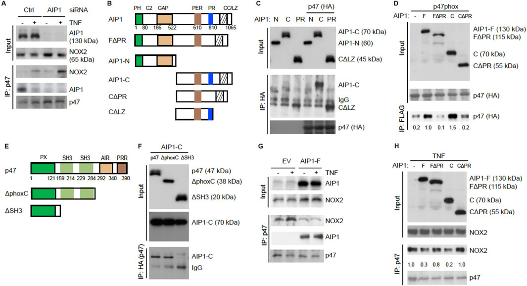
There was a mistake in Figure 5H as published. The NOX2 blot in the IP:p47 bracket was misplaced. A revised Figure 5 with the corrected Figure 5H appears below.

Figure 5. AIP1 blocks NOX2 activity in EC by disrupting the formation of an active NOX2 complex. (A) Association of AIP1 with p47phox. HAEC were transfected with control siRNA or AIP1 siRNA. Twenty four hour post-transfection, cells were either left untreated or treated with TNFα (10 ng/ml) for 15 min. AIP1-p47phox and NOX2-p47phox complexes were determined by a co-immunoprecipitation assay followed by Western blot as indicated. (B) Schematic diagram for AIP1 structural domains and expression constructs. PH, PH domain; C2, PKC conserved domain; GAP, GTPase-activating protein domain; PER, period-like domain; PRR, proline-rich region; CC/LZ, coiled coil/leucine-zipper domain. Various AIP1 N-terminal truncates (F1PH and AIP1-N) and C-terminal truncates (AIP1-C; C-PR, C-1PR) constructed with a Flag-tagged at the N-terminus are shown. (C,D) AIP1 via its PRR binds to p47phox. Various AIP1 truncates were co-transfected with HA-tagged p47phox into HAEC cells as indicated. Associations of AIP1 truncates with p47phox were determined by co-immunoprecipitation with anti-HA (for p47phox) followed by Western blot with anti-Flag (for AIP1 truncates). Immunoprecipitated AIP1 truncates are indicated. (E) Schematic diagram for the p47phox structural domains and expression constructs. PX, phosphoinositide-binding structural domain; SH3, Src homology 3 domain that binding to proline-rich region (PRR); AIR, autoinhibitory region; PPR, proline-rich region (PRR). 1phoxC: a mutant with the deletion of both AIR and PPR; 1SH3: a mutant with the deletion of the two SH3 domains. (F) p47phox via the SH domains bind to AIP1. HA-tagged p47phox truncates were co-transfected with FLAG-tagged AIP1-C into HAEC as indicated. Associations of p47phox truncates with AIP1-C were determined by co-immunoprecipitation with anti-HA (p47phox) followed by Western blot with anti-Flag (AIP1-C). AIP1-C and IgG in the immunoprecipitation are indicated. (G,H) AIP1 prevents/disrupts NOX2-p47phox complex formation. HAEC were infected with lentivirus with empty vector (EV), AIP1-F (E) and with a truncate (F), and cells were left untreated or treated with TNFα (10 ng/ml for 15 min). Cell lysates were subjected to co-immunoprecipitation assays with anti-p47phox followed by Western blot with anti-NOX2. All experiments were repeated three times.
The original article has been updated.
Publisher’s note
All claims expressed in this article are solely those of the authors and do not necessarily represent those of their affiliated organizations, or those of the publisher, the editors and the reviewers. Any product that may be evaluated in this article, or claim that may be made by its manufacturer, is not guaranteed or endorsed by the publisher.
Keywords: AIP1, NOX2, reactive oxygen species, vascular remodeling, neointimal hyperplasia
Citation: Zhang J, Chen C, Li L, Zhou HJ, Li F, Zhang H, Yu L, Chen Y and Min W (2025) Correction: Endothelial AIP1 regulates vascular remodeling by suppressing NADPH Oxidase-2. Front. Physiol. 16:1641354. doi: 10.3389/fphys.2025.1641354
Received: 04 June 2025; Accepted: 22 July 2025;
Published: 04 August 2025.
Edited and reviewed by:
Geoffrey A. Head, Baker Heart and Diabetes Institute, AustraliaCopyright © 2025 Zhang, Chen, Li, Zhou, Li, Zhang, Yu, Chen and Min. This is an open-access article distributed under the terms of the Creative Commons Attribution License (CC BY). The use, distribution or reproduction in other forums is permitted, provided the original author(s) and the copyright owner(s) are credited and that the original publication in this journal is cited, in accordance with accepted academic practice. No use, distribution or reproduction is permitted which does not comply with these terms.
*Correspondence: Yuxin Chen, eXV4aW5fY2hlbjIwMTVAMTYzLmNvbQ==; Wang Min, d2FuZy5taW5AeWFsZS5lZHU=
†These authors have contributed equally to this work
 Jiqin Zhang1,2†
 Chaofei Chen
 Fenghe Li
 Luyang Yu
 Yuxin Chen
 Wang Min