 Laena Pernomian
 Vanessa de Fátima Borges
 Gerson Jhonatan Rodrigues
 Cristina Espinosa-Diez
- 1Cardiovascular Translational Research Center, Department of Cell Biology and Anatomy, School of Medicine, University of South Carolina, Columbia, SC, United States
- 2Cedars Sinai Medical Center, Los Angeles, CA, United States
- 3Federal University of São Carlos, SãoCarlos, São Paulo, Brazil
- 4Center for Molecular Medicine and Genetics, School of Medicine, Wayne State University Detroit, Detroit, MI, United States
Editorial on the Research Topic
Phenotypic transitions and endothelial dysfunction in cardiovascular diseases: mechanisms, therapeutic targets, and modulation
The vascular system is essential for organ function and tissue homeostasis, with endothelial cells regulating vascular tone, permeability, angiogenesis, and blood interactions. Disruption of this balance causes endothelial dysfunction, a central driver of cardiovascular and metabolic diseases. This is aggravated by maladaptive phenotypic transitions, particularly endothelial-to-mesenchymal transition (EndMT), which, though beneficial in repair, promotes fibrosis, remodeling, and plaque instability when dysregulated, contributing to atherosclerosis, diabetes, and hypertension (Kovacic et al., 2019). Understanding these transitions remains a challenge. This Research Topic explores the interplay between endothelial dysfunction and vascular cell phenotypes in disease progression (Figure 1).
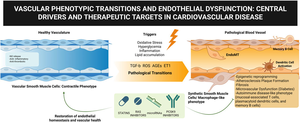
Figure 1. Vascular phenotypic transitions and endothelial dysfunction. Created with Biorender.com.
Within the context of atherosclerosis, endothelial dysfunction represents an initiating and central event, as highlighted by Yang et al., who offer an integrative perspective bridging traditional pharmacotherapy with emerging novel therapeutic approaches. Their review emphasizes how endothelial and vascular cell phenotypic changes drive the disease process, through EndMT-derived fibroblast accumulation (Brokopp et al., 2011), extracellular matrix deposition and inflammation (Chen et al., 2015), and vascular smooth muscle cell (VSMC) switching (Chappell et al., 2016). Yang et al. illustrate how targeting maladaptive cell states through statins, renin-angiotensin system inhibitors, microRNAs, and reprogramming strategies may represent a paradigm shift for long-term vascular health.
A similar emphasis on endothelial dysfunction is evident in the context of diabetes. Here, Liu et al. expand on the multifactorial pathways, including oxidative stress, insulin resistance, and chronic hyperglycemia, that converge to impair endothelial cell function (Shah and Brownlee, 2016). By showing how these stressors reduce nitric oxide bioavailability, disrupt intercellular junctions, and trigger epigenetic modifications, their review underscores the endothelial cell as the primary target of diabetic vascular damage. Importantly, they also highlight how therapeutic strategies, including compounds from traditional Chinese medicine, may help preserve or restore endothelial function, positioning these cells as central targets for reducing cardiovascular risk in diabetes.
The contribution by Song et al. further extends these insights into the domain of plaque regression, challenging the notion of atherosclerosis as an irreversible condition. Their review emphasizes that regression is not simply the reversal of plaque buildup but rather a coordinated process involving lipid lowering, endothelial repair, and vascular cell reprogramming. Endothelial progenitor cells emerge as key agents of vascular repair, while VSMC phenotypic plasticity is highlighted as both a pathological driver and a therapeutic opportunity. By introducing conceptual parallels with oncology, such as targeting genomic instability, Song et al. propose innovative avenues for reshaping the therapeutic landscape of cardiovascular disease.
Beyond these vascular conditions, the clinical study by Jaatinen et al. explores ischemia with non-obstructive coronary arteries (INOCA), where endothelial dysfunction manifests in the coronary microvasculature. Their findings of altered immune responses point to a possible autoimmune contribution to microvascular dysfunction in INOCA. This aligns with prior evidence linking endothelial dysfunction and immune dysregulation in autoimmune diseases (Moschetti et al., 2022; Cecere et al., 2024), further reinforcing the idea that vascular pathology emerges from a convergence of endothelial injury, phenotypic transitions, and immune activation.
These studies illustrate the role of endothelial dysfunction and vascular cell phenotypic transitions in a wide spectrum of cardiovascular diseases. Endothelial cells, along with phenotypically plastic of vascular cells, constitute both key drivers of vascular injury and promising therapeutic targets. These findings highlight the importance of moving beyond symptom control to strategies that restore vascular homeostasis by correcting dysfunctional cellular states, thereby opening the door to transformative therapies for cardiovascular diseases.
Author contributions
LP: Conceptualization, Writing – original draft, Writing – review and editing. VB: Writing – review and editing. GR: Writing – review and editing. CE-D: Writing – review and editing.
Funding
The author(s) declare that no financial support was received for the research and/or publication of this article.
Conflict of interest
The authors declare that the research was conducted in the absence of any commercial or financial relationships that could be construed as a potential conflict of interest.
Generative AI statement
The author(s) declare that no Generative AI was used in the creation of this manuscript.
Any alternative text (alt text) provided alongside figures in this article has been generated by Frontiers with the support of artificial intelligence and reasonable efforts have been made to ensure accuracy, including review by the authors wherever possible. If you identify any issues, please contact us.
Publisher’s note
All claims expressed in this article are solely those of the authors and do not necessarily represent those of their affiliated organizations, or those of the publisher, the editors and the reviewers. Any product that may be evaluated in this article, or claim that may be made by its manufacturer, is not guaranteed or endorsed by the publisher.
References
Brokopp C. E., Schoenauer R., Richards P., Bauer S., Lohmann C., Emmert M. Y., et al. (2011). Fibroblast activation protein is induced by inflammation and degrades type I collagen in thin-cap fibroatheromata. Eur. Heart J. 32 (21), 2713–2722. doi:10.1093/eurheartj/ehq519
Cecere A., Perazzolo Marra M., Zanatta E., Civieri G., Iliceto S., Tona F. (2024). Coronary microvascular dysfunction in autoimmune rheumatic diseases: beyond coronary flow velocity reserve. Front. Cardiovasc Med. 11, 1372703. doi:10.3389/fcvm.2024.1372703
Chappell J., Harman J. L., Narasimhan V. M., Yu H., Foote K., Simons B. D., et al. (2016). Extensive proliferation of a subset of differentiated, yet plastic, medial vascular smooth muscle cells contributes to neointimal formation in mouse injury and atherosclerosis models. Circ. Res. 119 (12), 1313–1323. doi:10.1161/CIRCRESAHA.116.309799
Chen P. Y., Qin L., Baeyens N., Li G., Afolabi T., Budatha M., et al. (2015). Endothelial-to-mesenchymal transition drives atherosclerosis progression. J. Clin. Invest 125 (12), 4514–4528. doi:10.1172/JCI82719
Kovacic J. C., Dimmeler S., Harvey R. P., Finkel T., Aikawa E., Krenning G., et al. (2019). Endothelial to mesenchymal transition in cardiovascular disease: JACC state-of-the-art review. J. Am. Coll. Cardiol. 73 (2), 190–209. doi:10.1016/j.jacc.2018.09.089
Moschetti L., Piantoni S., Vizzardi E., Sciatti E., Riccardi M., Franceschini F., et al. (2022). Endothelial dysfunction in systemic lupus erythematosus and systemic sclerosis: a common trigger for different microvascular diseases. Front. Med. (Lausanne) 9, 849086. doi:10.3389/fmed.2022.849086
Keywords: endothelial dysfuction, vascular phenotypic transitions, cardiovascular diseases, therapeutic strategies, vascular damage
Citation: Pernomian L, Borges VdF, Rodrigues GJ and Espinosa-Diez C (2025) Editorial: Phenotypic transitions and endothelial dysfunction in cardiovascular diseases: mechanisms, therapeutic targets, and modulation. Front. Physiol. 16:1720883. doi: 10.3389/fphys.2025.1720883
Received: 08 October 2025; Accepted: 14 October 2025;
Published: 23 October 2025.
Edited and reviewed by:
Irena Levitan, University of Illinois Chicago, United StatesCopyright © 2025 Pernomian, Borges, Rodrigues and Espinosa-Diez. This is an open-access article distributed under the terms of the Creative Commons Attribution License (CC BY). The use, distribution or reproduction in other forums is permitted, provided the original author(s) and the copyright owner(s) are credited and that the original publication in this journal is cited, in accordance with accepted academic practice. No use, distribution or reproduction is permitted which does not comply with these terms.
*Correspondence: Laena Pernomian, bGFlbmEucGVybm9taWFuQHVzY21lZC5zYy5lZHU=