 Jinyu Wang1*†
 Sheng Li2*†
- 1School of Basic Medical Science, Lanzhou University, Lanzhou, China
- 2The First People’s Hospital of Lanzhou City, Lanzhou, China
Cancer is one of the most important public health issues worldwide. Radiation therapy (XRT), chemotherapy, and targeted therapy are some of the main types of cancer therapy. Metals are used extensively in cancer diagnosis and therapy, and rare earth elements occupy an important niche in these areas. In recent years, an increasing number of studies have focused on the application of lanthanides in cancer diagnosis and therapy. However, no research has analyzed the current status and future trends of lanthanides in treating cancer. We downloaded data from publications from the Web of Science Core Collection. We used VOSviewer 1.16.16 software and Excel 2016 to analyze literature information, including publication years, journals, countries, institutes, authors, keywords, and co-cited references. A total of 7,849 publications were identified. The first study on the association of rare earth elements with cancer was published in 1945. However, before 1979, the number of publications per year was no more than 10. After 1980, the number of yearly publications increased. The United States was the most productive country (2,726, 34.73%), and the institution with the most frequent contributions was the Chinese Academy of Sciences (211, 2.69%). We observed close collaboration between countries and between institutes. The 7,839 publications were published in 1,579 journals, and Radiology was both the most productive journal (183, 2.33%) and cited journal (5,863 citations). A total of 33,987 authors investigated rare earth elements and cancer. Only 0.45% of the authors published more than 10 publications, and 79.07% of the authors published only one publication. Of the top 10 high-yield authors, seven were from developed countries and three were from China. However, among the top 10 co-cited authors, there was only one high-yield author. The main research topics in the application of lanthanide complex-doped nanomaterials in the diagnosis and treatment of cancer include magnetic resonance imaging contrast agents, photodynamic therapy, anticancer drug delivery, the efficacy and safety of yttrium-90 radioimmunotherapy and chemoembolization for the treatment of HCC, gadolinium magnetic resonance imaging (MRI) contrast agent for cancer diagnosis, and cerium oxide nanoparticles. In recent years, especially since 2016, the research frontiers are emerging in cerium oxide nanoparticles and photodynamic therapy. Studies related to the application of rare earth elements and cancer have significantly increased over the past 20 years. The United States contributed the most articles in the field, followed by China and Germany, and cooperation among countries was frequent. The Chinese Academy of Sciencess, Northwestern University, and Stanford University were the three most productive institutions, and cooperation among institutions was frequent. Many high-quality journals have published relevant research, but there are few highly productive journals.
Highlights
Question: What are the current status and future trends of lanthanides in treating cancer?
Pertinent findings: In this evidence mapping and scientometric analysis, we found that studies related to the application of rare earth elements and cancer have significantly increased over the past 20 years. The United States contributed the most articles in the field, followed by China and Germany, and cooperation among countries was frequent. The Chinese Academy of Sciencess, Northwestern University, and Stanford University were the three most productive institutions, and cooperation among institutions was frequent. Many high-quality journals have published relevant research, but there are few highly productive journals.
Implications for patient care: The application of lanthanide complex-doped nanomaterials in the diagnosis and treatment of cancer includes magnetic resonance imaging contrast agents, photodynamic therapy, anticancer drug delivery, the efficacy and safety of yttrium-90 radioimmunotherapy and chemoembolization for the treatment of HCC, gadolinium magnetic resonance imaging (MRI) contrast agents for the diagnosis of cancer, and cerium oxide nanoparticles are the main research topics in this field.
Introduction
Cancer is one of the most important public health issues worldwide (1) million cancer deaths in 2020 worldwide (2). It is reported that the oldest evidence of cancer was found in the remains of a 4,200-year-old Egyptian woman (3). Because cancer has a long history, there have been many different medical diagnostic and therapeutic approaches and many biological and clinical advances have been made (4). Radiation therapy (XRT), chemotherapy, and targeted therapy are some of the main types of cancer therapies (5). Metals are used extensively in cancer diagnosis and therapy, and rare earth elements occupy an important niche in these areas (5).
Rare earth elements are also known as lanthanides, which indicate elements with atomic numbers ranging from 57 to 71 and have been of interest since the 19th century (5, 6). Because the biological properties of rare earth element ions are similar to those of calcium ions, rare earth elements are used in medicine research (6). Cerium is one of the major rare earth elements, and cerium oxalate was the earliest therapeutic application of rare earth elements in medicine as an antiemetic agent (6). The preparation of active rare earth ion complexes was first described in the mid-19th century and used until the middle of the 1900s (6). From the beginning of the 20th century, rare earth elements have been used in different fields of medicine; e.g., salts of rare earth metal ions were used for the treatment of tuberculosis, lanthanum chloride was used for its anti-atherosclerotic effects, and lanthanum carbonate for the treatment of hyperphosphatemia (7–9).
Lanthanide coordination compounds play an important role in cancer diagnosis and therapy, and they can also be used as antibacterial agents (6). Gadolinium complexes, including gadopentetic acid and gadoteric acid, are widely used in imaging diagnostics, especially in magnetic resonance imaging (MRI) contrast agents for cancer imaging (10). Lanthanide radioisotopes (e.g., 177 Lu) have been used in cancer imaging and therapy (11). Other lanthanides such as Nd, Sm, and Yb, are good factors for in vivo luminescence imaging (12). Moreover, lanthanide oxide nanoparticles, nanodrums, and nanocrystals are considered promising potential imaging and anti-cancer agents (13).
In recent years, an increasing number of studies have focused on the applications of rare earth elements in cancer diagnosis and therapy (5, 6, 13–15). However, no research has analyzed the current status and future trends of rare earth elements in cancer. Our study aimed to analyze the distribution of years, authors, countries, institutions, and journals on the research of rare earth elements in cancer, identify the cooperation between countries and between institutions, and explore existing topics of interest and future prospects.
Materials and methods
Source of literature
We systematically searched the Science Citation Index-Expanded of the Web of Science (WoS) Core Collection from inception to November 4, 2021. The detailed search strategy was as follows: TS = “rare earth element” OR lanthanide OR cerium OR scandium OR Yttrium OR Lutetium OR Ytterbium OR Thulium OR Erbium OR Holmium OR Dysprosium OR Terbium OR Gadolinium OR europium OR Samarium OR Promethium OR Neodymium OR Praseodymium, and TS = cancer OR tumor OR carcinoma OR neoplasm. In our study, we considered any type of study, and there were no restrictions on publication year, language, or data category. To avoid the bias caused by the daily database updates, we conducted all searches on the same day. We exported records in CSV format from the database and all records would contain title, language, journal, author, year of publication, affiliation, keywords, document type, abstract, and counts of citation.
Statistical analysis
VOSviewer software version 1.6.16 was used to generate social network maps and perform cluster analysis, which analyzed frequently used keywords, top productive research institutions, authors, and countries, co-cited authors, co-cited journals, and co-cited references. Network maps consist of many links and nodes. Co-cited authors are authors that have been cited together, co-cited references are references means that references are co-cited in a set of publications. Different nodes represent different elements, including keywords, institutions, and countries. The larger the size of the nodes, the greater the frequency or number of publications. Different nodes were connected with different links, which represented relationships of collaboration, co-occurrence, or co-citations. The colors of the nodes and lines represented different clusters or years. Microsoft Excel 2019 was used to analyze the publication trends. A trinomial polynomial model was applied to forecast the growth of publications in the following year. We have standardized and adjusted some words during the analysis, e.g., we reclassified articles from England, Northern Ireland, Scotland, and Wales to the United Kingdom and articles from Hong Kong, Macau, and Taiwan to China.
Parameter setting
The parameters of VOSviewer were the counting method (fractional counting) and data type (bibliographic data). We ignored documents with a large number of authors; the maximum number of authors per document was 25.
Results
Language and document types of publication
A total of 7,849 publications were identified in the Science Citation Index-Expanded of the WoS, which were published in 15 languages. More than 7,600 publications (97.97%) were published in English, followed by unspecified (95, 1.21%), German (83, 1.06%), French (46, 0.59%), Chinese (18, 0.23%), Russian (13, 0.17%), Spanish (11, 0.14%), Korean (7, 0.09%), Italian (4, 0.05%), Japanese (4, 0.05%), Portuguese (2, 0.03%), Turkish (2, 0.03%), Cezch (1, 0.01%), Dutch (1, 0.01%), and Hungarian (1, 0.01%). The 7,849 included studies were divided into 19 document types. Articles were the main document type, accounting for 80.83% of the included studies. The remaining document types were other (2547, 32.45%), meeting abstracts (2560, 17.23%), reviews (543, 6.92%), case reports (400, 5.10%), clinical trials (349, 4.45%), editorial materials (127, 1.62%), letters (116, 1.48%), unspecified (50, 0.64%), early access (39, 0.50%), corrections (20, 0.26%), books (9, 0.12%), news (6, 0.08%), data sets (3, 0.04), reference materials (2, 0.03%), retracted publications (3, 0.03%), and reports (1, 0.01%).
Annual publications and growth forecast
The first study that included applications of rare earth elements to cancer diagnosis and treatment was published in 1945 (Figure 1). However, before 1979, the number of publications per year was no more than 10. After 1980, the number of publications increased and reached 100 publications in 1986, 229 publications in 2009, 343 publications in 2013, 423 publications in 2017, and 528 publications in 2019. From 2009 to 2019, 4,930 studies on the association of rare earth elements with cancer were published, accounting for 62.81% of the included studies.
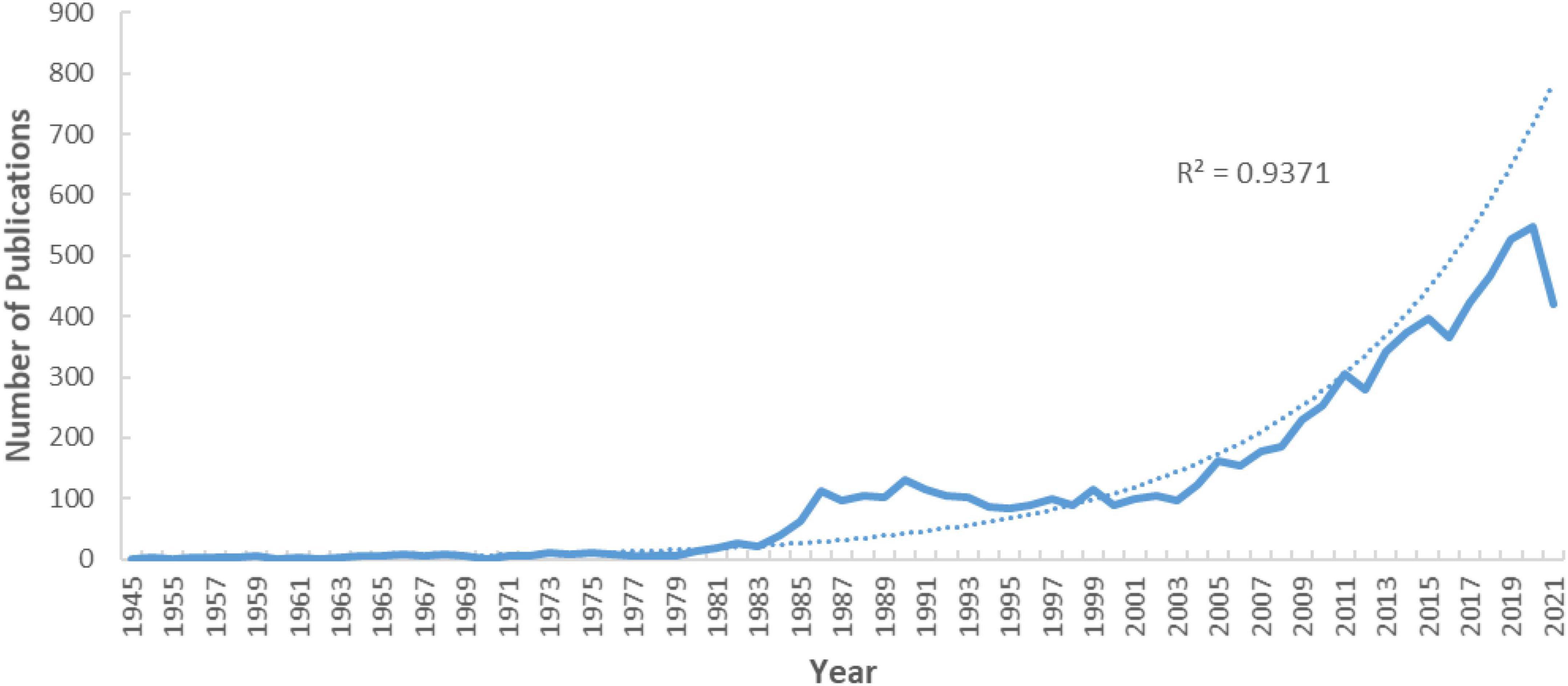
Figure 1. Publication years and growth forecast.
We also used the three-term polynomial model to assess the relationship between the growth of publications and publication year (Figure 1). We observed a significant correlation between the year and the number of publications with a high coefficient of determination (R2 = 0.9371). From the fitting curve, we can predict that the annual articles will continue to grow in the coming years.
Countries and institutions
In total, 101 countries were involved in research on the association of rare earth elements with cancer. The United States published the most publications, accounting for 34.73%, followed by China (1246, 16.10%), Germany (509, 6.48%), Italy (391, 4.98%), France (367, 4.76%), Japan (366, 4.66%), India (282, 3.59%), South Korea (228, 2.90%), Netherlands (209, 2.66%), and Canada (207, 2.64%) (Table 1). Figure 2 presents the analysis of the social relations of countries with more than five publications. Sixty countries that published more than five articles could be clustered into nine categories showing active cooperation.
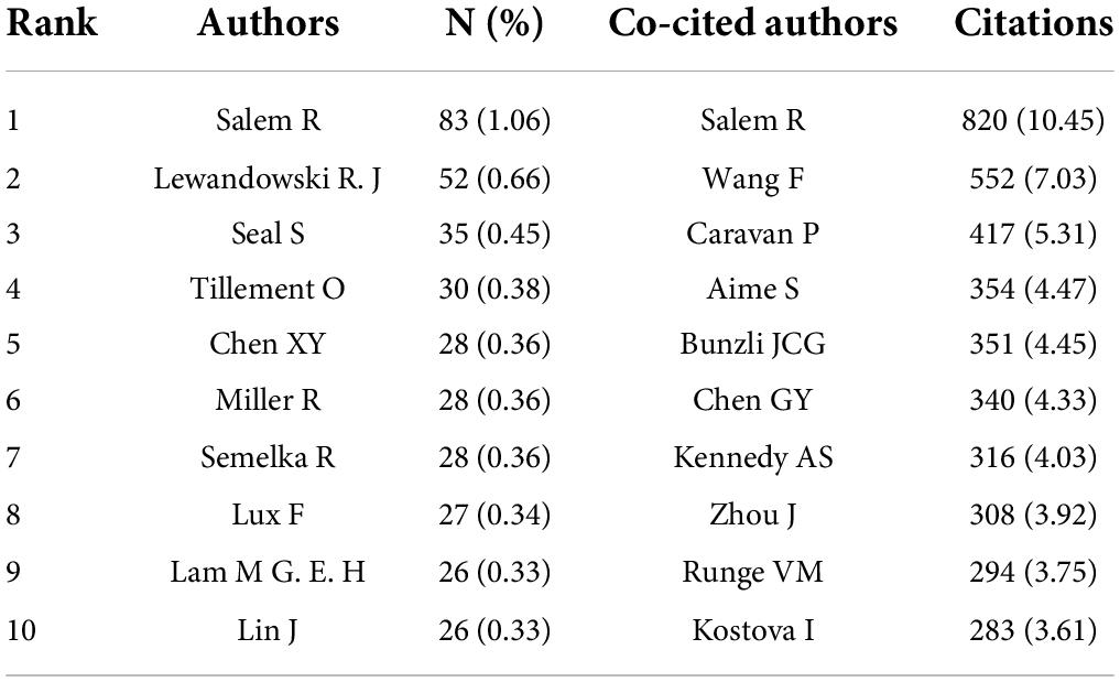
Table 1. The top 10 authors and co-cited authors in the research on the association of rare earth elements with cancer [n (%)].
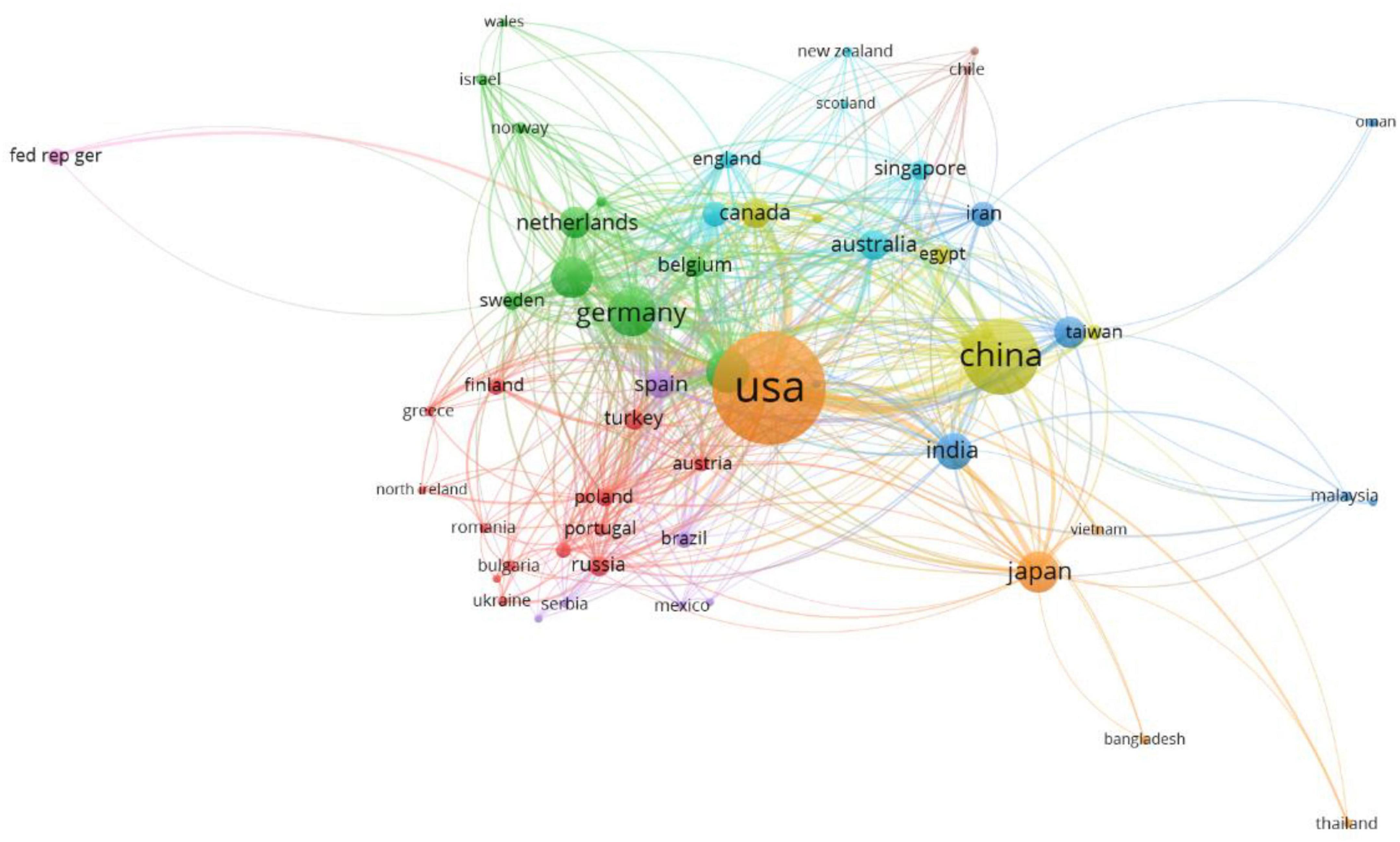
Figure 2. The network map of countries.
In general, 5,548 institutions contributed to research on the association of rare earth elements with cancer. Table 2 shows the top 10 institutions, and the Chinese Academy of Sciences ranked first (211, 2.69%), followed by Northwestern University (128, 1.63%), Stanford University (89, 1.13%), Fudan University (77, 0.98%), University of Texas (76, 0.97%), Mayo Clinic (71, 0.90%), National Cancer Institute (63, 0.80%), University of California Los Angeles (62, 0.79%), University of Pittsburgh (59, 0.75%), and the University of Pennsylvania (58, 0.74%). The collaborations of the institutions are presented in Figure 3. From the Figure, we can see that the cooperation among the main institutions was close.
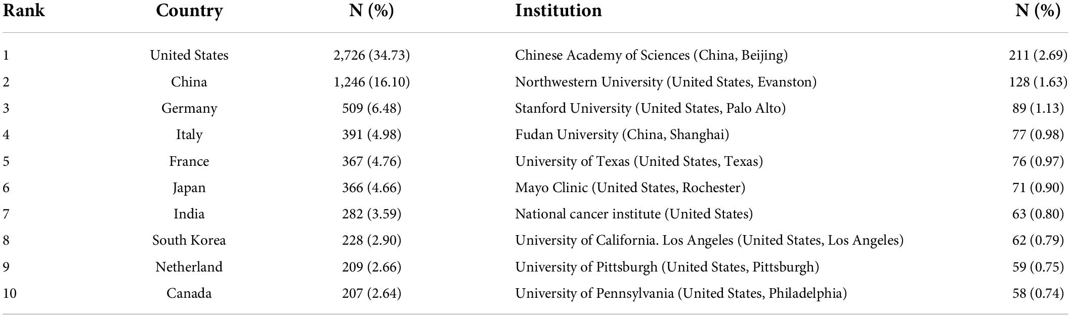
Table 2. The top 10 countries and institutions contributed to publications in the research on the association of rare earth elements with cancer [n (%)].
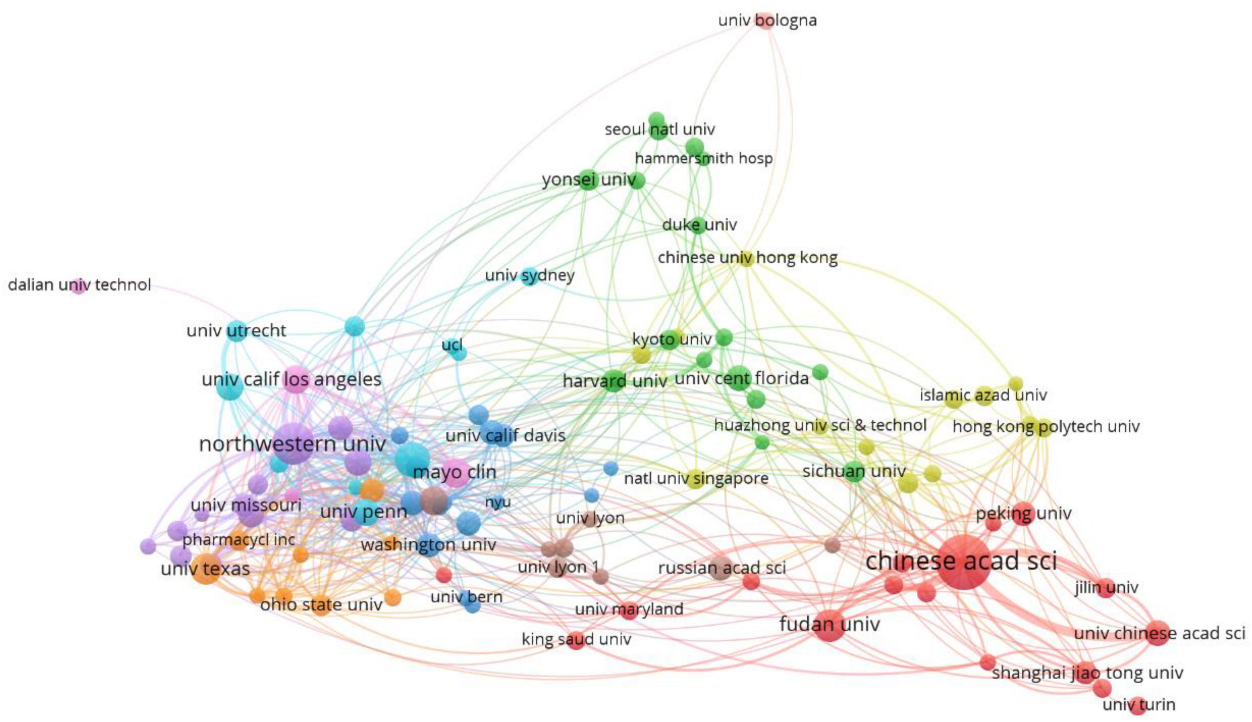
Figure 3. The network map of institutions.
Authors and co-cited authors
A total of 33,987 authors participated in the research on the association of rare earth elements with cancer, and the top 10 authors and co-cited authors are presented in Table 1. Salem R ranked first with 83 publications (1.06%), followed by Lewandowski (52, 0.66%), Seal S (35, 0.45%), and Tillement O (30, 0.38%). The remaining authors published fewer than 30 studies. The top three co-cited authors were Salem R (820, 10.45%), Wang F (552, 7.03%), and Caravan P (417, 5.31%). Other authors were cited fewer than 400 times. Figure 4 shows the collaborations of authors with more than 10 articles.
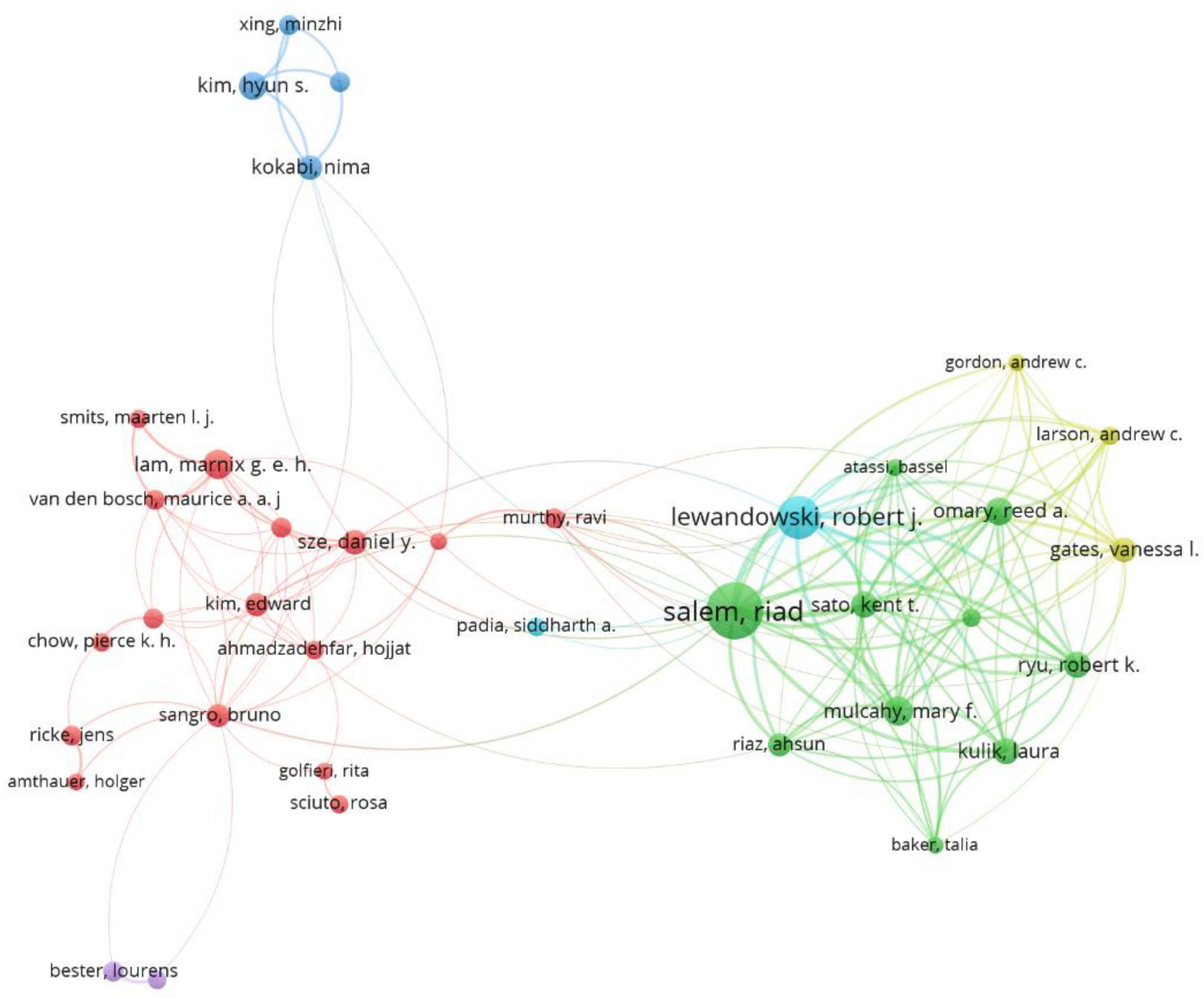
Figure 4. The network map of authors.
Journals and co-cited journals
A total of 7,849 identified publications were published in 1,579 journals, and the top 10 journals and co-cited journals are presented in Table 3. Radiology with 183 studies published the most articles, followed by the Journal of Nuclear Medicine (173, 2.20%), and the Journal of Vascular and Interventional Radiology (139, 1.77%). Seven of the top 10 journals were from the United States, and the other three journals were from Netherlands and Germany, respectively. The impact factors of the four journals were greater than 10. Radiology was also the most co-cited journal with 5,863 citations, followed by the Journal of the American Chemical Society (5,611 citations), and Biomaterials (4,127 citations). Of the top 10 co-cited journals, seven were from the United States and one from the United Kingdom, Netherlands, and Germany.
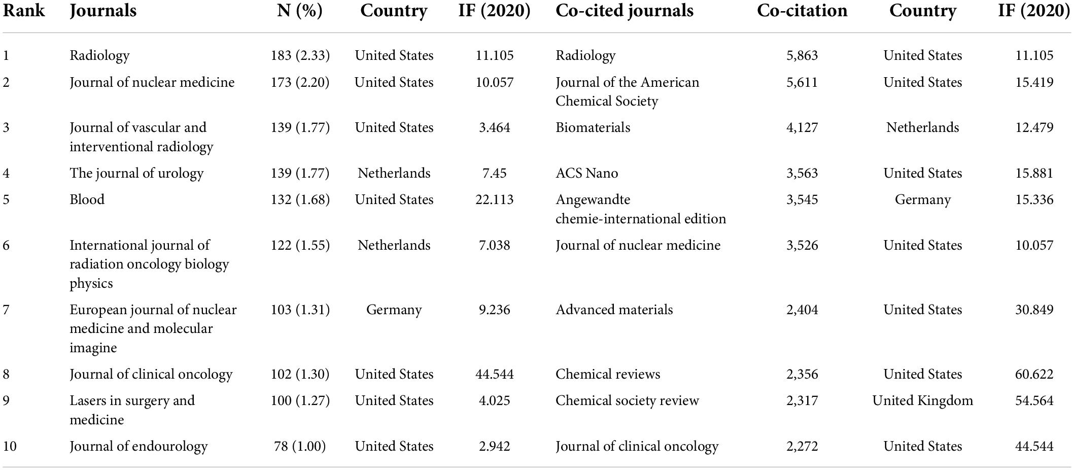
Table 3. The top 10 journals and co-cited journals in the research on the association of rare earth elements with cancer [n (%)].
Co-occurrence keywords
Keywords with a higher frequency could accurately indicate the main topic of a field. In our study, Gadolinium is the most often rare earth element, followed by Yttrium, Lanthanum, Cerium, and Holmium. Additionally, Table 4 presents the top 10 rare earth elements in the research on the association of rare earth elements with cancer. A total of 17,770 keywords appeared, but only 146 keywords had over 150 frequencies. Figure 5 and Table 5 present the results of the analysis according to co-occurrence frequency, cancer, nanoparticles, therapy, gadolinium, Magnetic Resonance Imaging, radioembolization, cells, contrast agents, in vivo, luminescence, and cerium oxide nanoparticles were the most common keywords.
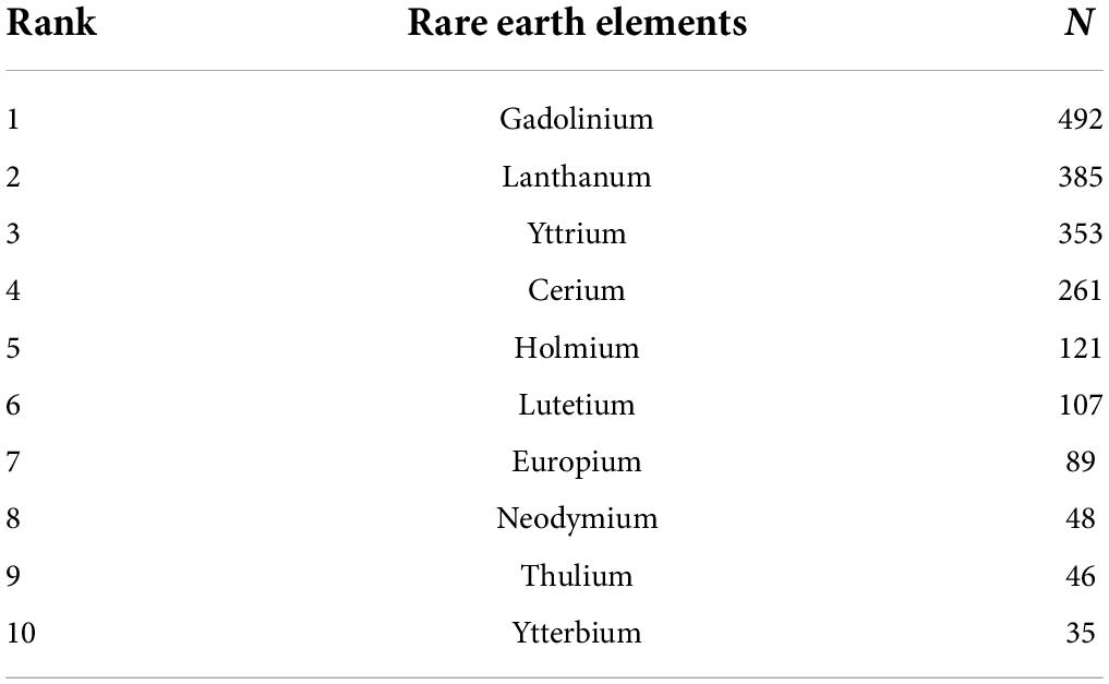
Table 4. The top 10 rare earth elements in the research on the association of rare earth elements with cancer.
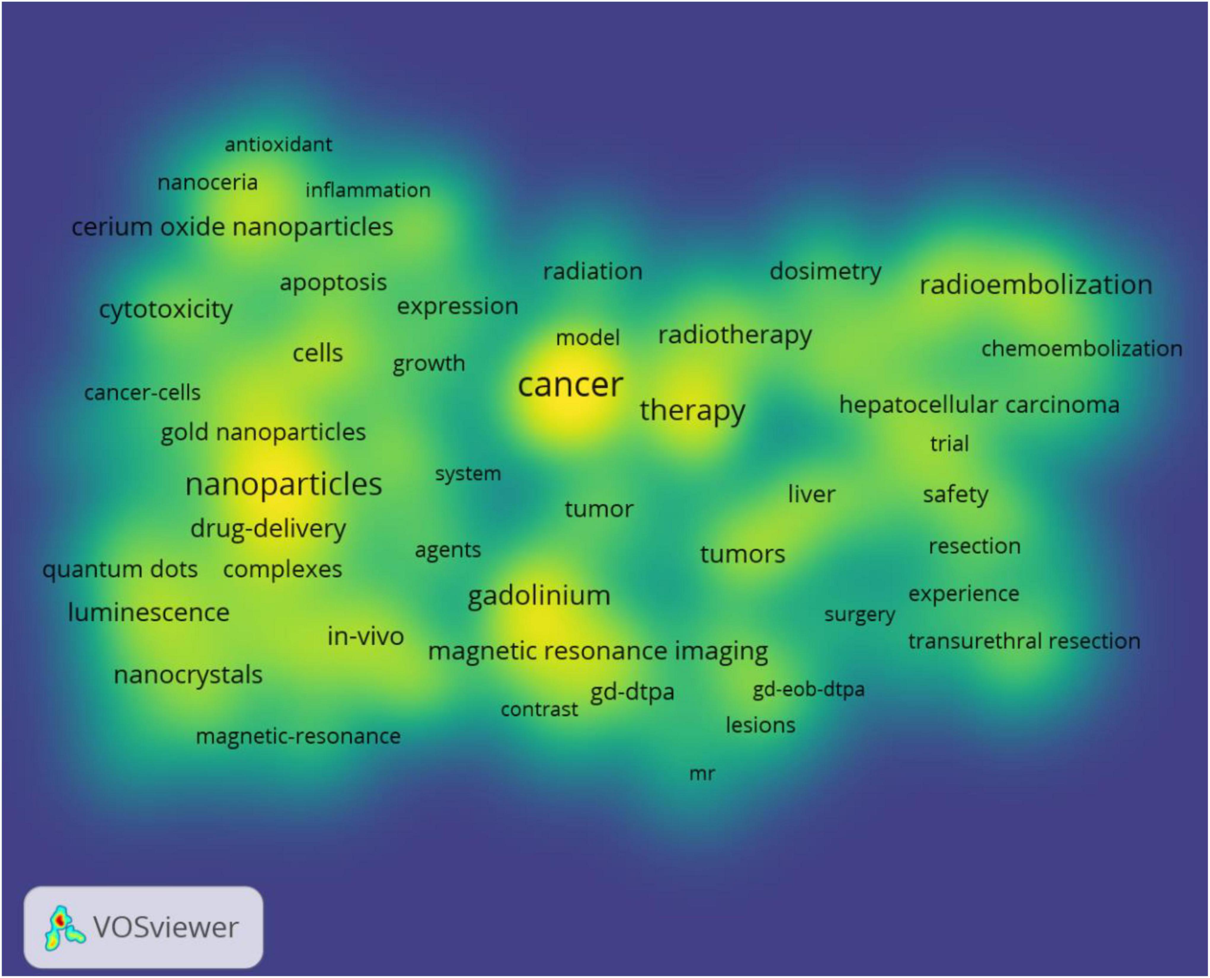
Figure 5. The density map of keywords.
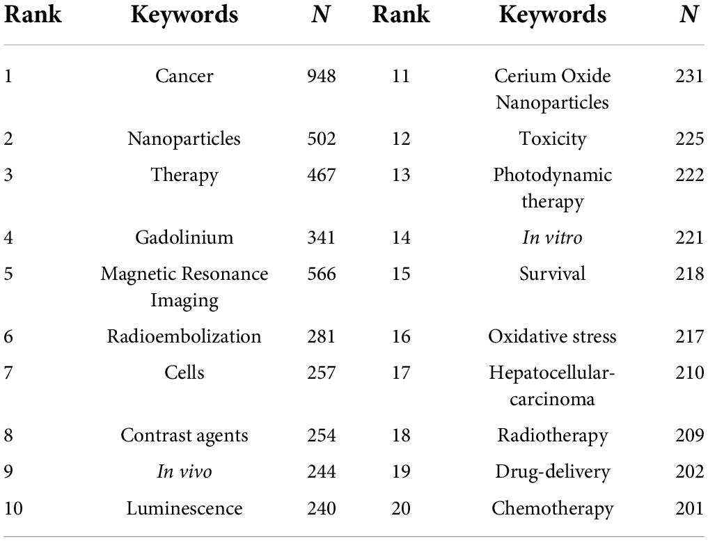
Table 5. The top 20 keywords in terms of frequency in the research on the association of rare earth elements with cancer.
Figure 6 shows the clustering analysis of co-occurrence keywords with frequencies greater than 100. In addition, 58 keywords were divided into four categories. Cluster 1 included 18 items and mainly focused on lanthanide complexes, nanoparticles, contrast agents, nanocrystals, and nanocrystals. Cluster 2 included 17 items and mainly focused on chemotherapy, hepatocellular carcinoma, radioimmunotherapy, yttrium-90, safety, and efficacy. Cluster 3 included 13 items and mainly focused on contrast agents, gadolinium, and magnetic resonance imaging. Cluster 4 included 10 items and mainly focused on cerium oxide nanoparticles, cytotoxicity, radiation, and gold nanoparticles. Figure 7 presents the overlay map of keywords with more than 100 frequencies, which showed that from 2016, the research frontiers mainly focused on oxidative stress, cerium oxide nanoparticles, quantum dots, drug delivery, nanoparticles, luminescence, and photodynamic therapy. Additionally, Figure 7 also showed that although Gadolinium is the most often element, it is not the element of research frontiers. After 2016, Cerium became a research frontier.
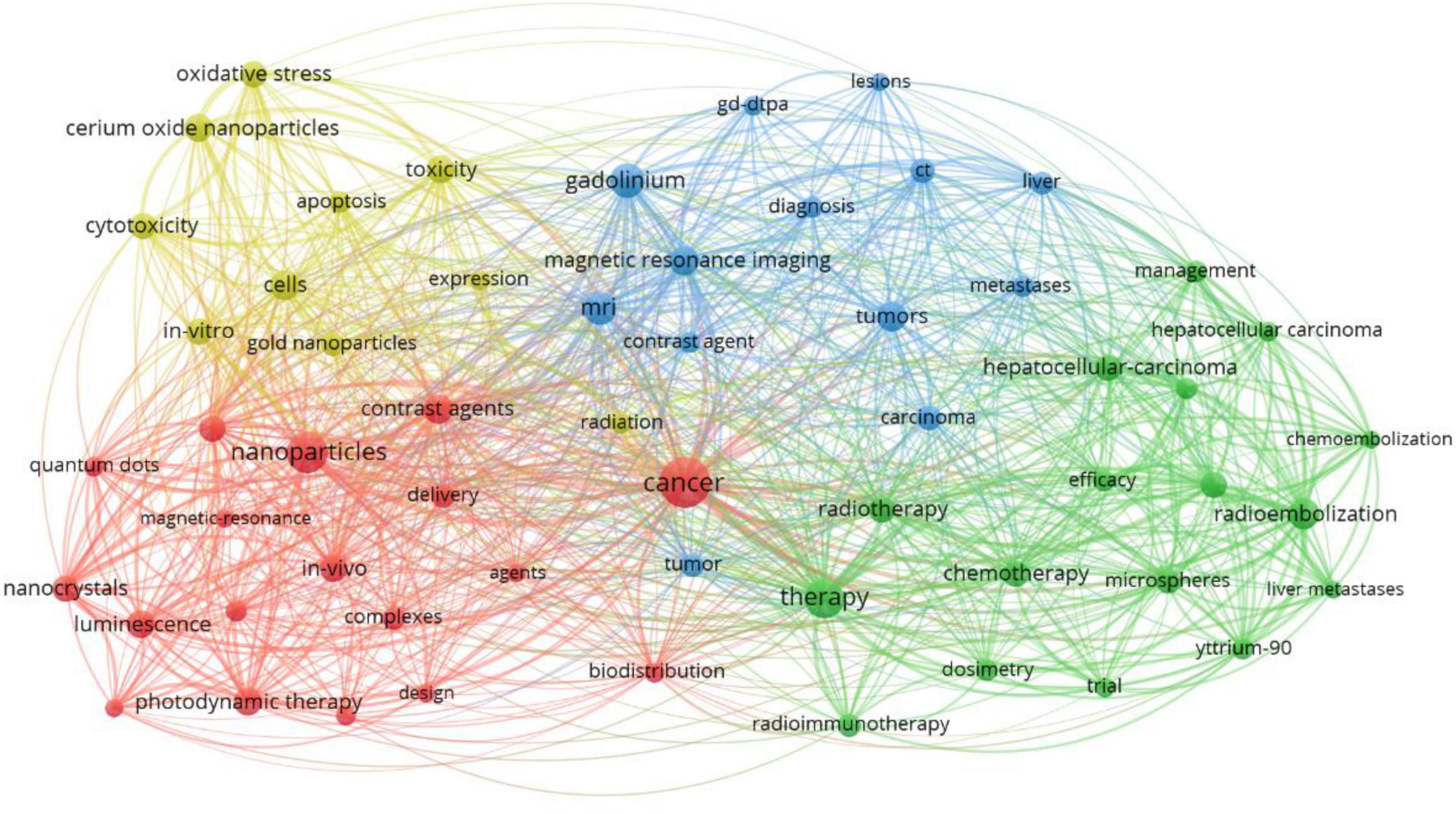
Figure 6. The network map of keywords.
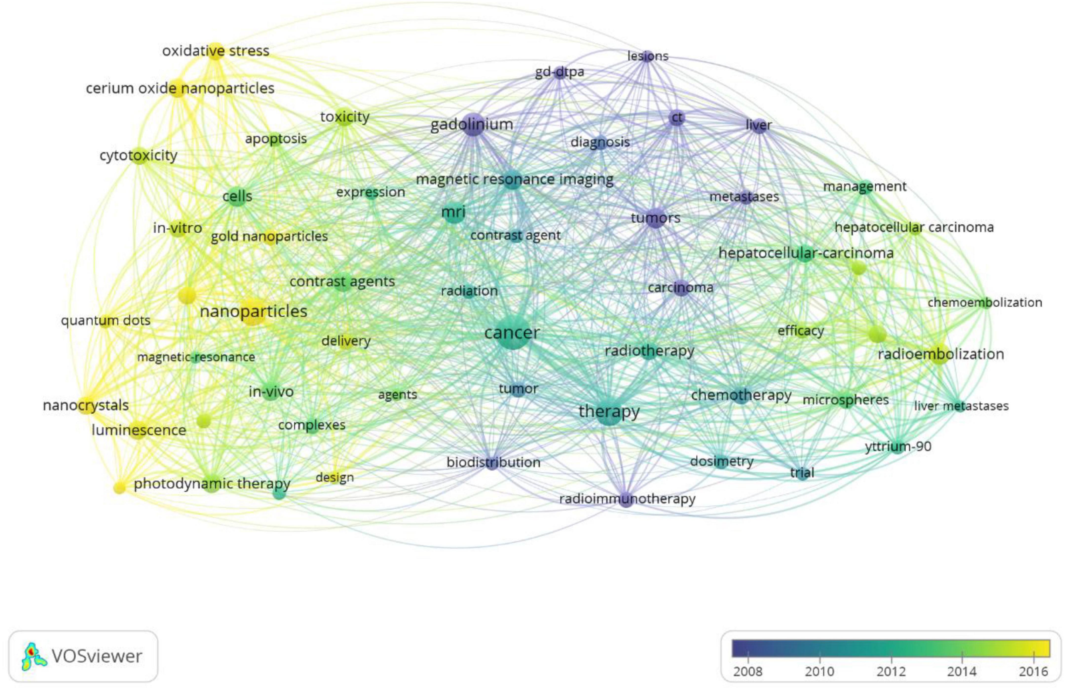
Figure 7. The overlay map of keywords.
Co-cited references
Table 6 presents the top 10 co-cited references in terms of research on the association of rare earth elements with cancer. One study was co-cited more than 200 times, two articles were co-cited between 120 and 140 times, five studies were co-cited 114–119 times, and two articles were co-cited 104 times.
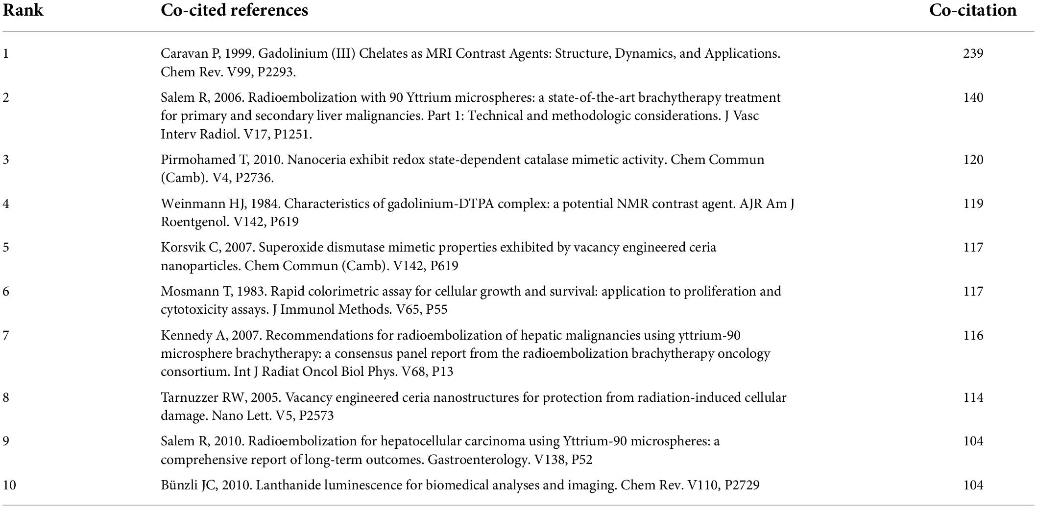
Table 6. The top 10 co-cited references in terms of the research on the association of rare earth elements with cancer [n (%)].
Discussion
We systematically searched the Web of Science and identified 7,849 articles. The first relevant research was published in 1945; however, in the 30 years after, there were only 31 articles related to the application of rare earth elements in cancer, which indicates that the application of rare earth elements in cancer developed slowly during this period. After 1980, the number of publications increased; the possible reasons include that since the beginning of the 20th century, rare earth elements were used in medicine (6–9). Over 77.96% of the articles were published between 2000 and 2021. With the continuous improvement of methods for the extraction and separation of lanthanide salts, an increasing number of researchers, countries, and scholars have focused on utilizing rare earth elements in cancer imaging and treatment, which has led to more abundant relevant research in recent years. A significant positive relationship was found between the year and the number of publications, and it is expected that research on the application of rare earth elements in cancer will continue to grow in the coming years.
A total of 33,987 authors investigated rare earth elements and cancer. Only 0.45% of the authors published more than 10 publications, and 79.07% of the authors published only one publication, which indicates that although an increasing number of authors participated in research on the application of rare earth elements in cancer, there are few high-yield authors. Of the top 10 high-yield authors, seven were from developed countries and three were from China. However, among the top 10 co-cited authors, there was only one high-yield author. Relevant authors should strengthen cooperation to innovate research methods and improve the quality of the studies.
Among the top 10 countries contributing to relevant publications, only one was a developing country, which indicates that research on the application of rare earth elements in cancer is relatively insufficient and backward. Therefore, developing countries need to cooperate with developed countries to learn advanced technology methods and research to promote the development of relevant research worldwide. Moreover, available funding for conducting relevant research is also needed for researchers in developing countries to increase the research output on rare earth elements and cancer. Among the top 10 institutions, eight were from the United States, and most were American universities. The remaining two were from China.
A total of 7,849 articles were published in 1,579 journals, but only 9.69% of journals published more than 10 papers, and 51.60% of journals only published one relevant paper, which reveals that many journals contributed to the publication of the application of rare earth elements and cancer, but journals insisted that publication of relevant articles was insufficient. Of the top 10 journals, six were from the United States and the mean impact factors of the six journals were greater than 16.41, and the impact factors of the four from the United States were greater than 10. Journals from the United States publish studies of rare earth elements and cancer most frequently, and the impact factors of these high-yield journals are also high. Among the top 10 co-cited journals, only three were also highly productive journals; Radiology was the journal with the highest number of relevant articles and was also the most co-cited journal. The impact factors of all of the top 10 co-cited journals were greater than 10, indicating that studies published in high-impact factor journals were cited more often. A possible reason for this phenomenon might be that higher-impact journals have higher requirements for topic selection and quality of published articles.
In bibliometrics, a network graph of keyword co-occurrences can reflect topics of interest (16). In this study, 17,770 keywords were generated, and 146 keywords were appearing over 50 times, revealing that a higher frequency of a fewer number of keywords could better indicate the main topic of interest in a field. We used the VOS viewer to perform cluster analysis for co-occurrence keywords and four topics were generated: cluster 1 contained 18 keywords, which mainly focused on lanthanide complex-doped nanomaterials applied in the diagnosis and treatment of cancer, including magnetic resonance imaging contrast agents, photodynamic therapy, and anticancer drug delivery. Nanomaterials with unique surfaces and internal structures showed improved circulation and reduced toxicity (17). Up-conversion nanoparticles can convert near-infrared (NIR) light at long wavelengths into visible (Vis) and even UV light with shorter wavelengths (18), and thus have many advantages including minimal photodamage, deep tissue penetration, absence of autofluorescence, weak photobleaching, and good chemical stability (19–21). Nanomaterials have been confirmed to have excellent application potential in cancer therapy (17). Cluster 2 included 17 keywords, mainly related to the efficacy and safety of yttrium-90 radioimmunotherapy and chemoembolization for the treatment of hepatocellular carcinoma (HCC). HCC is one of the most common and deadly cancers worldwide (22, 23). More than 70% of patients with HCC cannot undergo curative surgery; locoregional therapies, including chemoembolization, radioembolization, and radioimmunotherapy, are generally used to control the progress of the disease in patients with HCC globally (24–27). Previous studies showed that Yttrium-90 radioembolization and radioimmunotherapy are associated with better clinical outcomes in patients with HCC (28, 29). Cluster 3 contained 13 keywords focused on gadolinium magnetic resonance imaging (MRI) contrast agents for the diagnosis of cancer. Lanthanide plays an important role in the field of medicine, especially in cancer diagnosis and treatment. In recent years, gadolinium complexes have been widely used in MRI for diagnostic purposes and monitoring treatment progress (30–33). Cluster 4 included 10 keywords mainly concerned with cerium oxide nanoparticles in the role of enhancing radiation-induced and chemotherapy-induced cancer apoptosis. When conventional treatments including surgery, radiation, and cytotoxic chemotherapies are ineffective in cancer treatment, nanomedicines are one of the most recently developed treatment options (33–37). Cerium oxide nanoparticles consist of a cerium core surrounded by an oxygen lattice, which plays an important role in antioxidant behavior. Previous studies have indicated that cerium oxide nanoparticles have extensive potential as a therapeutic agent for the treatment of cancer because cerium oxide nanoparticles possess innate cytotoxicity to cancer cells, anti-invasive properties, and the ability to sensitize cancer cells to radiation-induced cell death while protecting the surrounding normal tissues (38–41).
Strengths and limitations
To the best of our knowledge, this study is the first bibliometric analysis of the application of rare earth elements for cancer. We clearly show the current status and future trends of relevant research by conducting frequency analysis, cluster analysis, and hotspot analysis.
There are some limitations to our study. First, because WOS is the most important source of data for bibliometric analysis in science (42), we only searched WOS in this study, which could result in insufficient and unrepresentative studies that were included in the current study. Second, most of the identified studies were published in English, which may have led to language bias. Third, since there are many authors and keywords in this study, although we have standardized and adjusted during the analysis, the omission may still exist.
Conclusion
Studies related to the application of rare earth elements and cancer have significantly increased over the past 20 years. The United States contributed the most articles in the field, following China and Germany, and cooperation among countries was close. The Chinese Academy of Sciencess, Northwestern University, and Stanford University were the top three productive institutions, and cooperation among institutions was active. Many high-quality journals have published relevant research, but there are few highly productive journals. The application of lanthanide complex-doped nanomaterials in the diagnosis and treatment of cancer includes magnetic resonance imaging contrast agents, photodynamic therapy, anticancer drug delivery, the efficacy and safety of yttrium-90 radioimmunotherapy and chemoembolization for the treatment of HCC, gadolinium magnetic resonance imaging (MRI) contrast agents for the diagnosis of cancer, and cerium oxide nanoparticles are the main research topics in this field. In recent years, especially since 2016, research frontiers include cerium oxide nanoparticles and photodynamic therapies.
Data availability statement
The original contributions presented in this study are included in the article/supplementary material, further inquiries can be directed to the corresponding author/s.
Author contributions
SL and JW conceived and designed the study, screened and selected the articles, interpreted the data, and drafted the manuscript. Both authors had full access to all the data in the study and had final responsibility for the decision to submit for publication.
Funding
This study was supported by the key research and development program of Gansu Province (20YF8FA075).
Conflict of interest
The authors declare that the research was conducted in the absence of any commercial or financial relationships that could be construed as a potential conflict of interest.
Publisher’s note
All claims expressed in this article are solely those of the authors and do not necessarily represent those of their affiliated organizations, or those of the publisher, the editors and the reviewers. Any product that may be evaluated in this article, or claim that may be made by its manufacturer, is not guaranteed or endorsed by the publisher.
References
1. Siegel RL, Miller KD, Jemal A. Cancer statistics, 2020. CA Cancer J. Clin. (2020) 70:7–30. doi: 10.3322/caac.21590
2. Sung H, Ferlay J, Siegel RL, Laversanne M, Soerjomataram I, Jemal A, et al. Global cancer statistics 2020: GLOBOCAN estimates of incidence and mortality worldwide for 36 cancers in 185 countries. CA Cancer J Clin. (2021) 71:209–49. doi: 10.3322/caac.21660
4. Russell LM, Liu CH, Grodzinski P. Nanomaterials innovation as an enabler for effective cancer interventions. Biomaterials. (2020) 242:119926. doi: 10.1016/j.biomaterials.2020.119926
5. Teo RD, Termini J, Gray HB. Lanthanides: applications in cancer diagnosis and therapy. J Med Chem. (2016) 59:6012–24. doi: 10.1021/acs.jmedchem.5b01975
6. Kaczmarek MT, Zabiszak M, Nowak M, Jastrzab R. Lanthanides: schiff base complexes, applications in cancer diagnosis, therapy, and antibacterial activity. Coordinat Chem Rev. (2018) 370:42–5. doi: 10.1016/j.ccr.2018.05.012
7. Ban S, Suzuki S, Kubota K, Ohshima S, Satoh H, Imada H, et al. Gastric mucosal status susceptible to lanthanum deposition in patients treated with dialysis and lanthanum carbonate. Ann. Diagn. Pathol. (2017) 26:6–9. doi: 10.1016/j.anndiagpath.2016.10.001
8. Fricker SP. The therapeutic application of lanthanides. Chem. Soc. Rev. (2006) 35:524–33. doi: 10.1039/b509608c
9. Zhang C, Wang S, Zhao S, Zhang X. Effect of lanthanum carbonate on coronary artery calcification and bone mineral density in maintenance hemodialysis patients with diabetes complicated with a dynamic bone disease. Medicine. (2017) 96:45–52. doi: 10.1097/MD.0000000000008664
10. Magerstädt M, Gansow OA, Brechbiel MW, Colcher D, Baltzer L, Knop RH, et al. Gd(DOTA): an alternative to Gd(DTPA) as a T1,2 relaxation agent for NMR imaging or spectroscopy. Magn. Reson. Med. (1986) 3:808–12. doi: 10.1002/mrm.1910030517
11. Larson SM, Carrasquillo JA, Cheung N-KV, Press OW. Radioimmunotherapy of human tumours. Nat. Rev. Cancer. (2015) 15:347–60. doi: 10.1038/nrc3925
12. Zhang T, Zhu X, Wong W-K, Tam H-L, Wong W-Y. Light-harvesting ytterbium(III)-porphyrinae-BODIPY conjugates: synthesis, exciation-energy transfer, and two-photon-indused near-infrared-emission studies. Chem. Eur. J. (2013) 19:739–48. doi: 10.1002/chem.201202613
13. Park JY, Chang Y, Lee GH. Multi-modal imaging and cancer therapy using lanthanide oxide nanoparticles: current status and perspectives. Curr. Med. Chem. (2015) 22:569–81. doi: 10.2174/0929867322666141128162843
14. He B, Wang J, Lin J, Chen J, Zhuang Z, Hong Y, et al. Association between rare earth element cerium and the risk of oral cancer: a case-control study in Southeast China. Front Public Health. (2021) 9:647120. doi: 10.3389/fpubh.2021.647120
15. Gao Y, Chen K, Ma J, Gao F. Cerium oxide nanoparticles in cancer. Oncotargets Ther. (2014) 7:835–40. doi: 10.2147/OTT.S62057
16. Liang YD, Li Y, Zhao J, Wang XY, Zhu HZ, Chen XH. Study of acupuncture for low back pain in recent 20 years: a bibliometric analysis via CiteSpace. J Pain Res. (2017) 10:951–64. doi: 10.2147/JPR.S132808
17. Shi J, Kantoff PW, Wooster R, Farokhzad OC. Cancer nanomedicine: progress, challenges and opportunities. Nat. Rev. Cancer. (2017) 17:20–37. doi: 10.1038/nrc.2016.108
18. Chen G, Qiu H, Prasad PN, Chen X. Upconversion nanoparticles: design, nanochemistry, and applications in theranostics. Chem. Rev. (2014) 114:5161–214. doi: 10.1021/cr400425h
19. Chatterjee D, Rufaihah A, Zhang Y. Upconversion fluorescence imaging of cells and small animals using lanthanide doped nanocrystals. Biomaterials. (2008) 29:937–43. doi: 10.1016/j.biomaterials.2007.10.051
20. Idris N, Li Z, Ye L, Sim EK, Mahendran R, Ho PC, et al. Tracking transplanted cells in live animal using upconversion fluorescent nanoparticles. Biomaterials. (2009) 30:5104–13. doi: 10.1016/j.biomaterials.2009.05.062
21. Zhang F, Shi Q, Zhang Y, Shi Y, Ding K, Zhao D, et al. Fluorescence upconversion microbarcodes for multiplexed biological detection: nucleic acid encoding. Adv. Mater. (2011) 23:3775–9. doi: 10.1002/adma.201101868
22. Llovet JM, Bru C, Bruix J. Prognosis of hepatocellular carcinoma: the BCLC staging classification. Semin Liver Dis. (1999) 19:329–38. doi: 10.1055/s-2007-1007122
23. Forner A, Reig ME, de Lope CR, Bruix J. Current strategy for staging and treatment: the BCLC update and future prospects. Semin Liver Dis. (2010) 30:61–74. doi: 10.1055/s-0030-1247133
24. European Association for the Study of the Liver. EASL clinical practice guidelines: management of hepatocellular carcinoma. J Hepatol. (2018)) 69:182–236.
25. Marrero JA, Kulik LM, Sirlin CB, Zhu AX, Finn RS, Abecassis MM, et al. Diagnosis, staging, and management of hepatocellular carcinoma: 2018 practice guidance by the American association for the study of liver diseases. Hepatology. (2018) 68:723–50. doi: 10.1002/hep.29913
26. Omata M, Cheng AL, Kokudo N, Kudo M, Lee JM, Jia J, et al. Asia-Pacific clinical practice guidelines on the management of hepatocellular carcinoma: a 2017 update. Hepatol Int. (2017) 11:317–70. doi: 10.1007/s12072-017-9799-9
27. Korean Liver Cancer Association, National Cancer Center. 2018 Korean Liver Cancer Association-National Cancer Center Korea practice guidelines for the management of hepatocellular carcinoma. Korean J Radiol. (2019) 20:1042–113. doi: 10.3348/kjr.2019.0140
28. Kim MA, Jang H, Choi NR, Nam JY, Lee YB, Cho EJ, et al. Yttrium-90 radioembolization is associated with better clinical outcomes in patients with hepatocellular carcinoma compared with conventional chemoembolization: a propensity score-matched study. J Hepatocell Carcinoma. (2021) 7:1565–77. doi: 10.2147/JHC.S335879
29. Ludwig AD, Labadie KP, Seo YD, Hamlin DK, Nguyen HM, Mahadev VM, et al. Yttrium-90-labeled anti-glypican 3 radioimmunotherapy halts tumor growth in an orthotopic xenograft model of hepatocellular carcinoma. J Oncol. (2019) 15:4564707. doi: 10.1155/2019/4564707
30. Rashid HU, Martines MAU, Jorge J, de Moraes PM, Umar HU, Khan MN, et al. Cyclenbased Gd3+ complexes as MRI contrast agents: relaxivity enhancement and ligand design. Bioorgan. Med. Chem. (2016) 4:5663–84. doi: 10.1016/j.bmc.2016.09.069
31. Bombieri G, Artali R, Mason SA, Mortillaro GJ, Mcintyre G, Aime S, et al. Innersphere water and hydrogen bonds in lanthanide DOTAM complexes. A neutron diffraction study, Inorg. Chim. Acta. (2018) 470:433–8. doi: 10.1016/j.ica.2017.09.021
32. Babicˇ A, Vorobiev V, Xayaphoummine C, Lapicorey G, Chauvin AS, Helm L, et al. Self-assembled nanomicelles as MRI blood-pool contrast agent. Chem. Eur. J. (2018) 24:1348–57. doi: 10.1002/chem.201703962
33. Phukan B, Mukherjee C, Varshney R. A new heptadentate picolinate-based ligand and its corresponding Gd(III) complex: the effect of pendant picolinate versus acetate on complex properties. Dalton Trans. (2018) 47:135–42. doi: 10.1039/C7DT04150K
34. Lee SK, Kim GS, Wu Y, Kim DJ, Lu Y, Kwak M, et al. Nanowire substrate-based laser scanning cytometry for quantitation of circulating tumor cells. Nano Lett. (2012) 12:2697–704. doi: 10.1021/nl2041707
35. Dicheva BM, ten Hagen TL, Li L, Schipper D, van Rhoon GC, Seynhaeve LB, et al. Cationic thermosensitive liposomes: a novel dual targeted heat-triggered drug delivery approach for endothelial and tumor cells. Nano Lett. (2013) 13:2324–31. doi: 10.1021/nl3014154
36. Kanapathipillai M, Mammoto A, Mammoto T, Kang JH, Jiang E, Ghosh K, et al. Inhibition of mammary tumor growth using lysyl oxidase-targeting nanoparticles to modify extracellular matrix. Nano Lett. (2012) 12:3213–7. doi: 10.1021/nl301206p
37. Zhao G, Rodriguez BL. Molecular targeting of liposomal nanoparticles to tumor microenvironment. Int J Nanomedicine. (2013) 8:61–71. doi: 10.2147/IJN.S37859
38. Wang Y, Yang F, Zhang HX, Zi XY, Pan XH, Chen F, et al. Cuprous oxide nanoparticles inhibit the growth and metastasis of melanoma by targeting mitochondria. Cell Death Dis. (2013) 4:e783. doi: 10.1038/cddis.2013.314
39. Asati A, Santra S, Kaittanis C, Perez JM. Surface-charge-dependent cell localization and cytotoxicity of cerium oxide nanoparticles. ACS Nano. (2010) 4:5321–31. doi: 10.1021/nn100816s
40. Wason MS, Colon J, Das S, Seal S, Turkson J, Zhao J, et al. Sensitization of pancreatic cancer cells to radiation by cerium oxide nanoparticle-induced ROS production. Nanomedicine. (2013) 9:558–69. doi: 10.1016/j.nano.2012.10.010
41. Waris G, Ahsan H. Reactive oxygen species: role in the development of cancer and various chronic conditions. J Carcinog. (2006) 5:14. doi: 10.1186/1477-3163-5-14
Keywords: rare earth elements, cancer, evidence mapping, scientometric analysis, VOS viewer
Citation: Wang J and Li S (2022) Applications of rare earth elements in cancer: Evidence mapping and scientometric analysis. Front. Med. 9:946100. doi: 10.3389/fmed.2022.946100
Received: 14 June 2022; Accepted: 15 July 2022;
Published: 10 August 2022.
Edited by:
Monica Catarina Botelho, Instituto Nacional de Saúde Doutor Ricardo Jorge (INSA), PortugalReviewed by:
Andy Wai Kan Yeung, University of Hong Kong, Hong Kong SAR, ChinaFrank Mayta-Tovalino, National University of San Marcos, Peru
Copyright © 2022 Wang and Li. This is an open-access article distributed under the terms of the Creative Commons Attribution License (CC BY). The use, distribution or reproduction in other forums is permitted, provided the original author(s) and the copyright owner(s) are credited and that the original publication in this journal is cited, in accordance with accepted academic practice. No use, distribution or reproduction is permitted which does not comply with these terms.
*Correspondence: Jinyu Wang, d2FuZ2ppbnlAbHp1LmVkdS5jbg==; Sheng Li, bGlzaGVuZzc2QHNvaHUuY29t
†These authors have contributed equally to this work