 Miriam Haidukowski1
 Eliana Casamassima1
 Maria Teresa Cimmarusti1
 Maria Teresa Branà1Francesco Longobardi2
 Pasquale Acquafredda3
 Antonio Logrieco1*
 Claudio Altomare1
- 1Department of Biology, Agriculture and Food Science, Institute of Sciences of Food Production, National Research Council (CNR), Bari, Italy
- 2Department of Chemistry, University of Bari, Bari, Italy
- 3Department of Earth and Geo-Environmental Sciences, University of Bari, Bari, Italy
Aflatoxin B1 (AfB1) is a carcinogenic mycotoxin that contaminates food and feed worldwide. We determined the AfB1-adsorption capability of non-viable Pleurotus eryngii mycelium, an edible fungus, as a potential means for removal of AfB1 from contaminated solutions. Lyophilized mycelium was produced and made enzymatically inert by sterilization at high temperatures. The material thus obtained was characterized by scanning electron microscopy with regard to the morpho-structural properties of the mycotoxin-adsorbing surfaces. The active surfaces appeared rough and sponge-like. The AfB1-mycelium system reached equilibrium at 37°C, 30 min, and pH 5–7, conditions that are compatible with the gastro-intestinal system of animals. The system remained stable for 48 h at room temperature, at pH 3, pH 7, and pH 7.4. A thermodynamic study of the process showed that this is a spontaneous and physical adsorption process, with a maximum of 85 ± 13% of removal efficiency of AfB1 by P. eryngii mycelium. These results suggest that biosorbent materials obtained from the mycelium of the mushroom P. eryngii could be used as a low-cost and effective feed additive for AfB1 detoxification.
Introduction
The contamination of food with mycotoxins is a worldwide problem with impact on the health of humans and animals and on the economy of many countries, especially in sub-tropical and temperate areas. The problem is caused by the spoilage of agricultural products by microscopic filamentous fungi, mostly belonging to species and strains in the genera Aspergillus, Fusarium, and Penicillium, which in favorable environmental conditions are able to produce toxic secondary metabolites that accumulate into food and feedstuffs. Aflatoxin B1 (AfB1) is the most toxic mycotoxin and is classified in the “group 1” substances (carcinogenic to humans) by the International Agency for Research on Cancer (World Health Organization and International Agency for Research on Cancer, 1993). AFB1 has potent hepatotoxic, carcinogenic, and mutagenic effects on humans and animals and is produced mainly by isolates of the species Aspergillus flavus and A. parasiticus. AFB1 occurrence is a major problem in a number of crops, including cereals, groundnuts, legumes, and cotton seeds, which can be contaminated at any stage from field to storage. Human exposure to AFB1 can result from ingestion of contaminated food or from consumption of meat, eggs, and dairy products from animals that have been fed with contaminated feed (Rushing and Selim, 2019).
Much attention is devoted to measures of prevention and monitoring that aim at the reduction of contamination levels in commodities. Nevertheless, often the contamination occurs despite careful application of prevention means, and it is necessary to put into action decontamination measures to avoid the complete loss of the produce and mitigate the risk of mycotoxins in food and feed. Different chemical and physical methods for decontamination and detoxification have been developed, but their use is often limited by high cost, lack of information on nature and toxicity of degradation products and, above all, loss of nutritional, organoleptic, and visual qualities (Boudergue et al., 2009).
For protection of animals from aflatoxicosis, the use of adsorbent materials which are able to bind with high efficiency the mycotoxins in feeds is being receiving growing interest (Williams et al., 2004). The adsorbents reduce the bioavailability of mycotoxins in the gastro-intestinal tract and thus their diffusion into the bloodstream and transport to the target organs (Kabak et al., 2006; Kolosova and Stroka, 2011). Aluminosilicates are the most used adsorbents, followed by activated carbon and special polymers (Huwig et al., 2001; Vila-Donat et al., 2018). The EU has approved the use of various adsorbent materials as food additives, for example the use of bentonite as a feed additive for all animal species is regulated by the Commission Implementing Regulation (EU) No. 1060/2013. The efficiency of these mycotoxin ligands differs considerably, depending on the chemical structures of both the adsorbent and the toxin (Fu and Viraraghavan, 2001; Crini, 2006). Also, the use of these materials may have a negative impact on the quality of decontaminated feeds. For this reason, scientific interest is partly shifting toward the use of less expensive, though effective, and environmentally friendly materials such as microbial biomasses (Low et al., 2008).
Some studies have demonstrated the ability of some strains of lactic and bifidus bacteria to efficiently bind AfB1 (El-Nezami et al., 1998; Peltonen et al., 2001), through a chemical-physical phenomenon related to the features of structural elements of the bacterial wall such as peptiglycans and polysaccharides (Kabak et al., 2006). However, few materials have been studied for this purpose and to our knowledge there are no studies concerned with the use of non-viable fungal mycelium as mycotoxin adsorbent. The mycelium of fungi has noteworthy adsorbing properties, mostly due to the ability of the polysaccharides constituting the cell wall to form hydrogen, ionic, or hydrophobic interactions with organic and inorganic molecules (Huwig et al., 2001). These properties are the subject of research with practical applications in different contexts, including bioremediation of soils and wastewater from heavy metals and organic pollutants (Gavrilescu, 2004; Gadd, 2009). Hence, the use of fungal mycelium also as biosorbent for mycotoxins appears conceivable.
The present work is focused on a new adsorbent material made from mushroom mycelium. This novel biosorbent is different from those already on the market, since it is palatable and has nutritional value. Particularly, we report on the characterization of the biosorbent properties of the mycelium of Pleurotus eriyngii (DC.) Quél. (king oyster mushroom). This fungus combines several advantageous features. Species of Pleurotus can be grown easily and are cultivated worldwide. They can be grown on a variety of lignocellulosic materials, including wastes which are produced through agricultural, forest, and food-processing activities. They grow faster than other cultivable mushrooms and cultivation has no particular technical hurdles (Sánchez, 2010); therefore, large biomasses of Pleurotus spp. can be obtained at sensible cost. Besides being easily cultivable and edible, Pleurotus spp. exhibit some properties of biotechnological interest. These fungi are high producers of extracellular ligninolytic enzymes, namely phenol oxidases (mainly laccases) and peroxidases (lignin peroxidase and Mn peroxidase) (Sánchez, 2010). Because of the low substrate specificity of these ligninolytic enzymes, applications of Pleurotus have been investigated for bioremediation purposes, e.g., decontamination of wastewater and water sediments from phenolic endocrine disruptors (Loffredo et al., 2013), degradation of dyes (Kalmiş et al., 2008) and mycotoxins (Alberts et al., 2009; Branà et al., 2017) and of other recalcitrant environmental pollutants (Rigas et al., 2009; Purnomo et al., 2010). In addition, mycelia of Pleurotus spp. have been reported to have binding and sequestering capabilities for heavy metals (recently reviewed by Kapahi and Sachdeva, 2017).
Fungal cell walls have already received attention as biosorbents for bioremediation of polluted soils and wastewaters (Wang and Chen, 2009). We here propose a novel application intended for feed industry. In particular, we have investigated the capability of non-viable mycelium of P. eryngii to bind AfB1; in addition, the effects of physical and chemical conditions on the binding efficiency were studied through a Design of Experiment (DOE) methodology. In most of the works concerned with the adsorption process, an approach that takes into account one factor at a time is used, while there are few studies that use a factorial design model to evaluate the relative importance and the interaction of different operative factors on the biosorption process (Manal, 2007). The design determines which factors have significant effects on the response, as well as the cases in which the effect of a factor varies with the level of another (Brasil et al., 2006), using the least possible number of experiments. The determination of interactions between factors is the key for optimization of complex processes. In the absence of such a study, important interactions might remain undetermined and the optimization becomes difficult to achieve (Brasil et al., 2006). For this reason, our study firstly evaluated the effects of different factors on the adsorption process and then we proceeded with the assessment of the stability of the mycelium-mycotoxin system and the identification of the experimental conditions that achieve the highest efficiency in mycotoxin binding of AfB1 and its removal from a solution. Our results show that in the optimized process, non-viable mycelium of the fungus P. eryngii is able to absorb up to 85% of AfB1 at temperature (37°C) and pH (5 and 7) conditions that are compatible with animal physiology, and a possible development of fungal mycelium-based biosorbent as feed additive can be conceived.
Materials and Methods
Reagents and Standards
The standard solution of AfB1 at 1 mg/ml was prepared by dissolving the solid commercial mycotoxin (SigmaAldrich, Milan, Italy) in toluene/acetonitrile (9,1, v/v). The stock solution was diluted, at a concentration of 10 μg/ml and quantified according to AOAC Official Method 971.22 (AOAC, 2000). The stock solution was evaporated at 50°C in an air stream and dissolved in appropriate buffers (pH 5 or 7) at a concentration of 500 ng/ml. The calibration solutions were obtained by diluting at 0.6, 1.2, 2.4, 5.7, 11.0, 23.0, 57.0 ng/ml. The solutions were stored at −20°C and warmed to room temperature before use. All solvents (grade HPLC) were purchased by VWR. International S.r.l (Milan, Italy), water was of Milli-Q quality (Millipore, Bedford, MA, USA). Regenerated cellulose membrane filters (RC 0.2 μm) were obtained from Phenomenex (Bologna, Italy). The filter paper used was Whatman # 4 (Whatman, Maidstone, UK). The 0.1 mol/L phosphate buffer (PBS) was prepared by dissolving the tablets (SigmaAldrich, Milan, Italy) in water and adjusted to pH 7 or to pH 7.4 with sodium hydroxide. The 0.01 mol/L acetate buffer (pH 5) was prepared by dissolving tri-hydrate sodium acetate (SigmaAldrich, Milan, Italy) in water adjusted to pH 5 with acetic acid. The 1 mmol/L citrate buffer (pH 3) was prepared by dissolving tri-sodium citrate 2-hydrate in water and adjusted to pH 3 with citric acid.
Preparation of the Pleurotus eryngii Mycelium
The isolate P. eryngii ITEM 13681 that was used in this study was obtained from the collection of Institute of Sciences of Food Production (ITEM Collection, http://www.ispa.cnr.it/Collection/, Bari, Italy). The culture was grown in purity on malt extract agar (MEA, Oxoid, Basingstoke, UK) slants, for 30 days at 28°C, which were used as sources of inoculum for subsequent cultures in malt extract broth (MEB). Five mycelial plugs (8 mm diameter) were transferred onto Roux flasks filled with 200 ml of MEB and incubated under static conditions for 20 days at 28°C. After incubation, the mycelium was separated from the culture broth by filtration through filter paper by applying vacuum and then washed four times with 25 ml of sterile distilled water. The biomass collected was then inactivated by autoclaving at 121°C for 20 min, lyophilized for 3 days, varying the temperature from −20 to 20°C and maintaining the pressure of 0.030 mbar, and finally ground with a mortar and then sieved to collect a fine powder (particle size ≤500 μm).
Dosage of Laccase Activity
A 100 mmol/L sodium malonate buffer (pH 4.5) was prepared by dissolving sodium malonate hydrate in distilled water. The solution was adjusted to pH 4.5 with 100 mmol/L malonic acid. An amount of 0.1 gram of ground autoclaved mycelium was extracted with 5 ml of 100 mmol/L phosphate buffer (PBS) at pH 7.3 and incubated for 60 min, at 25°C in a rotary shaker at 150 rpm. The extract obtained was filtered and used for the enzymatic assay. The laccase activity was determined spectrophotometrically by oxidation of 2,2′-azino-bis-3-ethylbenzothiazoline-6-sulfonate (ABTS) at 37°C (Li et al., 2008). The reaction mixture (1.5 ml) contained 0.75 ml of sodium malonate buffer (100 mM, pH 4.5), 0.075 ml of ABTS (2 mM in water solution), 0.655 ml of H2O, and 0.02 ml of enzyme extract. The oxidation of the ABTS was evaluated spectrophotometrically (Varian Cary 50) by the increase of absorbance at 420 nm. One laccase unit was defined as the quantity of enzyme able to oxidize 1 μmol of ABTS in 1 min, given a molar extinction coefficient ε420 = 36,000 M−1 cm−1.
Analysis of Aflatoxin B1
The chromatographic analysis of the AfB1 was performed by high-performance liquid chromatography (HPLC, Agilent Technology Series 1,260) associated to a fluorescence detector (FLD). Before the injection, the mycotoxin was derivatized by a photochemical post-column derivatization reaction (UVE™ LCTech GmbH, Obertaufkirchen, Germany). A Synergi 4U MAX-RP 80A reverse phase column (150 mm × 4.6 mm, 4.0 μm) (Phenomenex, Torrance, California, USA) was used, preceded by a pre-column (MAX-RP, 4 mm × 3.0 mm, Phenomenex) thermostatically controlled at 40°C. The mobile phase consisted of water-acetonitrile, 60:40, with a flow rate set at 1 ml/min. The fluorometric detector was set at the wavelengths of 365 nm (excitation) and 435 nm (emission). Under these analytical conditions, the retention time of the AfB1 was about 6 min. AfB1 was quantified by measuring the peak area and comparing it with the calibration curve obtained with standard solutions. The quantification limit of the method (LOQ) was 0.6 ng/ml, based on a 10:1 signal-to-noise ratio.
Determination of Major Variables Affecting Adsorption and Optimization of the Process
A factorial design was employed to reduce the total number of experiments needed to achieve the optimization of the system. The design adopted determined which factors have significant effects on the response and how the effect of one factor varies with the levels of the other factors (interactions). A full factorial design 24 was adopted. All the experiments were done in duplicate, the experiments were arranged in random blocks to avoid systematic errors, and the experiments were performed in two different working days. The variables studied were pH of solution (5 and 7), time of interaction t (30 and 120 min), mass of adsorbent (50 and 500 mg), and concentration of AfB1 (50 and 500 ng/ml). In order to evaluate both the stability of AfB1 in the buffer solutions under the experimental conditions and any nonspecific interaction of the toxin with the buffer components or the test tube surface, we prepared blank controls. A blank control consisted of a standard working solution of AfB1 in the absence of adsorbent material, which was treated in the same way as the experimental treatments. In addition, negative controls (solution containing the adsorbent material in the absence of AfB1) were set up during each test to assess the absence of potential matrix constituents that could interfere with the chromatographic analysis. Reduction of AfB1 in the treatments was compared to the blank control. The values of p from the analysis of variance (ANOVA) were used to check the significance (p < 0.05) of the effect of different parameters and of the interactions between variables (Kavak, 2009). Optimization of the process was carried out, considering the two most significant parameters obtained from the previous analysis: mass of adsorbent (m) and concentration of AfB1. A completely randomized factorial experimental design 32 was used for optimization, in which the mass of adsorbent (400, 700, 1,000 mg) and AfB1 concentration (50, 525, 1,000 ng/ml) were investigated at three levels and five center points. Blank controls and negative controls were set up for this experiment as described above.
Aflatoxin Adsorption Experiments
Different amounts of powdered mycelium were transferred into 15-ml test tubes and 8 ml of AfB1 solution at different concentrations at pH 5 or 7 were added. The suspensions were mixed on a vortex to ensure homogeneity and placed in an orbital shaker at 250 rpm, in the dark, at different temperatures and for different periods of time. Subsequently, the samples were centrifuged for 10 min at 10397 × g at 25°C, the supernatant was recovered, and the pellet was washed twice with the same buffer used for suspension. The supernatant and the washing solutions were collected and analyzed by HPLC/FLD. For each experiment, a control was prepared using AfB1 standard solution in buffer without adsorbent material, in order to evaluate the stability of mycotoxins in the buffer solution under the experimental conditions or the occurrence of any nonspecific toxin interaction with the surface of the tubes. A negative control consisting of the buffer solution without the adsorbent material and AfB1 was also analyzed to evaluate the absence of potential matrix constituents able to interfere with the chromatographic analysis of the toxin. The experiments were carried out in triplicate.
The percentage of adsorption (Ads%) was calculated using the following equation:
where C0 was the initial concentration of AfB1 in solution and Ce was the mycotoxin concentration measured in the supernatant and the washing solutions after the adsorption.
Desorption
Aliquots of P. eryngii powdered mycelium were weighed and subjected to the treatment to assess the adsorption of AfB1. After recovery of the supernatant and the washing solutions, the remaining pellet was treated with 8 ml of either citrate buffer (pH 3) or phosphate-buffered saline buffer (PBS, pH 7.4). The tubes were kept at room temperature in the dark for 48 h and subsequently centrifuged for 10 min at 9500 × g at 25°C. The supernatant was recovered and analyzed by HPLC/FLD. The experiments were carried out in triplicate.
The percentage of desorption was determined by comparing the quantity of mycotoxin released (qdes) and that adsorbed (qa) on mycelium, according to the following equation:
The mycotoxin released (qdes) per gram of biomass was calculated from the concentration of mycotoxin after desorption (Cdes):
where V was the volume of the solution and m was the weight of the biosorbent.
To test the biosorbent for re-usability, the AfB1 adsorbed was then extracted with methanol. The tubes were kept at 40°C in the dark for 1 h and subsequently centrifuged for 10 min at 8422 × g at 25°C. The supernatant was recovered and analyzed by HPLC/FLD.
Adsorption Isotherms
The adsorption isotherms were determined to study the effect of the amount of adsorbent (isotherm I) and of the AfB1 concentration (isotherm II) on the mycotoxin binding. Equilibrium experiments were set up according to the result of a preliminary screening, using 30 min of contact time at pH 7. For isotherm I, the concentration of AfB1 was 200 ng/ml and the amount of mycelium varied from 600 to 1,200 mg. For isotherm II, the amount of mycelium was 250 mg and the concentration of AfB1 varied from 200 to 2000 ng/ml.
The amount of adsorbed mycotoxin (qa) ng of mycotoxin absorbed per milligram of fungal mycelium (ng/mg) was calculated as the difference between the concentration of mycotoxin in the test solution (C0) and the concentration of mycotoxin recovered from the supernatant of (Ce), according to the following equation:
where V was the volume of solution (ml) and m was the mass of fungal mycelium (mg).
The adsorption isotherms were obtained by plotting the values of the amount of mycotoxin adsorbed in mg/g at equilibrium (qa) as a function of the amount of residual mycotoxin in solution in ng/ml at equilibrium (Ce), and reporting the percentage of adsorption as a function of the dosage of the adsorbent in mg/ml. The data were fitted by the Langmuir and Freundlich isotherm models (Freundlich, 1906; Langmuir, 1916).
A dimensionless constant known as the separation factor (KR) derived from the Langmuir (KL is the Langmuir constant) equation was used to assess the favorability of adsorption:
The Gibbs free energy change (ΔG0, kJ/mol), the standard enthalpy (ΔH0, kJ/mol), and the standard entropy (ΔS0, kJ/mol·K) were calculated according to Kavak, 2009.
SEM Characterization
For scanning electron microscope (SEM) investigations, the samples were previously fixed on an aluminum stub with a carbon-based, electrically conductive, double-sided adhesive disc and then sputtered with a 30-nm-thick carbon film using an Edwards Auto 306 thermal evaporator.
Images of the samples were taken with a secondary electrons (SE) detector mounted on a SEM of LEO, model EVO50XVP. Operating conditions of the SEM were: 7.5 kV accelerating potential, 500 pA probe current, and 9 mm working distance.
Statistics
Adsorption/desorption experiments were performed in triplicate. The results obtained were subjected to one-way ANOVA with a significance level of p < 0.05. The data-processing software used were Excel 2016 (Microsoft Corporation, Redmond, Washington, USA) and OriginPro 2017 (OriginLab Corporation, Northampton, Massachusetts, USA). The statistical software used was STATGRAPHICS® centurion XVII (Statpoint Technologies, Inc. The Plains, Virginia, USA).
Results
Laccase Activity in the Autoclaved Mycelium
In order to rule out the occurrence of enzymatic degradation in the reduction of AfB1 concentration in the solutions exposed to the adsorbent, ground mycelium of P. eryngii was autoclaved to obtain denaturation of the proteins and subsequently extracted with PBS (pH 7.3); the extract was then analyzed for laccase activity. No laccase activity was found in the P. eryngii mycelium subjected to the heat treatment. This allowed to clarify that the removal of AfB1 in the working solutions treated with autoclaved P. eryngii mycelium was not due to enzymatic degradation.
Identification of Major Variables Affecting Adsorption
The ANOVA was employed to analyze the role of different variables (pH, time, mass of the adsorbent, and concentration of AfB1) on the adsorption process. The main factors and interaction effects are shown in Table 1. Only two factors, that is, mass of adsorbent and concentration of AfB1, were significantly different from 0 at the 95.0% confidence level (p < 0.05). Time, pH, and interaction between factors were not statistically significant. The Pareto chart of standardized effects at p = 0.05 is presented in Figure 1. The same two factors (mass of adsorbent and concentration of AfB1) showed a statistically significant effect (p = 0.05), with absolute values higher than 2.3.
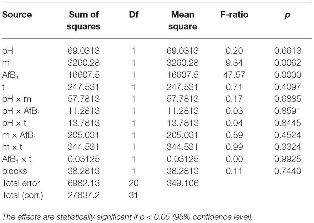
Table 1. Effect of pH, mass of mycelium (m), AfB1 concentration (AfB1), and time (t), and interactions thereof on AfB1 adsorption by P. eryngii mycelium.

Figure 1. Pareto chart of the standardized effect for AfB1 adsorption. A is the pH, B is the adsorbent mass, C is the mycotoxin concentration, and D is the time. The effect of one factor is statistically significant (p < 0.05) if its absolute value is higher than 2.3 (sector of the chart at the right of the vertical line).
Optimization of Adsorption
Mass of adsorbent (m) and concentration of AfB1 were identified as effective factors of adsorption and their effect was optimized by a factorial experiment in which the two variables were investigated at three levels. The 32 factorial design matrix and the results of the experiments are shown in Table 2.
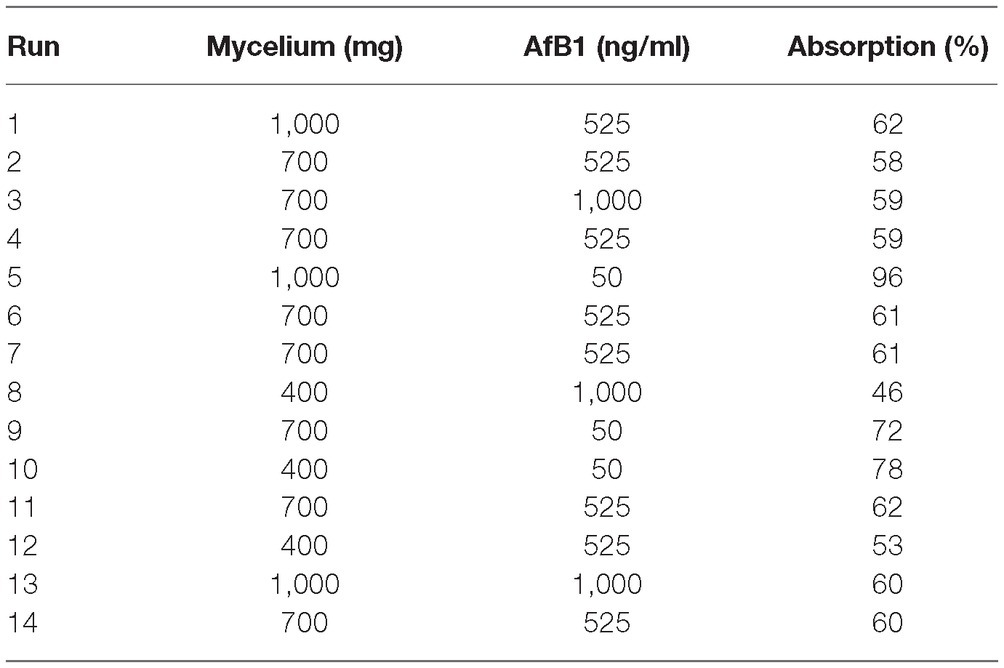
Table 2. The coded values for experimental design and the results.
The model expressed by Eq. (1), where the variables are expressed in their original units, represents the removal efficiency of AfB1 (Ads %) as a function of m and AfB1.
The model equation is useful in indicating the direction in which the variables should be changed in order to optimize the AfB1-removal efficiency of the adsorbent. The results of ANOVA are presented in Table 3. The statistical significance of each coefficient was determined by values of p: the smaller the values of p, the more significant is the coefficient. This implies that the first-order main effects of mass of adsorbent and mycotoxin concentration are more significant than their quadratic main effect. However, the quadratic main effect of AfB1 concentration is more significant than other second main effect.
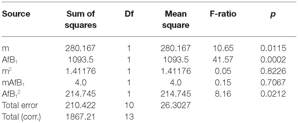
Table 3. Statistical significance of coefficients assessed by ANOVA.
The fit of the model was checked by the determination of the coefficient (R2). In this case, the value of the determination coefficient (R2 = 0.8873) indicated that the 11.27% of the total variable was not explained by the model.
Figure 2 shows the effect of the initial concentration of AfB1 and the quantity of the mycelium on mycotoxin removal efficiency.
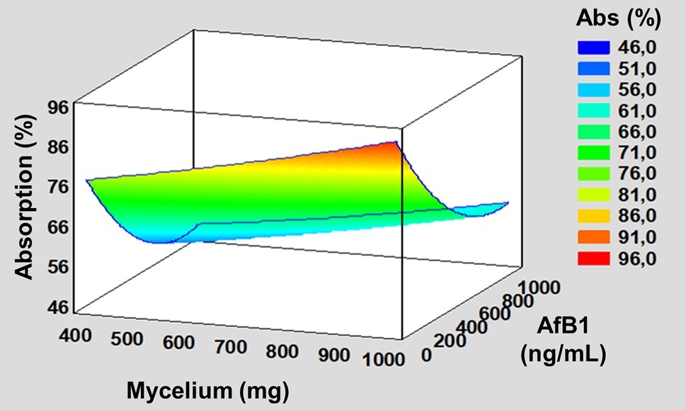
Figure 2. Estimate response surface plot for the effect of mass of adsorbent and mycotoxin concentration on the AfB1 removal.
The working conditions at the optimum point for removal efficiency of AfB1 were determined as follows:
Application of the optimum parameter m = 1,000 mg and AfB1 = 50 ng/ml to our model resulted in a theoretical optimum removal efficiency of AfB1 by Pleurotus mycelium of 90.07%. The experimentally determined removal efficiency for the same levels of “m” and “AfB1” was 85 ± 13% showing a satisfactory goodness-to-fit of the model.
SEM Analysis
A SEM micrograph of the P. eryngii mycelium is shown in Figure 3. The surface appears rough and sponge-like. The approximate pore size of 5–15 μm was measured from SEM analysis.
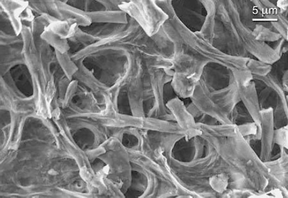
Figure 3. SEM secondary electron micrograph of P. eryngii mycelium.
Adsorption Isotherms
Several adsorption isotherm models have been used to describe experimental adsorption data. The Langmuir and Freundlich models are the most frequently employed models. In this work, both models were used to describe the effect of mycotoxin concentration (Figure 4) and the effect of adsorbent dosage (Figure 5).
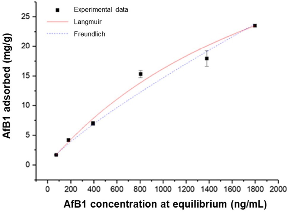
Figure 4. Effect of mycotoxin concentration on AfB1 adsorption by mycelium. Equilibrium adsorption isotherms were obtained at constant temperature (37°C) and pH (7) by testing a fixed amount of mycelium with increasing mycotoxin concentration.
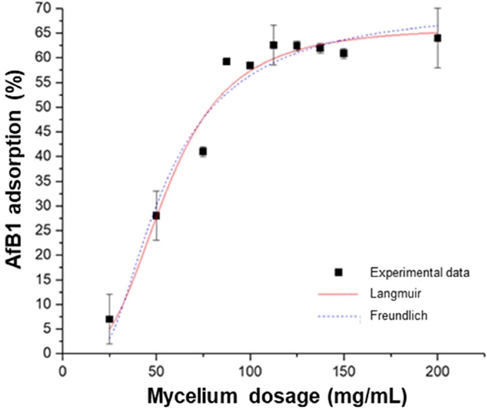
Figure 5. Effect of adsorbent dosage on AfB1 adsorption by mycelium. Equilibrium adsorption isotherms were obtained at constant temperature (37°C) and pH (7) by testing a fixed amount of mycotoxin with increasing adsorbent dosage.
The linear regression analysis was applied to assess the goodness of the fits and to calculate the parameters involved in the adsorption mechanism (Table 4). The results obtained by comparing R2 and SSres showed that, for both the effect of adsorbent quantity and the effect of AfB1 concentration, the isotherm that fits the experimental data is the Langmuir isotherm. This suggests that the AfB1 adsorption mechanism is monolayer and that occurs at a finite (fixed) number of definite equivalent sites. The model describes a homogeneous adsorption in which each molecule has enthalpy and activation energy of the constant process and is graphically characterized by a plateau, such as a saturation point where each molecule occupies a site and there can be no further adsorption.
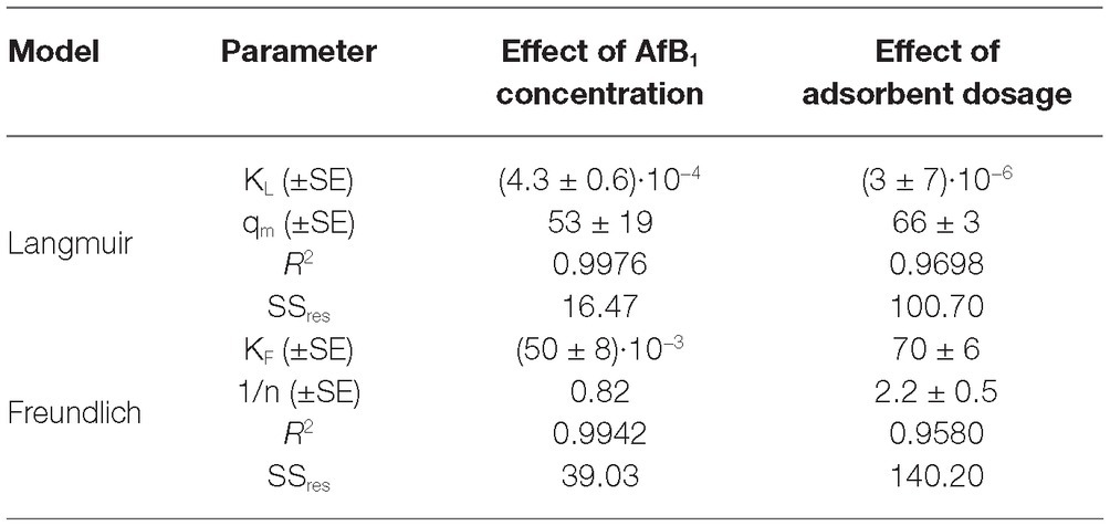
Table 4. Isotherm model parameters for the adsorption of AfB1 by P. eryngii mycelium.
The Langmuir model can be used to predict whether the adsorption system is favorable or unfavorable by calculating the dimensionless constant KR (Weber and Chakravorti, 1974). For favorable adsorption, the KR value should fall in the range 0–1. The adsorption is considered unfavorable when KR > 1, the isotherm is linear when KR = 1, and the adsorption is irreversible when KR = 0. In this study, the values of KR for AfB1 adsorption on P. eryngii mycelium are comprised between 0 and 1, which suggests a favorable process for the system.
Thermodynamic Parameters
The effect of temperature on the adsorption of AfB1 by P. eryngii mycelium was investigated. The uptake of AfB1 was found to increase when temperature increased: 66 ± 3% at 22°C, 67 ± 0% at 37°C, and 85 ± 3% at 50°C The increase of adsorption at increasing temperature indicates an endothermic nature of the adsorption process, as confirmed also by a positive ΔH0.
Molar free energy change of the adsorption process (ΔG0), standard enthalpy change (ΔH0), and standard entropy change (ΔS0) are shown in Table 5.
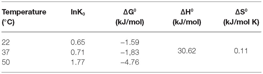
Table 5. Thermodynamic parameters for the AfB1 adsorption.
The negative ΔG0 values are indicative of a spontaneous adsorption process. The ΔG0 values decreased as the temperature was raised, which is an indication of a physical adsorption nature of the process. Generally the free energy variation for the physical adsorption is between −20 and 0 kJ/mol, while in chemisorption, the range is −80 to 400 kJ/mol (Kavak, 2009). Besides the physical nature of the process, the experimental data show that the adsorption process needs to be activated by a moderately high temperature. This implies that the process is reversible and that the material can be regenerated by an appropriate treatment.
Desorption Experiments
To verify the stability of the system over time, the percentage of desorption of the mycotoxin adsorbed on P. eryngii mycelium was assessed at room temperature and at the pH values of 3 and 7.4.
Desorption studies showed a very low desorption after 48 h at 25°C, at all the pH values tested. The percentage of desorption was 10 ± 4% at pH 3 and 7 ± 4% at pH 7.4. These results indicate a good stability of the system.
However, treatment with methanol resulted in a complete desorption of AfB1 from P. eryngii mycelium (recovery percentage 108 ± 6%). This result supports a possible re-utilization of the adsorbent after use, by regeneration of the adsorbing properties with an appropriate chemical treatment.
Discussion
AfB1 is one of the most important mycotoxins. It is produced by different species of Aspergillus, mainly A. flavus and A. parasiticus, in a number of agricultural products, including cereals, wine, spices, flavor products, peanuts, and soy. In this research work, we studied a method for the removal of AfB1 from a solution by absorption, a promising detoxification technology that is growing in industrial interest and economic prospect. In particular, we investigated the AfB1-adsorbing capability of the fungal mycelium of P. eryngii, an edible mushroom. The mycelium was produced and then processed, making it enzymatically inert by sterilization at high temperatures and subsequent lyophilization. The material thus obtained was morphologically characterized by SEM and subjected to various batch tests to assess its performance as biosorbent.
The adsorbents for mycotoxins are high-molecular weight compounds that are able to bind mycotoxins in contaminated feeds without releasing them into the gastro-intestinal tract of the animal. In this way, the toxin-adsorbent complex passes through the animals’ intestine and is eliminated with the feces. This prevents or minimizes the exposure of the animal to mycotoxins (Kabak et al., 2006). The temperature and pH conditions during animal digestion vary according to the class they belong to. In particular for ruminants, which are polygastric animals (cattle, sheep), the bolus temperature is 38–40°C and the pH is 6.2–6.5. In the case of monogastrics, such as pigs, poultry, dogs, and cats, the pH varies during digestion from 4 to 6 and the temperature is between 38 and 40°C. For horses, the pH during digestion is 7.4–7.6 and the temperature is 37.5 –38.5°C. The biosorbent capability of P. eryngii mycelium was studied at the temperature of 37°C and at the pH values of 5 and 7, which are compatible with the temperature and pH of the gastro-intestinal apparatus of most farm animals (Cunningham and Klein, 2007). In contrast with the approach used in most of the studies on adsorbents, which is based on variation of one factor at a time, we applied a factorial design model (DOE) to evaluate the influence of the different operative factors on the biosorption process (Manal, 2007). Concentration of mycotoxin present in the solution and the quantity of adsorbent material were identified as determinants of the process. The pH of the solution was irrelevant in a range from 5 to 7 (range compatible with the pH of application). Zavala-Franco et al. (2018) studied the adsorbing capability of different biosorbents by an in vitro poultry digestive model. In that study, the same variables as in our work, that is mass of adsorbent and AfB1 concentration, were assumed to be the main variables affecting the system. Also, Phillips et al. (1988) and Diaz et al. (2002) reported that pH had no influence on AfB1-binding by inorganic adsorbents. The DOE method, that we adopted herein, is intended to describe the variation of outcomes under conditions that are hypothesized to reflect the variation. This mathematical approach was developed to extrapolate the information needed through the least number of independent experiments. The fact that the results of our study are consistent with those obtained by more traditional approaches corroborates the validity of the DOE approach.
The system works in the same way over a range of time that goes from 30 to 120 min. This allowed to obtain a system that reaches equilibrium in a very short time (30 min) and that was found to remain stable for 48 h at room temperature, at pH 3 and pH 7.4, giving a desorption of 10 ± 4% and 7 ± 4% respectively. In optimal conditions, the mycelium of P. eryngii reaches 85 ± 13% of Afb1 removal efficiency, values slightly lower than those achieved by other adsorbent materials, such as aluminosilicates (Phillips et al., 1988) and bentonites (Diaz et al., 2002), both of which can remove up to 95% of Afb1. However, the latter have the disadvantage of showing high inclusion rates for vitamins and minerals, while mycelium of P. eryngii can be used as an alternative adsorbent material that is effective without causing nutritional losses. In a recently reported study, the adsorbing capability of different biosorbents, i.e., banana peel, Pyracantha leaves, and Aloe powder, were compared to that of zeolite in a laboratory model that simulated the conditions of the poultry gastro-intestinal tract (Zavala-Franco et al., 2018). The adsorption values assessed were 70, 69, 46, and 28% for zeolite, Aloe powder, Pyracantha leaves, and banana peel, respectively. Although determined in a different experimental system and therefore hardly comparable, these values appear significantly lower than adsorption achieved with Pleurotus mycelium.
The value of ΔH0 of mycelium sorption is positive, indicating that the reaction is endothermic. The magnitude of ΔH0 gives an indication of the type of adsorption, which can be either physical or chemical (Della Gatta, 1985). In the first case, the energy requirement is small (<40 kJ/mol) allowing the equilibrium to be attained rapidly and the process to be easily reversible (Ringot et al., 2005). On the contrary, chemical adsorption involves higher enthalpy changes (>40 kJ/mol). In this study, the value of enthalpy is less than 40 kJ/mol, indicating a physical adsorption phenomenon. The positive and small value of ΔS0 reflects the little increasing randomness at the solid/liquid interface during the adsorption of AfB1 on P. eryngii mycelium. The reaction was reversible and optimization of the process resulted in 85 ± 13% of AfB1 removal.
The effectiveness of adsorption processes depends on the chemical structures of the adsorbent and the mycotoxin involved. The most important feature for adsorption is the physical structure of the adsorbent, that is, its total charge, the charge distribution, the pore size, and the accessible surface area. The properties of the adsorbed mycotoxins, such as polarity, solubility, shape, and charge distribution, also play a significant role (Huwig et al., 2001). To the best of our knowledge, there is no previously published study on the mycotoxin-binding capability of fungal mycelium, though the adsorbing capability of fungal biomass has been shown for several organic and mineral (heavy metals) pollutants (Ahmaruzzaman, 2008; Wang and Chen, 2009). To date, the biosorption mechanism of organic compounds and metal ions by fungal biomass has been studied largely in relation to chitin and its deacetylated derivative, chitosan. The carboxylate and/or phosphate ligands along with the hydroxy and amide functional groups on the fungal cell wall components, which form relatively weak bonds with adsorbed molecules, have been proposed to be involved. Our SEM observations showed that the cell walls of P. eryngii mycelium are highly porous, with a pore size of 5–15 μm, which significantly increases the exposure of the cell wall active surfaces and of the sites of binding, thus making the process more efficient.
Conclusions
Our results show that non-viable mycelium of the fungus P. eryngii is able to efficiently adsorb AfB1 in conditions (temperature and pH) compatible with the physiology of animals’ digestion. A study was conducted to identify the major factors involved in the process. The concentration of mycotoxin in the solution and the quantity of adsorbent material were identified as determinants of the process. The pH of the solution was irrelevant in a range from 5 to 7 (range compatible with the pH of possible application). In addition, the system worked with no significant variation in the time lapse 30–120 min. of exposure. This allowed to obtain a system that reached equilibrium in a short time (30 min) and that remained stable in both acidic and slightly alkaline conditions that are compatible with pH values of the gastro-intestinal trait of farm animals. The thermodynamic study of the process showed that it is a spontaneous process with ΔG0 = −2.73 kJ/mol (average of ΔG0 at three temperatures 22, 37 and 50°C), endothermic (ΔH0 = 30.62 kJ/mol and ΔS0 = 0.11 kJ/mol·K) and that it is a physical adsorption, regulated by weak and reversible interactions, whereby the material can be regenerated with an appropriate treatment such as quantitative extraction with methanol. Optimization of biosorption resulted in 85 ± 13% of removal efficiency by P. eryngii mycelium.
The mycelium of P. eryngii is a biological and edible material and this characterizes this adsorbent as completely different from the materials currently used in the industry. The ongoing proof of concept and validation studies in vitro rumen models and in vivo might open the path for practical use of new, efficient though low-cost fungal mycelium-based feed additives for mycotoxin-biosorbtion and mitigation of mycotoxin risk.
Data Availability
The datasets generated for this study are available on request to the corresponding author.
Author Contributions
CA and MH conceived the research. CA, MH, and AL wrote the manuscript. MB carried out the microbiological work. EC carried out the DOE study. PA performed the scanning electron microscope (SEM) study. All authors designed the experiments, analyzed the data, contributed to manuscript revision, read and approved the submitted version.
Funding
This work was financially supported by H2020-E.U.3.2-678781-MycoKey.
Conflict of Interest Statement
The authors declare that the research was conducted in the absence of any commercial or financial relationships that could be construed as a potential conflict of interest.
Acknowledgments
The skilled technical assistance of Roberto Schena is gratefully acknowledged.
Abbreviations
ΔG0, Gibbs free energy; ΔH0, Standard enthalpy; ΔS0, Standard entropy; ABTS, 2,2′-azino-bis-3-ethylbenzothiazoline-6-sulfonate; Ads%, Percentage of adsorption; AfB1, Aflatoxin B1; FLD, Fluorescence detector; LOQ, Quantification limit of the method; MEA, Malt extract agar; MEB, Malt extract broth; PBS, Phosphate buffer saline; SE, Secondary electrons; SEM, Scanning electron microscope.
References
Ahmaruzzaman, M. (2008). Adsorption of phenolic compounds on low-cost adsorbents: a review. Adv. Colloid Interf. Sci. 143, 48–67. doi: 10.1016/j.cis.2008.07.002
Alberts, J. F., Gelderblom, W. C. A., Botha, A., and Van Zyl, W. H. (2009). Degradation of aflatoxin B 1 by fungal laccase enzymes. Int. J. Food Microbiol. 135, 47–52. doi: 10.1016/j.ijfoodmicro.2009.07.022
AOAC (2000). Section 49.2.02 (AOAC method 971.22) preparation of standards. In official methods of analysis. 17th edn. (Gaithersburg, MD, USA: AOAC International).
Boudergue, C., Burel, C., Dragacci, S., Favrot, M. C., Fremy, J. M., Massimi, C., et al. (2009). Review of mycotoxin-detoxifying agents used as feed additives: mode of action, efficacy and feed/food safety. EFSA Support. Publ. 6:22E. doi: 10.2903/sp.efsa.2009.EN-22
Branà, M. T., Cimmarusti, M. T., Haidukowski, M., Logrieco, A. F., and Altomare, C. (2017). Bioremediation of aflatoxin B1-contaminated maize by king oyster mushroom (Pleurotus eryngii). PLoS One 12:e0182574. doi: 10.1371/journal.pone.0182574
Brasil, J. L., Ev, R. R., Milcharek, C. D., Martins, L. C., Pavan, F. A., dos Santos, A. A. Jr., et al. (2006). Statistical design of experiments as a tool for optimizing the batch conditions to Cr (VI) biosorption on Araucaria angustifolia wastes. J. Hazard. Mater. 133, 143–153. doi: 10.1016/j.jhazmat.2005.10.002
Crini, G. (2006). Non-conventional low-cost adsorbents for dye removal: a review. Bioresour. Technol. 97, 1061–1085. doi: 10.1016/j.biortech.2005.05.001
Cunningham, J. G., and Klein, B. G. (2007). Veterinary physiology. (Philadelphia: Saunders Elsevier).
Della Gatta, G. (1985). Direct determination of adsorption heats. Thermochim. Acta 96, 349–363. doi: 10.1016/0040-6031(85)80074-5
Diaz, D. E., Hagler, W. M., Hopkins, B. A., and Whitlow, L. W. (2002). Aflatoxin binders I: in vitro binding assay for aflatoxin B1 by several potential sequestering agents. Mycopathologia 156, 223–226. doi: 10.1023/A:1023388321713
El-Nezami, H., Kankaanpaa, P., Salminen, S., and Ahokas, J. (1998). Ability of dairy strains of lactic acid bacteria to bind a common food carcinogen, aflatoxin B1. Food Chem. Toxicol. 36, 321–326. doi: 10.1016/S0278-6915(97)00160-9
Fu, Y., and Viraraghavan, T. (2001). Fungal decolorization of dye wastewaters: a review. Bioresour. Technol. 79, 251–262. doi: 10.1016/S0960-8524(01)00028-1
Gadd, G. M. (2009). Biosorption: critical review of scientific rationale, environmental importance and significance for pollution treatment. J. Chem. Technol. Biotechnol 84, 13–28. doi: 10.1002/jctb.1999
Gavrilescu, M. (2004). Removal of heavy metals from the environment by biosorption. Eng. Life Sci. 4, 219–232. doi: 10.1002/elsc.200420026
Huwig, A., Freimund, S., Käppeli, O., and Dutler, H. (2001). Mycotoxin detoxication of animal feed by different adsorbents. Toxicol. Lett. 122, 179–188. doi: 10.1016/S0378-4274(01)00360-5
Kabak, B., Dobson, A. D., and Var, I. I. L. (2006). Strategies to prevent mycotoxin contamination of food and animal feed: a review. Crit. Rev. Food Sci. Nutr. 46, 593–619. doi: 10.1080/10408390500436185
Kalmiş, E., Azbar, N., and Kalyoncu, F. (2008). Evaluation of two wild types of Pleurotus ostreatus (MCC07 and MCC20) isolated from nature for their ability to decolorize Benazol black ZN textile dye in comparison to some commercial types of white rot fungi: Pleurotus ostreatus, Pleurotus djamor, and Pleurotus citrinopileatus. Can. J. Microbiol. 54, 366–370. doi: 10.1139/w08-025
Kapahi, M., and Sachdeva, S. (2017). Mycoremediation potential of Pleurotus species for heavy metals: a review. Bioresour. Bioprocess. 4:32. doi: 10.1186/s40643-017-0162-8
Kavak, D. (2009). Removal of boron from aqueous solutions by batch adsorption on calcined alunite using experimental design. J. Hazard. Mater. 163, 308–314. doi: 10.1016/j.jhazmat.2008.06.093
Kolosova, A., and Stroka, J. (2011). Substances for reduction of the contamination of feed by mycotoxins: a review. World Mycotoxin J. 4, 225–256. doi: 10.3920/WMJ2011.1288
Langmuir, I. (1916). The constitution and fundamental properties of solids and liquids. Part I. solids. J. Am. Chem. Soc. 38, 2221–2295. doi: 10.1021/ja02268a002
Li, A., Zhu, Y., Xu, L., Zhu, W., and Tian, X. (2008). Comparative study on the determination of assay for laccase of Trametes sp. Afr. J. Biochem. Res. 2, 181–183.
Loffredo, E., Castellana, G., Traversa, A., and Senesi, N. (2013). Comparative assessment of three ligninolytic fungi for removal of phenolic endocrine disruptors from freshwaters and sediments. Environ. Technol. 34, 1601–1608. doi: 10.1080/09593330.2012.760654
Low, B. T., Ting, Y. P., and Deng, S. (2008). Surface modification of Penicillium chrysogenum mycelium for enhanced anionic dye removal. Chem. Eng. J. 141, 9–17. doi: 10.1016/j.cej.2007.10.004
Manal, F. (2007). Biosorption of cadmium and lead by Phragmites australis biomass using factorial experiment design. Global J. Biotechnol. Biochem. 10, 10–20.
Peltonen, K., El-Nezami, H., Haskard, C., Ahokas, J., and Salminen, S. (2001). Aflatoxin B1 binding by dairy strains of lactic acid bacteria and bifidobacteria. J. Dairy Sci. 84, 2152–2156. doi: 10.3168/jds.S0022-0302(01)74660-7
Phillips, T. D., Kubena, L. F., Harvey, R. B., Taylor, D. R., and Heildebaugh, N. D. (1988). Hydrated sodium calcium aluminosilicate: a high affinity sorbent for aflatoxin. Poult. Sci. 67, 243–247. doi: 10.3382/ps.0670243
Purnomo, A. S., Mori, T., Kamei, I., Nishii, T., and Kondo, K. (2010). Application of mushroom waste medium from Pleurotus ostreatus for bioremediation of DDT-contaminated soil. Int. Biodeterior. Biodegradation 64, 397–402. doi: 10.1016/j.ibiod.2010.04.007
Rigas, F., Papadopoulou, K., Philippoussis, A., Papadopoulou, M., and Chatzipavlidi, J. (2009). Bioremediation of lindane contaminated soil by Pleurotus ostreatus in non sterile conditions using multilevel factorial design. Water Air Soil Pollut. 197, 121–129. doi: 10.1007/s11270-008-9795-8
Ringot, D., Lerzy, B., Bonhoure, J. P., Auclair, E., Oriol, E., and Larondelle, Y. (2005). Effect of temperature on in vitro ochratoxin a biosorption onto yeast cell wall derivatives. Process Biochem. 40, 3008–3016. doi: 10.1016/j.procbio.2005.02.006
Rushing, B. R., and Selim, M. I. (2019). Aflatoxin B1: a review on metabolism, toxicity, occurrence in food, occupational exposure, and detoxification methods. Food Chem. Toxicol. 124, 81–100. doi: 10.1016/j.fct.2018.11.047
Sánchez, C. (2010). Cultivation of Pleurotus ostreatus and other edible mushrooms. Appl. Microbiol. Biotechnol. 85, 1321–1337. doi: 10.1007/s00253-009-2343-7
Vila-Donat, P., Marín, S., Sanchis, V., and Ramos, A. J. (2018). A review of the mycotoxin adsorbing agents, with an emphasis on their multi-binding capacity, for animal feed decontamination. Food Chem. Toxicol. 114, 246–259. doi: 10.1016/j.fct.2018.02.044 [Epub 2018/02/21].
Wang, J., and Chen, C. (2009). Biosorbents for heavy metals removal and their future. Biotechnol. Adv. 27, 195–226. doi: 10.1016/j.biotechadv.2008.11.002
Weber, T. W., and Chakravorti, R. K. (1974). Pore and solid diffusion models for fixed-bed adsorbers. AICHE J. 20, 228–238. doi: 10.1002/aic.690200204
Williams, J. H., Phillips, T. D., Jolly, P. E., Stiles, J. K., Jolly, C. M., and Aggarwal, D. (2004). Human aflatoxicosis in developing countries: a review of toxicology, exposure, potential health consequences, and interventions. Am. J. Clin. Nutr. 80, 1106–1122. doi: 10.1093/ajcn/80.5.1106
World Health Organization and International Agency for Research on Cancer, (1993). Some naturally occurring substances: food items and constituents, heterocyclic aromatic amines and mycotoxins. IARC Monographs on the Evaluation of the Carcinogenic Risk of Chemicals to Humans, 56.
Zavala-Franco, A., Hernández-Patlán, D., Solís-Cruz, B., López-Arellano, R., Tellez-Isaias, G., Vázquez-Durán, A., et al. (2018). Assessing the aflatoxin B1 adsorption capacity between biosorbents using an in vitro multicompartmental model simulating the dynamic conditions in the gastrointestinal tract of poultry. Toxins 10:484. doi: 10.3390/toxins10110484
Keywords: biosorption, aflatoxin, Pleurotus eryngii, feed additive, king oyster mushroom
Citation: Haidukowski M, Casamassima E, Cimmarusti MT, Branà MT, Longobardi F, Acquafredda P, Logrieco A and Altomare C (2019) Aflatoxin B1-Adsorbing Capability of Pleurotus eryngii Mycelium: Efficiency and Modeling of the Process. Front. Microbiol. 10:1386. doi: 10.3389/fmicb.2019.01386
Edited by:
Eugenia Bezirtzoglou, Democritus University of Thrace, GreeceReviewed by:
Anatoly V. Zherdev, Research Center of Biotechnology of the Russian Academy of Sciences, RussiaCarlos Augusto Fernandes Oliveira, University of São Paulo, Brazil
Zhaowei Zhang, Oil Crops Research Institute (CAAS), China
Copyright © 2019 Haidukowski, Casamassima, Cimmarusti, Branà, Longobardi, Acquafredda, Logrieco and Altomare. This is an open-access article distributed under the terms of the Creative Commons Attribution License (CC BY). The use, distribution or reproduction in other forums is permitted, provided the original author(s) and the copyright owner(s) are credited and that the original publication in this journal is cited, in accordance with accepted academic practice. No use, distribution or reproduction is permitted which does not comply with these terms.
*Correspondence: Antonio Logrieco, antonio.logrieco@ispa.cnr.it