 Sushmita Patwardhan
 Francesco Smedile1,2
 Donato Giovannelli
 Costantino Vetriani
- 1Department of Marine and Coastal Sciences, Rutgers University, New Brunswick, NJ, United States
- 2National Research Council, Institute for Coastal Marine Environment, Messina, Italy
- 3Department of Biology, University of Naples “Federico II,” Naples, Italy
- 4National Research Council, Institute for Marine Biological and Biotechnological Resources, Ancona, Italy
- 5Earth-Life Science Institute, Tokyo Institute of Technology, Tokyo, Japan
- 6Department of Biochemistry and Microbiology, Rutgers University, New Brunswick, NJ, United States
Tor Caldara is a shallow-water gas vent located in the Mediterranean Sea, with active venting of CO2 and H2S. At Tor Caldara, filamentous microbial biofilms, mainly composed of Epsilon- and Gammaproteobacteria, grow on substrates exposed to the gas venting. In this study, we took a metaproteogenomic approach to identify the metabolic potential and in situ expression of central metabolic pathways at two stages of biofilm maturation. Our findings indicate that inorganic reduced sulfur species are the main electron donors and CO2 the main carbon source for the filamentous biofilms, which conserve energy by oxygen and nitrate respiration, fix dinitrogen gas and detoxify heavy metals. Three metagenome-assembled genomes (MAGs), representative of key members in the biofilm community, were also recovered. Metaproteomic data show that metabolically active chemoautotrophic sulfide-oxidizing members of the Epsilonproteobacteria dominated the young microbial biofilms, while Gammaproteobacteria become prevalent in the established community. The co-expression of different pathways for sulfide oxidation by these two classes of bacteria suggests exposure to different sulfide concentrations within the biofilms, as well as fine-tuned adaptations of the enzymatic complexes. Taken together, our findings demonstrate a shift in the taxonomic composition and associated metabolic activity of these biofilms in the course of the colonization process.
Introduction
Marine gas vents transport volatile elements and compounds from the geosphere to the hydrosphere by seepage through sediments and bedrocks (Suess, 2014). At these sites, geological processes, biogeochemical reactions and the activity of microorganisms act together to alter the composition of volatile products. The release of such reduced volatiles into the oxygenated water column generates a redox disequilibrium that can be harnessed by benthic prokaryotic communities to convert chemical energy into ATP. Hydrothermal and gas vents occur both in shallow water (depth <200 meters) and in the deep-sea (depth >200 meters; Tarasov et al., 2005). Shallow-water hydrothermal and gas vents are associated with submarine volcanoes, arc and back-arc volcanoes and occur globally in the proximity of active plate margins and intraplate hotspots (reviewed in Price and Giovannelli, 2017).
The subduction of the African plate below Europe has resulted in the formation of the Mediterranean Ridge and deep subduction basins as well as active volcanic arcs in the Tyrrhenian and Aegean Seas (Dando et al., 1999). The terrestrial volcanic systems in these areas have been well-studied, both because of the long history of devastation caused by their eruptions and because of their geothermal potential. The submarine parts of the system have received relatively little attention until the last decade, and are still poorly studied compared to some of the mid-ocean ridge systems (Price and Giovannelli, 2017). Volcanic arc hydrothermal systems release large volumes of volatiles, because of both degassing of the subducted slab and the mantle, and the decomposition of carbonates in the overlying marine sediments (Barry et al., 2019). Since most of the known venting in the Mediterranean is from shallow vents, the majority of the outlets are of the gasohydrothermal type, emitting large volumes of carbon dioxide.
The microbiology of the shallow-water vent systems of the Aegean and Tyrrhenian basins has been mainly investigated in sediments (Gugliandolo and Maugeri, 1993; Sievert et al., 1999, 2000; Giovannelli et al., 2013; Maugeri et al., 2013; Price et al., 2013; Kerfahi et al., 2014). However, besides sediment communities, substrate-attached chemosynthetic microbial biofilms are widespread and, in their role of primary producers, relevant in these ecosystems (Kalanetra et al., 2004; Reigstad et al., 2011; Miranda et al., 2016). Here, we investigated two types of biofilm communities at a shallow-water gas vent located at Tor Caldara, Italy.
Tor Caldara is a natural reserve located near the town of Anzio, about 60 km south of Rome. Submarine active venting of gases, including CO2 originating from a deep magma source, occurs near the coast of the Tyrrhenian sea at Tor Caldara, in association with the quiescent volcanic system of Colli Albani, which is located inland around 40 km north-east of the venting site (Carapezza and Tarchini, 2007). The absence of a thermal anomaly at Tor Caldara is attributed to the infiltration of cold meteoric water (Carapezza et al., 2012). In a previous study, we investigated the chemical composition of the gases at Tor Caldara and the taxonomic diversity of the associated filamentous biofilm communities (Patwardhan et al., 2018). The gases at Tor Caldara are mainly composed of CO2 (avg. of 76.67 mol%) and H2S (avg. of 23.13 mol%) with minor contribution of CH4 (avg. of 0.18 mol%), CO (avg. of 0.0080 mol%) and H2 (avg. of 0.00072 mol%). Young and established filamentous biofilms—the former collected on glass slides deployed at the venting site at Tor Caldara and the latter attached to the native rocky substrates—were dominated by members of the classes Epsilonproteobacteria (aka Campylobacteria, synonym pending validation) and Gammaproteobacteria, respectively, albeit in different proportions. On average, sulfur-oxidizing Epsilonproteobacteria of the genus Sulfurovum accounted for 57.6% of the active young biofilms, while sulfur-oxidizing Gammaproteobacteria of the genus Thiomicrospira as well as sequences related to the Thiothrix CF-26 group constituted more than 60% of the active established biofilm community (Patwardhan et al., 2018). This observed transition from Epsilon- to Gammaproteobacteria during the maturation of the biofilms revealed an ecological succession between the young and established communities. Previous studies of sulfidic environments, including caves, shallow-water and deep-sea hydrothermal vents, reported a spatial segregation between these two classes of bacteria and correlated this distribution pattern with geochemical data, leading to the hypothesis that Epsiloproteobacteria can cope with higher concentrations of hydrogen sulfide than Gammaproteobacteria (Engel et al., 2004; Macalady et al., 2008; Reigstad et al., 2011; Giovannelli et al., 2013; O’Brien et al., 2015; Miranda et al., 2016; Meier et al., 2017). Based on these studies, and on the observed succession observed at Tor Caldara, we tested the hypothesis that Epsilon- and Gammaproteobacteria are adapted to different concentrations of sulfide. Sulfide gradient experiments with laboratory strains showed that, conservatively, Epsilonproteobacteria can tolerate sulfide concentrations up to 20 times higher than Gammaproteobacteria, possibly facilitating their role as pioneer colonizers of sulfidic environments (Patwardhan et al., 2018).
To conserve energy, prokaryotes oxidize reduced inorganic sulfur compounds via a number of different pathways. The Sox pathway is widespread and occurs in both anaerobic as well as aerobic phototrophic and chemolithoautotrophic microorganisms. The well-characterized Sox enzyme complex in the alphaproteobacterium, Paracoccus pantotrophus, has four periplasmic proteins: SoxYZ, SoxXA, SoxB, and SoxCD. These proteins work in concert to oxidize thiosulfate, hydrogen sulfide, sulfur as well as sulfite all the way to sulfate. In some sulfur-oxidizing bacteria (SOB) such as Rhodobacter capsulatus, sulfide is oxidized to sulfur by the membrane bound sulfide quinone oxidoreductase (SQR) enzyme. Additionally, in few SOBs, sulfide is also oxidized to sulfur by the periplasmic sulfide dehydrogenase (FccAB) (Visser et al., 1997; Muβmann et al., 2007).
In this study, we used an integrated metagenomic/metaproteomic approach to compare the metabolic potential and the in situ expression of central metabolic pathways of the established and young filaments at Tor Caldara. We identified and quantified key genes and proteins involved in the carbon, sulfur and nitrogen cycles and compared them between the young and established biofilms to establish how different metabolic pathways were expressed in the two communities. We also reconstructed three metagenome-assembled genomes (MAGs) belonging to the classes Epsilonproteobacteria and Gammaproteobacteria.
Materials and Methods
Study Site
Tor Caldara is a natural reserve located approximately 60 km south of Rome and 40 km south-west of Colli Albani, Italy (Patwardhan et al., 2018). The study site (41°29′ 9″ N 12°35′ 23″ E) is a coastal submarine gas vent located at a depth of approximately 3 meters. The site is characterized by vigorous venting of gases that escape from the sandy seabed. The sediment in the venting area is distinctly darker in color than the control sediment because of possible sulfide deposition. Conspicuous growth of white microbial filaments is seen on the rocks directly exposed to the gas venting.
Sample Collection
Sampling of filaments and collection and analysis of gases were performed as described previously (Patwardhan et al., 2018). Briefly, established filaments (EF from here on) growing on rocks exposed to the gas venting were collected by a SCUBA diver in August 2016. To study the early stages of colonization, young biofilms (YF from here on) were collected on sterile glass slides mounted on an aluminum rod and exposed to the venting area for four days. Biofilms samples were then stored in RNA Later at –80°C for further nucleic acid and protein extraction. Representative samples of the two communities (one EF and one YF) were further used for downstream applications.
Nucleic Acid Extraction, Metagenomic Library Preparation and Analysis
Using data from previous analyses of the composition of the Tor Caldara microbial communities as a guideline (Patwardhan et al., 2018), we selected one YF and one EF biofilm sample for metaproteogenomic analyses. DNA was extracted from YF and EF biofilm biomass stored in RNA Later following a phenol:chloroform extraction protocol. Briefly, 0.8 g of sample were added to 850 μl of extraction buffer (50 mM Tris–HCl, 20 mM EDTA, 100 mM NaCl; pH 8.0) supplemented with100 μl of lysozyme (100 mg/ml) and incubated at 37°C for 30 min. This mix was then supplemented with 5 μl of proteinase K (20 mg/ml), incubated at 37°C for 30 min, and subsequently supplemented with 50 μl SDS (20%) and further incubated at 65°C for 1 hr. Nucleic acids were extracted by performing a series of phenol:chloroform:isoamylalcohol (25:24:1) and chloroform:isoamyl alcohol (24:1) extractions. Multiple samples were co–extracted to reduce potential bias. The final supernatant was precipitated in 3 M sodium–acetate and isopropanol, washed twice with 70% ice–cold ethanol and re–suspended in ultra–pure sterile water. The library was prepared at Molecular Research LP (Shallowater, TX), using the Nextera DNA Sample preparation kit (Illumina) following the manufacturer’s user guide. The initial concentration of DNA was evaluated using the Qubit® dsDNA HS Assay Kit (Life Technologies). Initial library preparation resulted in very large size library (>2,500 bp), therefore, library preparation was repeated after DNA inhibitor removal using the DNEasy PowerClean Pro Cleanup Kit (Qiagen). Fifty nanogram DNA was used to prepare the library. The sample underwent the simultaneous fragmentation and addition of adapter sequences. These adapters are utilized during a limited-cycle (5 cycles) PCR in which unique indices were added to the sample. Following the library preparation, the final concentration of the library was measured using the Qubit® dsDNA HS Assay Kit (Life Technologies), and the average library size was determined using the Agilent 2100 Bioanalyzer (Agilent Technologies). The library was diluted (to 10 pM) and sequenced paired end (2 × 2 50 bp, 10 million paired sequences per sample) for 500 cycles on a shared multiplexed run using an Illumina Novaseq 6000 instrument. Following the removal of the adapters, at the sequencing facility, the sequences were quality checked using FastQC v.0.11.51. A total of 6,698,798 and 10,125,188 paired end sequences were used for downstream analysis for the EF and YF samples respectively. Sequences encoding rRNA genes were extracted using Metaxa2 v2.1 using default parameters (Bengtsson-Palme et al., 2015) and annotated using Silva ngs v.1.3.9 (Quast et al., 2013). The paired end sequences were then assembled using Megahit v1.1.3 (Li et al., 2015) with default parameters, and assembly was checked using QUAST v3.4 (Gurevich et al., 2013). The EF assembly had a total of 99,137 contigs with the largest contig being 212,020 bp long, whereas the YF assembly had a total of 87,836 contigs with the largest contig being 261,286 bp long. Assembled metagenomes were submitted to DOE Joint Genome Institute’s Integrated Microbial Genome Metagenomic Expert Review (IMG/ER) pipeline for Open Reading Frame (ORF) identification as well as functional and taxonomic annotation (Markowitz et al., 2012). The EF and YF metagenomic assemblies had a total of 308,874 and 362,063 sequences, respectively. The sequences of both metagenomes had COG (51%), KO (41%) and KEGG (25%) annotations. Post annotation, using default parameters for bowtie2 v.2.3.3.1, the reads were mapped back to the assembly (Langmead et al., 2009). Using HTSeq v0.9.1, the number of reads mapping to each gene was counted (Anders et al., 2015). Absolute read counts mapped to each gene were normalized for varying gene lengths and difference in sequencing depth between samples. These normalized gene abundances were expressed as TPM (Transcripts Per Million; Wagner et al., 2012). Sequences are available through the NCBI Short Read Archive database with accession number PRJNA498803.
Protein Extraction and Metaproteomic Analysis
The total protein fraction was extracted from approximately 1 g of the YF and EF biofilms. Hundred microliter of 2X Laemmli buffer and 50 mM DTT was added to each sample, which was then sonicated, heated at 95°C for 10 min, and centrifuged, with supernatant saved. The precipitate was further extracted by addition of 100 μL of 8M urea, sonicated and centrifuged. Both supernatant were combined and protein concentration was measured using the Pierce 660nm Protein Assay Reagent (ThermoFisher). Thirty microliter out of Two hundred microliter of each sample were run into NuPAGE 10% Bis-Tris Gel (1.5 mmX10 well, Invitrogen). The gel was stained with Coomassie brilliant Blue R250. Standard in-gel tryptic digest was performed as described in Xie et al. (2015). Label-free liquid chromatography-tandem mass spectrometry (LC-MS/MS) was carried out on the two samples, which were analyzed by nano LC-MS/MS using a Q-Exactive HF mass spectrometer interfaced with a Ultimate 3000 RSLCnano chromatography system (Thermofisher). Samples were loaded onto a fused silica trap column (Acclaim PepMap 100, 75 μm × 2 cm, ThermoFisher). After washing for 5 min at 5 μl/min with 0.1% TFA, the trap column was brought in line with an analytical column (Nanoease MZ peptide BEH C18, 130A, 1.7 μm, 75 μm × 250 mm, Waters) for LC-MS/MS. Peptides were fractionated at 300 nL/min using a segmented linear gradient 4–15% B in 30min (A: 0.2% formic acid; B: 0.16% formic acid, 80% acetonitrile), 15–25%B in 40 min, 25–50%B in 44 min, and 50–90%B in 11min. Mass spectrometry data were acquired using a data-dependent acquisition procedure with an MS1 scan (resolution 120,000) followed by MS/MS (resolution 30,000; HCD relative collision energy 27%) on the 20 most intense ions with a dynamic exclusion duration of 20 s. The open-source X!Tandem software (Craig and Beavis, 2004) was used to match spectral data to peptide sequences of the corresponding metagenomes for functional and taxonomical annotation. The minimum acceptable peptide expectation scores were set at 10–2; the overall peptide false positive rate (Gupta et al., 2011) was 0.07%. A total of 138,387 and 97,607 peptides were obtained in the metaproteomes of EF and YF, respectively. Protein counts were normalized to the total proteins obtained from each sample. Normalized counts for proteins of interest were manually compared between the two samples. These normalized counts were expressed as a percent of total proteins observed in each sample. Raw metaproteomic data were deposited into the ProteomeXchange database with accession number PXD0233572.
Whole Genome Reconstruction From Metagenomes
Quality checked reads from both samples were co-assembled using Megahit v1.1.3 with default parameters and QUAST v3.4 was used to check the quality of the co-assembly. The co-assembled contigs from the two metagenomes were then binned using MaxBin2.0 (Wu et al., 2014) to recover individual genomes. Completeness, taxonomic affiliation and contamination of recovered bins was checked using CheckM v1.0.7 (Parks et al., 2015). The resulting three metagenome-assembled genomes (MAGs TCMF1, TCMF2, and TCMF9) were annotated in RAST (Rapid Annotation using Subsystem Technology; Aziz et al., 2008) and are available with IDs: TCMF1: 6666666.318663; TCMF2: 6666666.318627; TCMF9: 6666666.318829).
Phylogenetic Analyses
Sulfide quinone oxidoreductase (SQR) amino acid sequences obtained in this study and from GenBank (via Blast) were aligned with ClustalX v 2.0 (Thompson et al., 1997) and manually adjusted using Seaview (Galtier et al., 1996). Phylogenetic distances were calculated using the LG amino acid replacement matrix (Le and Gascuel, 2008) and the maximum likelihood method was used to evaluate tree topologies in PhyML 3.0 (Guindon et al., 2010). Branch support was estimated using the approximate likelihood ratio test (aLRT; Anisimova and Gascuel, 2006) and by bootstrap analysis with 1,000 resamplings. Both estimates gave comparable results.
Results
Microbial Community Composition of the Established and Young Filaments
Previous 16S rRNA gene and transcript analyses of the Tor Caldara biofilms showed that there was very little taxonomic variation within replicates of the YF and EF communities, respectively, and that the observed shift in community composition between the YF and EF biofilms was controlled predominantly by the biofilm age (Patwardhan et al., 2018). Based in these findings, two biofilms (one YF and one EF) were selected for further metaproteogenomic analyses. The bacterial diversity of EF and YF was evaluated following in silico extraction of the 16S rRNA gene sequences from the two metagenomes. The EF biofilm was dominated by Gammaproteobacteria (54%), followed by Epsilonproteobacteria (29%) and small proportions of Bacteroidia (4%) and Alphaproteobacteria (3%; Figure 1A). Within the Gammaproteobacteria, the genus Thiomicrospira (40%; now reclassified as Thiomicrorhabdus; Boden et al., 2017) and uncultured members belonging to the Thiotrichales (11%) were dominant (Figure 1B). A reverse trend was observed in the YF biofilm, with Epsilonproteobacteria (67%) dominating the community, followed by Gammaproteobacteria (23%), Bacteroidia (2%), and Alphaproteobacteria (1%; Figure 1A). In the YF biofilm, Sulfurovum (66%) was the most abundant genus within the Epsilonproteobacteria (Figure 1B).
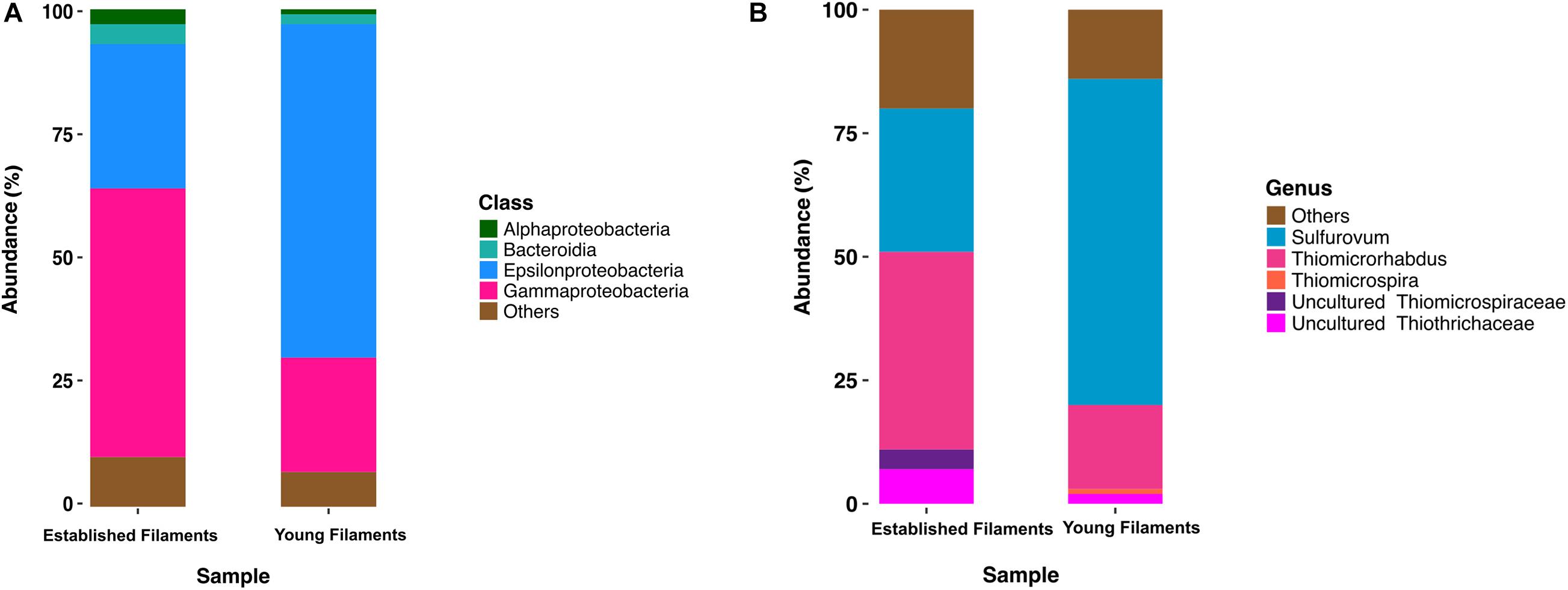
Figure 1. Class (A) and genus (B) level distribution of 16S rRNA gene and transcript sequences recovered from established and young filaments.
Comparative Metagenomics and Metaproteomics of the Established and Young Filaments
We compared the normalized abundances of key genes and proteins involved in central metabolic pathways, including carbon fixation, nitrogen, sulfur metabolism and (micro)aerobic respiration (Supplementary Tables 1, 2). Since shallow-water vents are enriched in heavy metals, genes and proteins involved in their detoxification were also identified and compared (Supplementary Tables 1, 2). In line with the 16S rRNA gene-inferred community composition, the majority of functional genes and proteins were affiliated with Gamma- and Epsilonproteobacteria (Figures 2–4). The metagenomes provided a snapshot of the metabolic potential of the microbial communities, and were also used to annotate the metaproteomes, which revealed the in situ expression of central metabolic pathways of the communities. The metagenomes were also used to obtain metagenome-assembled genomes (MAGs) of the most abundant members of the filamentous biofilms.
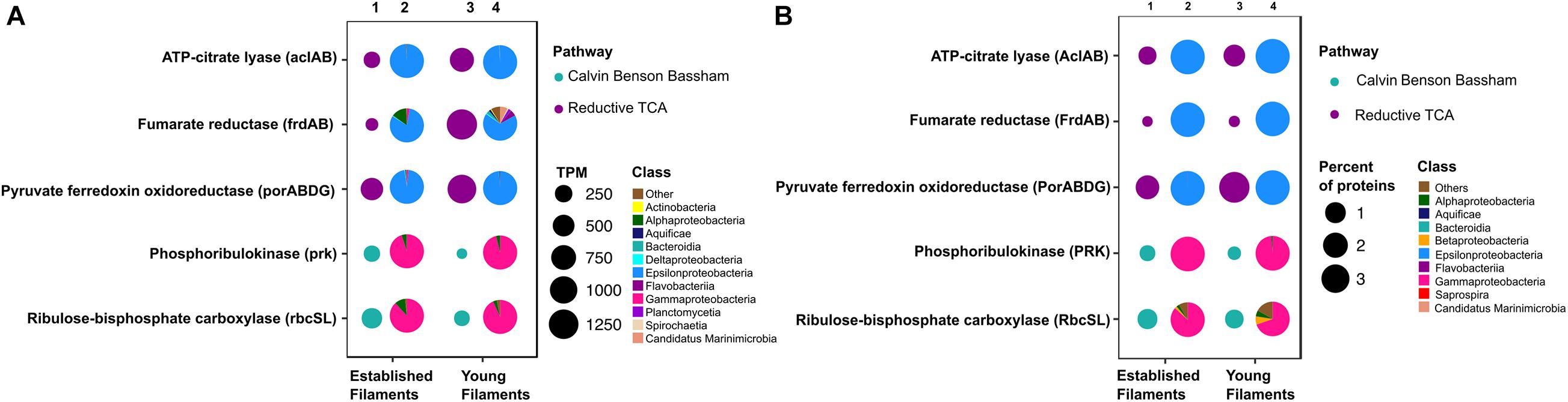
Figure 2. Profiles of genes and proteins for carbon fixation in established and young filamentous biofilms. (A) Transcripts per million (TPM; columns 1 and 3) and taxonomic affiliation of reads annotated to genes (columns 2 and 4); (B) Percentage (columns 1 and 3) and taxonomic affiliation of reads annotated to proteins (columns 2 and 4).
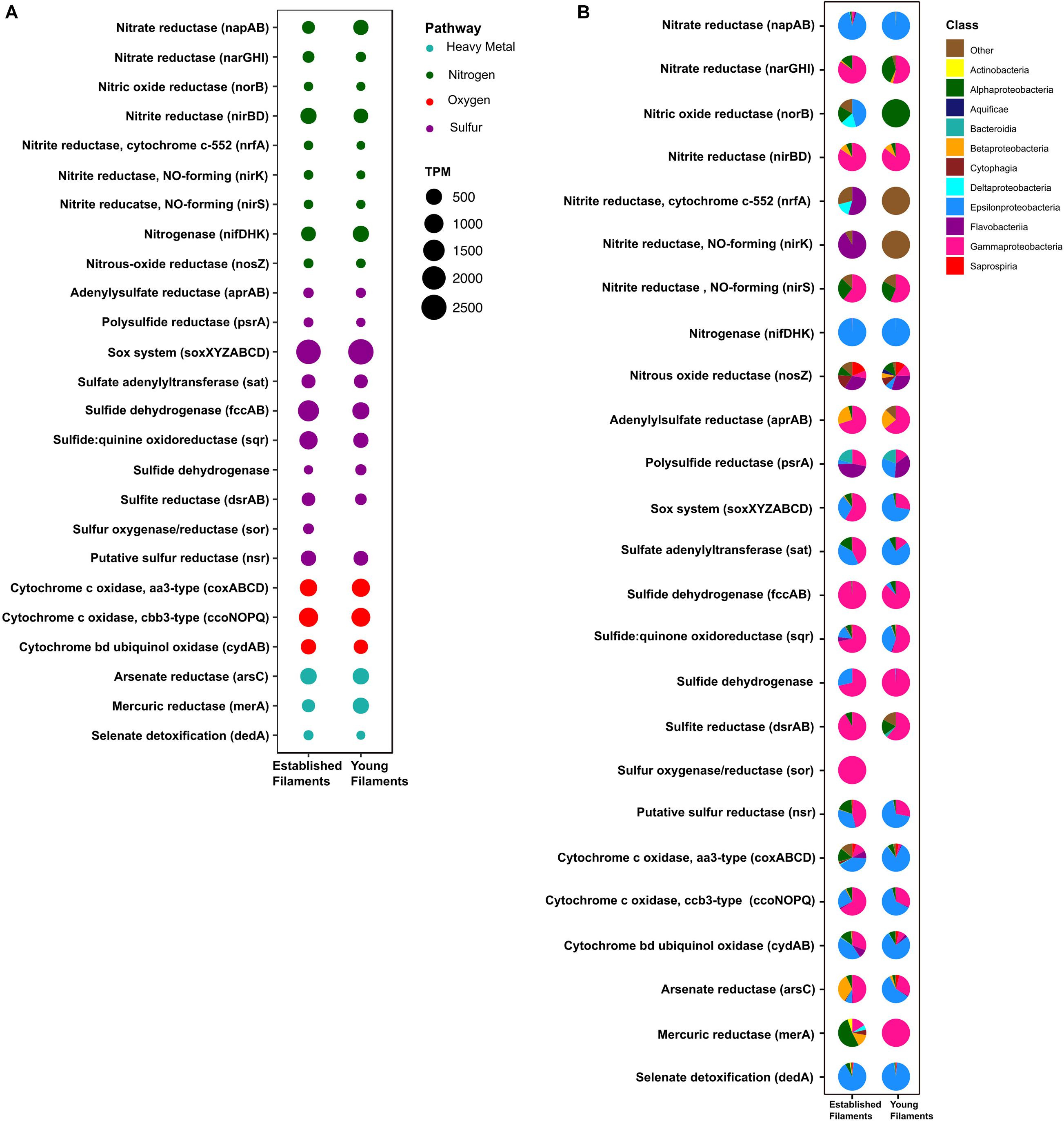
Figure 3. (A) Transcripts per million (TPM) and (B) taxonomic affiliation of reads annotated to key genes involved in nitrogen, sulfur, oxygen and heavy metal detoxification pathways for established and young filaments.
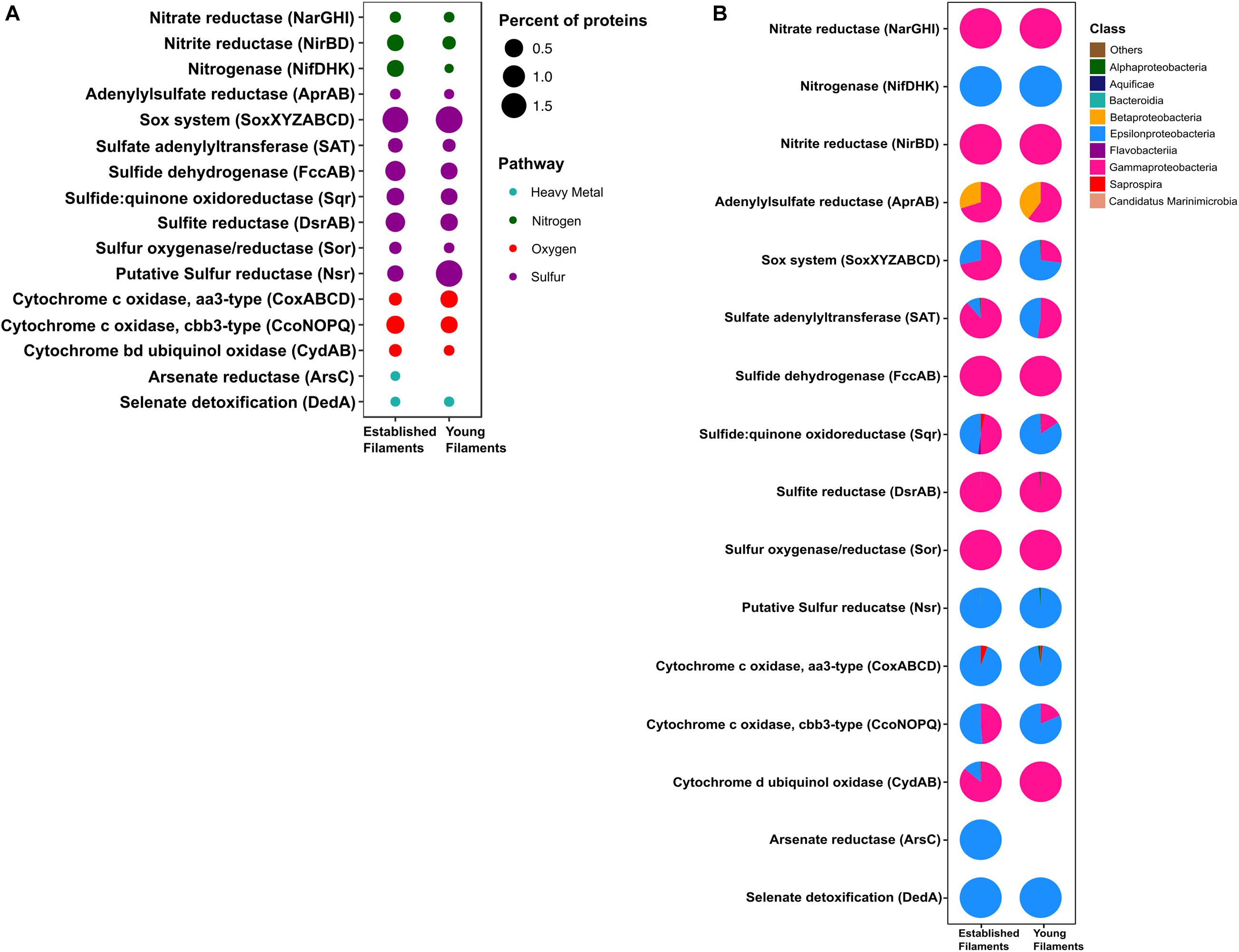
Figure 4. (A) Percentage and (B) taxonomic affiliation of key proteins involved in nitrogen, sulfur, oxygen and heavy metal detoxification pathways for established and young filaments.
Carbon Fixation
The metagenome and the metaproteome of both filamentous communities included genes and proteins involved in the reductive tricarboxylic acid (rTCA) and Calvin-Benson-Bassham (CBB) cycles for carbon fixation (Figure 2). The YF biofilm community had three times as many genes for the rTCA than the EF biofilm, while EF had twice as many genes for the CBB cycle than YF (Figure 2A, columns 1 and 3, and Supplementary Table 1). While proteins of the rTCA cycle contributed only to 2.2% of the total proteins in EF, they contributed to 5% of the total in YF (Figure 2B, columns 1 and 3, and Supplementary Table 2). Taxonomically, the rTCA proteins were all affiliated with Sulfurovum and Sulfurimonas within the Epsilonproteobacteria (Figure 2B, columns 2 and 4). In contrast, proteins of the CBB cycle comprised 1.1 and 0.7% of the total proteins in EF and YF, respectively (Figure 2B, columns 1 and 3, and Supplementary Table 2), with most of them taxonomically identified as Gammaproteobacteria and specifically affiliated to the genera Thiomicrospira, Thiothrix and Thioalkalivibrio (Figure 2B, columns 2 and 4). These bacteria are chemolithotrophs that derive energy from the oxidation of reduced sulfur species and are key players in marine geothermal environments (Sievert and Vetriani, 2012).
Nitrogen and Sulfur Metabolism
The metagenomes of the two biofilm communities encoded for genes involved in nitrate reduction, denitrification, nitrogen fixation, sulfur oxidation/reduction, oxygen respiration, and heavy metal detoxification (Figure 3A). However, the abundance of some of these genes differed between the two biofilm communities. For instance, the napAB (encoding two components of the periplasmic nitrate reductase complex) and nifDHK (nitrogenase) genes, involved in nitrate reduction and nitrogen fixation, respectively, were found to be enriched in the YF biofilm (Figure 3A and Supplementary Table 1) and were taxonomically affiliated with the Epsilonproteobacteria (Figure 3B). The narGHI (membrane bound nitrate reductase) and nirS (nitrite reductase) genes, as well as those for sulfur oxidation, fccAB (sulfide dehydrogenase), and sulfate reduction/sulfide oxidation, dsrAB (dissimilatory bisulfite reductase) were enriched in EF (Figure 3A and Supplementary Table 1). The sor gene (sulfur oxygenase/reductase) was found in EF but was absent in YF. Taxonomically, the nar, nir and sor genes were mostly affiliated with the Gammaproteobacteria (Figure 3B). The sqr (sulfide quinone oxidoreductase) gene was also enriched in the EF biofilm community and predominantly affiliated with the Gammaproteobacteria (Figures 3A,B). However, the proportion of Epsilonproteobacteria- affiliated sqr genes increased in the YF community (Figure 3B).
In the metaproteome, the Epsilonproteobacteria-affiliated nitrogenase (Nif) accounted for 0.35% of the total proteins in EF as opposed to 0.001% in YF (Figure 4A and Supplementary Table 2). The periplasmic nitrate reductase (Nap) was undetectable in both the samples, while both the membrane bound nitrate reductase (Nar) and the nitrite reductase (Nir), affiliated with the Gammaproteobacteria, were more abundant in EF (0.02 and 0.3%; Figure 4A). Proteins involved in the dissimilatory sulfite reductase (Dsr) pathway for either elemental sulfur oxidation or sulfite reduction and the APS pathway for indirect sulfite oxidation (Apr, SAT) were twice as abundant in the EF biofilm compared to the YF one (Figure 4A and Supplementary Table 2). Dsr and Apr in both metaproteomes were mostly affiliated with Gammaproteobacteria, while SAT was mostly affiliated with Gammaproteobacteria in the EF biofilm and with both Gammaproteobacteria and Epsilonproteobacteria in the YF community (Figure 4B). The sulfide dehydrogenase (Fcc) was twice more abundant in the EF than in the YF biofilm (Figure 4A) and it was affiliated with Gammaproteobacteria in both samples (Figure 4B and Supplementary Table 2). In contrast, the abundance of the sulfide quinone oxidoreductase (SQR) was comparable between the two biofilms (Figure 4A), but its taxonomic affiliation changed from comparably epsilon- and gammaproteobacterial in the EF biofilm to predominantly epsilonproteobacterial in the YF community (Figure 4B and Supplementary Table 2). The Sor protein, involved in the disproportionation of elemental sulfur (Kletzin, 1989), was classified as Thioalkalivibrio spp. and was enriched in the EF biofilm (Figure 4A and Supplementary Table 2). A putative NADPH-dependent sulfur reductase (NSR) affiliated with Sulfurovum spp. was six times more abundant in the total proteins of YF than EF (Figure 4A and Supplementary Table 2). The sox genes and the Sox proteins were almost equally abundant in both the YF and EF biofilms (Figures 3A, 4A); however, in the YF biofilm, both sox genes and Sox proteins were predominantly affiliated with the Epsilonproteobacteria, while in the EF biofilm they were mostly affiliated with the Gammaproteobacteria (Figures 3B, 4B). We detected only a few [Ni-Fe] hydrogenases in the metagenomes of the two biofilm communities, while none in the metaproteomes.
Oxygen Reduction
Three cytochrome oxidase gene clusters (coxABCD, ccoNOP and cydAB) were abundant and comparable between the two metagenomes, indicating that oxygen respiration played an important role in both biofilm communities (Figure 3A and Supplementary Table 1). In the YF biofilm, the majority of all three genes were taxonomically classified as Epsilonproteobacteria, while in the EF biofilm the taxonomic affiliation of coxABCD and cydAB revealed a higher diversity of the oxygen respiring community, except for ccoNOP, which was mostly affiliated with the Gammaproteobacteria (Figure 3B). Compared to proteins involved in nitrate/nitrite respiration, the aa3-type and cbb3-type cytochromes, mediating oxygen respiration, were highly enriched (seven times as much) in YF (Figure 4A and Supplementary Table 2). The majority of these proteins were classified as Epsilonproteobacteria (Figure 4B) and were specifically affiliated with Sulfurovum spp., with the exception of cytochrome cbb3 in EF, which was equally affiliated with Epsilon- and Gammaproteobacteria (Figure 4B). The cytochrome d (CydAB) was mostly affiliated with Gammaproteobacteria (Figure 4B).
Heavy Metal Detoxification
Elevated concentrations of potentially toxic heavy metals such as mercury, arsenic, selenium etc. are present at hydrothermal vents, leading to heavy metal resistance in the resident microbiota (Rathgeber et al., 2002; Vetriani et al., 2005; Price et al., 2013). Genes involved in the detoxification of arsenate (arsC), mercury (merA) and selenate (dedA) were recovered from both metagenomes (Figure 3A). The abundance of the arsC gene was comparable in both samples (Figure 3A and Supplementary Table 1), while this gene was affiliated mostly with Gammaproteobacteria and Epsilonproteobacteria in the EF the YF, respectively (Figure 3B). The merA gene (mercury reduction) was more abundant in the YF than the EF metagenome (Figure 3A and Supplementary Table 1), and was affiliated with the Gammaproteobacteria, while the dedA gene (selenate detoxification) was more abundant in the EF and it was mostly affiliated with the Epsilonproteobacteria (Figures 3A,B). In the metaproteome, only DedA, which has been shown to be involved in selenite resistance (Ledgham et al., 2005), was expressed in the YF biofilm, as opposed to both DedA and arsenate reductase (Ars) being expressed in EF (Figure 4A and Supplementary Table 2). Both enzymes were classified as Epsilonproteobacteria (Figure 4B). While present in both metagenomes, the mercuric reductase gene (merA) was not found in the metaproteomes of either biofilm communities (Figures 3A,B).
Metagenome-Assembled Genomes (MAGs)
Despite the fundamental role as primary producers of Gamma- and Epsilonproteobacteria in marine geothermal habitats, there is a dearth of representative cultures and their genomes. A total of 31 MAG bins were obtained from the co-assembly of the two metagenomes. Out of these, three (TCMF1, TCMF2, and TCMF9) were ≥85% complete with contamination of ≤8% and complied with the proposed criteria that define medium to high quality draft MAGs (Bowers et al., 2017). The 16S rRNA genes were recovered from bins TCMF2 and TCMF9 (Table 1).
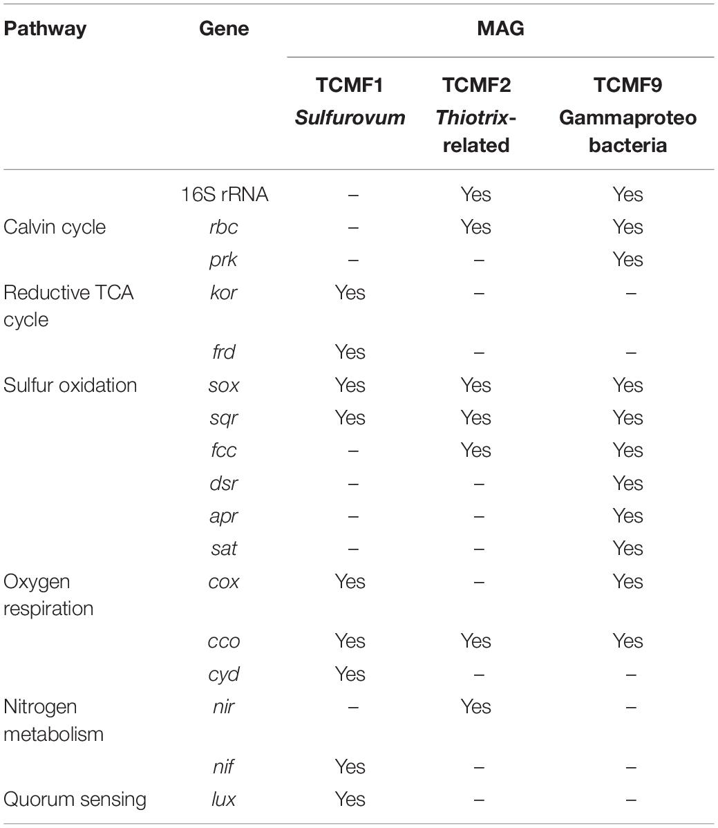
Table 1. Genes involved in main metabolic pathways recovered from MAGs.
The 16S rRNA gene of MAG TCMF2 showed >97% sequence similarity to 16S rRNA genes related to the Thiotrix-related CF-26 group recovered from microbial mats at shallow-water vents located on the South Tonga Arc (HQ153886) (Murdock et al., 2010) and at the Kuieshan Island (JQ611107). In our previous study of the Tor Caldara vents, dominant OTUs annotated as Thiotrix-related CF-26 from EF showed sequence similarity to the OTUs recovered from the same microbial mats, indicating that the TCMF2 MAG likely belongs to this group of uncultured Thiotrichales. Most of the genes involved in the CBB cycle, including RuBisCo (rbcSL), were present in MAG TCMF2. Further, the potential for thiosulfate oxidation was evidenced by the presence of sox genes, in addition to the sqr and fccAB genes involved in sulfide oxidation. This MAG also contained the genes for cbb3-type cytochrome oxidase and nirBD, involved in microaerobic and nitrite respiration, respectively (Table 1).
MAG TCMF9 showed 93% 16S rRNA gene similarity to a gammaproteobacterial endosymbiont clone of the gutless oligochaete worm, Olavius ilvae. O. ilvae is known to harbor sulfur-oxidizing gammaproteobacterial endosymbionts that fix CO2 via the CBB cycle (Ruehland et al., 2008). This MAG contained most of the genes involved in the CBB cycle, including RuBisCo (rbcSL) and phosphoribulokinase (prk). A whole suite of genes required for thiosulfate and sulfide oxidation such as the sox system (except soxC), sqr, and fccAB were present. This MAG also contained all three genes (dsrAB, aprAB, sat) involved in the reverse dsr pathway, indicating versatility for the oxidation of sulfur compounds. Three out of the four genes encoding the aa3-type cytochrome oxidase (cox) and all genes encoding the microaerobic cbb3-type cytochrome oxidase (cco) were present (Table 1). The majority of the genes involved in bacterial chemotaxis and flagellar motility were also present (data not shown).
Since the 16S rRNA gene was not recovered in MAG TCMF1, other single-copy ribosomal proteins were used to constrain its lineage. Both the 30S ribosomal protein S8 and S9 were greater than 90% identical to a Sulfurovum species. Additionally, the average amino acid identity (AAI) shared between TCMF1 and Sulfurovum strain NBC37 was 58%, which is well within the range that defines the genus boundary (Rodriguez-R and Konstantinidis, 2014). Based on this data, TCMF1 belonged to the Sulfurovum lineage within the Epsilonproteobacteria, which dominated the YF community (Figure 1B). Sulfurovum spp. are sulfur/hydrogen oxidizers that fix CO2 using the rTCA cycle (Inagaki, 2004; Mino et al., 2014; Giovannelli et al., 2016). Out of the three diagnostic genes for the rTCA cycle, kor and frd were present. Sox and sqr genes for sulfide/sulfur oxidation were also present. Interestingly, this MAG contained the nifDHK gene essential for nitrogen fixation, while the periplasmic nitrate reductase encoding gene, napAB, which is widespread in the Epsilonproteobacteria (Vetriani et al., 2014), was missing. Three out of the four genes encoding the aa3-type cytochrome oxidase (cox) and all genes encoding the microaerobic cbb3-type (cco) and bd cytochrome oxidase (cyd) were present, indicating the ability to respire oxygen within a broad range of concentrations. The luxS gene, involved in quorum sensing and conserved across all members of the Epsilonproteobacteria (Pérez-Rodríguez et al., 2015), was also present in TCMF1.
Given the presence of genes coding for sqr both in the metagenomes and in the recovered MAGs, we carried out phylogenetic analyses of representative SQR enzymes from the Tor Caldara biofilms and close relatives obtained from GenBank. This analysis placed the sequences into three discrete clusters separated from the Chlorobium lineage, from which SQR was originally characterized: one cluster was related to Sulfurovum spp., one was related to the Thiotricales (Thiomicrospira and Thiothrix spp.) and a third cluster included two groups of sequences: one related to Sulfurovum spp. and one related to the Thiotricales (Figure 5). In line with the taxonomic annotation derived from other genes, the SQR from MAG TCMF1 was related to Sulfurovum sequences, while the enzymes from MAGs TCMF2 and TCMF9 were related to Thiotricales-derived sequences (Figure 5).
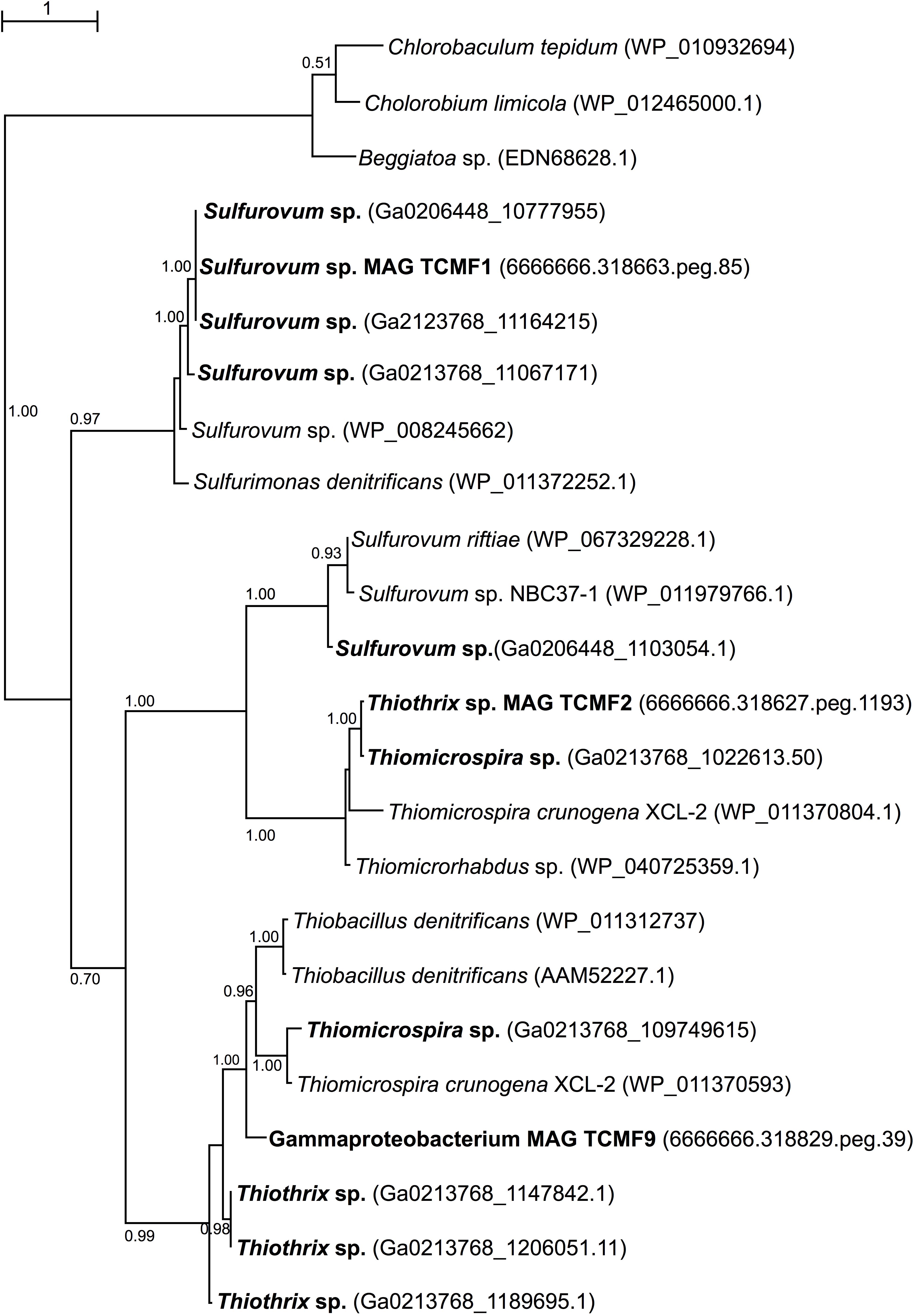
Figure 5. Maximum likelihood phylogenetic tree showing the position of the sulfide quinone oxireductase amino acid sequences, SQR, obtained in this study (indicated in boldface). Closely related sequences were obtained from GenBank. Approximate likelihood ratio test (aLRT) values for branch support are indicated. Bar, 1% estimated substitutions.
Discussion
Chemosynthetic microbial biofilm are commonly found in geothermal and sulfidic environments (Sievert and Vetriani, 2012), including cold seeps (Ristova et al., 2015), sulfidic caves (Macalady et al., 2006), thermal springs (Beam et al., 2016), mud volcanoes (Heijs et al., 2005) as well as deep-sea (Gulmann et al., 2015; O’Brien et al., 2015) and shallow-water hydrothermal vents (Miranda et al., 2016; Patwardhan et al., 2018). Members of Gammaproteobacteria and Epsilonproteobacteria dominate most of these biofilms and a spatial segregation between the two classes of bacteria has been correlated with the in situ sulfide concentration (Engel et al., 2004; Macalady et al., 2008; Crépeau et al., 2011; Gulmann et al., 2015; O’Brien et al., 2015; Miranda et al., 2016; Meier et al., 2017).
At Tor Caldara, a submarine gas vent in the Tyrrhenian Sea characterized by vigorous venting of CO2 and unusually a high concentration of H2S (avg. of 23.13 mol%; Patwardhan et al., 2018), white filamentous microbial communities grow profusely on the rocks near the gas venting. To investigate the composition and function of established and young biofilms, we collected native substrates (EF) and deployed glass slides (YF) in the vicinity of a vent. Within 4 days, filamentous biofilms colonized the slides.
Our survey of 16S rRNA genes and transcripts revealed a shift in the two main taxonomic groups during colonization, whereby sulfur-oxidizing Epsilonproteobacteria dominated the YF biofilms, while Gammaproteobacteria became prevalent in the EF community. Further, we showed that representative species of Epsilon- and Gammaproteobacteria are adapted to different sulfide concentrations (Patwardhan et al., 2018). While in most 16S rRNA gene-based surveys of Epsilonproteobacteria-dominated deep-sea hydrothermal vent sites the genera Sulfurovum and Sulfurimonas typically co-occur (e.g., Huber et al., 2007), we did not detect the latter within the Tor Caldara biofilms. With few exceptions, most Sulfurimonas spp. isolated from marine sulfidic habitats can use hydrogen and reduced sulfur species as an electron donors (Han and Perner, 2015). In contrast, most Sulfurovum spp. are strictly sulfur and sulfide-oxidizers. Further, the abundance of Sulfurovum relative to Sulfurimonas phylotypes at hydrothermal vents was found to correlate with increased oxygen concentration, suggesting that the former are more oxygen-tolerant (Meier et al., 2017). Therefore, it is possible that the absence of hydrogen in the Tor Caldara gases, along with the turbulent nature of these gas emissions that transiently expose the biofilms to the oxic water column, favors the occurrence of Sulfurovum vs. Sulfurimonas spp.
In this study, we integrated metagenomic and metaproteomic approaches to investigate the in situ expression of central metabolic pathways related to carbon fixation and energy conservation in the YF and EF biofilms. In line with our previous 16S rRNA amplicon survey, the metagenome-extracted 16S rRNA genes showed that Epsilonproteobacteria and Gammaproteobacteria dominated the YF and EF biofilms, respectively (Figures 1A,B).
The Relative Expression of the rTCA and CBB Cycles for Carbon Fixation Reflects the Taxonomic Composition of the YF and EF Biofilms
Chemoautotrophs are the primary producers at hydrothermal vents, forming the basis of the food web by fixing inorganic CO2 into organic material (Jannasch and Wirsen, 1979; Karl et al., 1980; Sievert and Vetriani, 2012). Till date, six pathways for carbon fixation are known: the Calvin-Benson-Bassham (CBB) reductive pentose phosphate cycle, the reductive tricarboxylic acid cycle (rTCA), the reductive acetyl coenzyme A pathway (Wood-Ljungdahl), the 3-hydroxypropionate bi-cycle, the 3-hydroxypropionate/4-hydroxybutyrate cycle, and the dicarboxylate/4-hydroxybutyrate cycle (Nakagawa and Takai, 2008; Berg, 2011). The rTCA and CBB cycles are most prevalent at diffuse-flow hydrothermal vents and, in these habitats, they are generally diagnostic of Epsilon- and Gammaproteobacteria, respectively (Nakagawa and Takai, 2008). Enrichment of proteins of the rTCA cycle affiliated with Epsilonproteobacteria, in addition to a similar signature at the gene level, confirmed that autotrophy using the rTCA cycle in Epsilonproteobacteria was the predominant mechanism for carbon fixation in the YF communities (Figures 2, 6 and Supplementary Tables 1, 2). The majority of the rTCA genes and proteins were classified as Sulfurovum spp. (Figure 2), which is known to be ubiquitous in vent environments (Inagaki, 2004; Dahle et al., 2013; Mino et al., 2014; Giovannelli et al., 2016). These findings underline the importance of Sulfurovum spp. in the YF community and are in accordance with data from the 16S rRNA diversity analysis (Patwardhan et al., 2018; Figure 1). While the EF community also encoded genes and expressed enzymes of the rTCA cycle, the CBB cycle was overrepresented in the EF biofilm at both the gene and protein levels, with contributions from various genera within the Gammaproteobacteria, in particular Thiomicrospira, Thioalkovibrio and Thiothrix-related spp. (Figures 2, 6 and Supplementary Tables 1, 2). This is also in line with our 16S rRNA survey, which showed these genera as dominant in the EF biofilm (Patwardhan et al., 2018; Figure 1). Thus, we observed a shift from rTCA-based carbon fixation (Epsilonproteobacteria) to CBB-based carbon fixation (Gammaproteobacteria) during biofilm maturation. The lower energy demand of the rTCA cycle, compared to the CBB cycle (Nakagawa and Takai, 2008; Hügler and Sievert, 2011), might provide an advantage to the Epsilonproteobacteria during early substrate colonization.
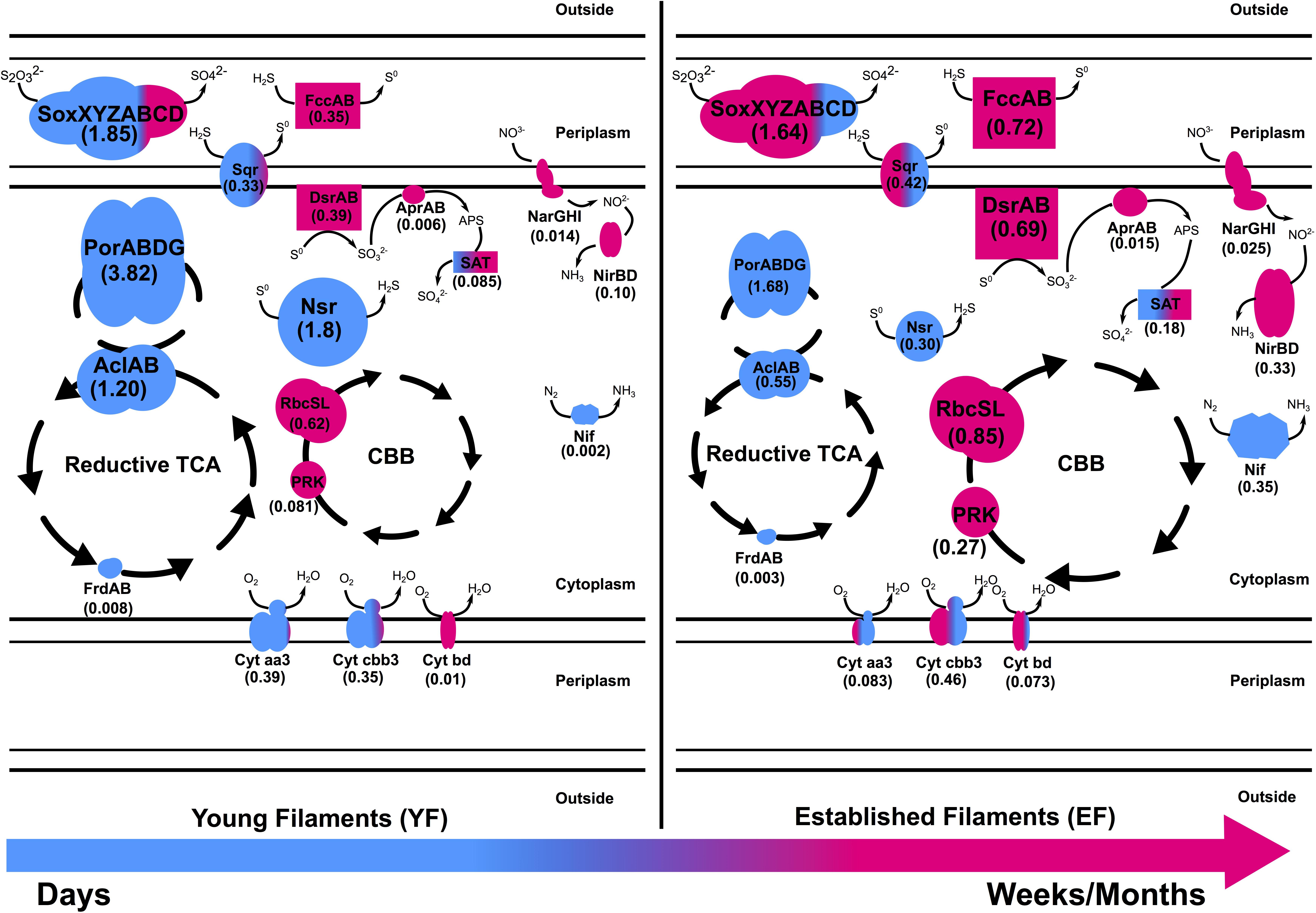
Figure 6. Metaproteomic-based reconstruction of the central metabolic pathways expressed by the Tor Caldara young and established biofilm communities. The size of the enzymatic complexes is proportional to their abundance in the metaproteome. The numbers indicate the normalized protein abundance (%). The colors indicate the taxonomic affiliation of the enzymes and reflect the relative proportions of the two predominant bacterial classes. Blue: Epsilonproteobacteria; Pink: Gammaproteobacteria.
The Expression of Different Pathways for Sulfide/Sulfur Oxidation in the YF and EF Biofilms May Reflect Adaptations to Fluctuating Redox Regimes
In shallow-water geothermal systems, the concentration of reduced sulfur compounds and other electron donors for microbial oxidations, along with the availability of oxidants, vary considerably over time (Yücel et al., 2013; Price and Giovannelli, 2017). Hence, chemolithoautotrophic microorganisms have adapted to such fluctuating redox regimes. In particular, reduced sulfur compounds represent an important energy source that drives energy-yielding reactions in geothermal environments (Nakagawa and Takai, 2008). The abundance and range of various genes and proteins involved in sulfur oxidation pathways presented here validated our initial findings about the importance of sulfur-oxidizing bacteria at Tor Caldara (Patwardhan et al., 2018). For instance, the Sox pathway for sulfur oxidation was one of the most abundant energy-yielding pathway recovered both in the metagenomes and metaproteomes (Figures 3, 4, 6 and Supplementary Tables 1, 2).
Thiosulfate oxidation catalyzed by the multienzyme complex Sox is widespread in sulfur-oxidizing chemoautotrophs (Friedrich et al., 2001, 2005; Yamamoto and Takai, 2011). This enzyme complex, consisting of four protein components, soxYZ, soxXA, soxB, and soxCD, catalyzes the oxidation of thiosulfate and sulfide to sulfate. The abundance of the Sox complex in both filament communities at the gene as well as the protein (YF:1.8%; EF:1.6%) level indicates that this is the predominant pathway used by the biofilm bacteria to conserve energy (Figures 3A, 4A and Supplementary Tables 1, 2). This is consistent with a metaproteogenomic study of chimney-associated microbial communities from deep-sea hydrothermal vents, which revealed that Sox represented one of the most highly expressed energy-yielding pathways (Pjevac et al., 2018). However, the taxonomic affiliation of the sox reads (Figure 3B) and Sox enzymes (Figure 4B) from the Tor Caldara biofilms shows a finer resolution, indicating that Epsilonproteobacteria are mostly expressing the Sox complex in the YF biofilm, while Gammaproteobacteria take over that same role in the EF biofilms (Figure 6).
A similar trend is evident in the profile of the sulfide:quinone oxidoreductase enzyme complex (SQR) involved in oxidation of sulfide to elemental sulfur, which is predominantly expressed by Epsilonproteobacteria in the YF filaments, while its contribution by Gammaproteobacteria increases in the EF filament community (Figures 4B, 6). In addition to SQR, in some chemolithotrophic bacteria oxidation of hydrogen sulfide to elemental sulfur is catalyzed by the flavocytochrome c-sulfide dehydrogenase (FccAB; Visser et al., 1997; Muβmann et al., 2007). The metaproteomic data presented here show that the FccAB enzyme is affiliated only with Gammaproteobacteria, and that its expression is higher in the EF biofilm than in the YF community (Figures 4A, 6). The bioenergetics of microbial sulfide oxidation might provide a framework to interpret the SQR and Fcc expression profiles in the Tor Caldara biofilms. Since the midpoint potential of the NAD+/NADH couple at pH 7 is approximately 50 mV more negative than the midpoint potential of the S0/H2S couple, chemolithotrophic SOBs require energy to transport electrons from hydrogen sulfide upwards to NAD+ by reverse electron flow. In SQR-catalyzed sulfide oxidation, the electrons from sulfide enter the transport chain at the level of quinones, generating the necessary electrochemical proton potential across the membrane for reverse electron transfer (Griesbeck et al., 2000). However, when Fcc is involved, the electrons enter the transport chain at the level of c-type cytochromes, whose midpoint potential is more electropositive than that of quinones. That increases the energetic burden for the reverse transfer of electrons upwards to NAD+ and implies that, in chemolithotrophic SOBs, sulfide oxidation by SQR provides more energy than sulfide oxidation by Fcc (Griesbeck et al., 2000). However, due to its high affinity for sulfide (Brune, 1995), Fcc is hypothesized to be expressed at low sulfide concentrations, supplementing the energetically more efficient SQR. Previous work on the Tor Caldara biofilms demonstrated that Epsilonproteobacteria are adapted to higher sulfide concentrations than Gammaproteobacteria and that they are the pioneer colonizers at this site (Patwardhan et al., 2018). We hypothesize that, early in the colonization process (YF biofilm), Epsilonproteobacteria might be driving down sulfide levels within the biofilms and that the increased expression of the high affinity FccAB in EF might provide an advantage to the Gammaproteobacteria in the subsequent stages of colonization (EF biofilm; Figure 6). The hypothesis that Fcc is more prevalent under low sulfide and more oxidized conditions (Brune, 1995; Griesbeck et al., 2000) supports our model. Overall, the co-expression of the energy efficient SQR and the high affinity Fcc may provide metabolic flexibility in response to fluctuating sulfide concentrations within the biofilm communities.
The reverse dissimilatory sulfate reductase pathway (rDSR) oxidizes stored intracellular sulfur to sulfate via sulfite (Friedrich et al., 2005). Consistent with findings in other filamentous Gammaproteobacteria (Muβmann et al., 2007; Chernousova et al., 2009; Sharrar et al., 2017), all three proteins involved in this pathway, namely, DsrAB, AprAB, and SAT, were affiliated with Gammaproteobacteria and were enriched in the EF biofilm (Figures 4, 6 and Supplementary Table 2). The expression of the rDSR pathway is in accordance with the stored intracellular elemental sulfur observed in electron micrographs of EF (Patwardhan et al., 2018), while its higher expression in the EF biofilm suggests that this community may experience transient depletions of hydrogen sulfide. This stored sulfur might also be disproportionated into sulfite, thiosulfate and sulfide as evidenced by the expression of gammaproteobacterial Sor (Figure 4) in the EF biofilm, and might provide a competitive advantage to the secondary colonizers (Kletzin, 1989; Veith et al., 2012). Since the Dsr pathway for S-oxidation is not encoded in the currently available genomes of Epsilonproteobacteria (Yamamoto and Takai, 2011), the epsiloproteobacterial SAT found in the metaproteome is probably involved in sulfate assimilation.
Sulfur oxidation was complemented with sulfur reduction evidenced by high expression of the a putative Sulfurovum-affiliated NADH-dependent sulfur reductase (NSR; Schut et al., 2007) in the YF (Figures 4, 6 and Supplementary Table 2). Despite sulfur respiration is known to be coupled to hydrogen oxidation in two Sulfurovum spp. (Yamamoto et al., 2010; Mino et al., 2014), we did not detect hydrogenases in the metaproteomes. Since hydrogen concentrations are low at Tor Caldara, the Sulfurovum spp. found there may be adapted to oxidize alternative energy sources, such as malate and/or formate (Campbell et al., 2006; Yamamoto et al., 2010; Keller et al., 2015).
Epsiloproteobacteria-Mediated Nitrogen Fixation and Gammaproteobacteria-Mediated Nitrate Reduction Are Prevalent in the EF Biofilm
Microbial nitrate respiration occurs via two main pathways, dissimilatory nitrate reduction to ammonia (DNRA) and denitrification. Both processes have been investigated in marine geothermal environments (Bourbonnais et al., 2012a, b; Pérez-Rodríguez et al., 2013; Vetriani et al., 2014). The first step in nitrate reduction is carried out by the enzyme nitrate reductase. Currently, two different types of respiratory nitrate reductases, the membrane-bound Nar and the periplasmic Nap, are known to occur in bacteria (Richardson et al., 2001). Nar is a low-affinity enzyme expressed under nitrate rich conditions, whereas Nap is a high-affinity enzyme whose expression is favored under low nitrate conditions (Potter et al., 1999; Mintmier et al., 2018). The genes encoding both Nar and Nap were present in the EF and YF communities, as well as the genes encoding various nitrite reductases (NirBD, NirK, NirS and NrfA), the nitric oxide reductase (NorB) and the nitrous oxide reductase (NosZ; Figure 3A and Supplementary Table 1). However, metaproteomic analyses showed that only the Gammaproteobacteria-affiliated Nar and Nir enzymes were expressed, while Nap and the enzymes involved in the denitrification pathway could not detected (Figure 4B). The coastal area at Tor Caldara is likely enriched in nitrate (possibly from runoff of lawn fertilizers), which might explain the expression of NarGHI, but not of NapAB (Figure 4A and Supplementary Table 2). The NirBD nitrite reductase, often involved in the reduction of nitrite to ammonia in DNRA (Reyes et al., 2017), was also found to be abundant. Both the Nar and Nir enzymes were enriched in the EF biofilm, indicating that nitrate respiration is prevalent in the established community.
Apart from nitrate respiration, nitrogen fixation also plays an important role in nitrogen cycling at vents (Rau, 1981; Mehta et al., 2003). Expression of epsilonproteobacterial NifDHK was observed in both filament communities but was highly enriched in the EF (Figures 4, 6 and Supplementary Table 2). While nitrogen fixation is not common in Epsilonproteobacteria, recent studies have shown that some members of this class have the genomic potential for it (Keller et al., 2015; Waite et al., 2017), and the ability to fix nitrogen was demonstrated experimentally in Lebetimonas spp. (Meyer and Huber, 2013). Nitrogen fixation is an energy expensive reaction and nitrogenases are usually expressed in nitrogen limiting conditions (Olivares et al., 2013). We hypothesize that, when the filamentous community transitions from a young to an established state, nitrate respiration carried out by Gammaproteobacteria might result in a depletion in nitrogen within the biofilm microenvironment. Hence, the ability to fix nitrogen might give the Epsilonproteobacteria an advantage while competing for a fixed nitrogen source against the secondary colonizers in the established biofilm.
Enzymes for Aerobic and Microaerobic Respiration Are Differentially Expressed in the YF and EF Biofilms
Most of the well-characterized bacterial oxidases belong to the heme-copper superoxidase family. There are two types of oxidases that use cytochrome c as a substrate: cytochrome c oxidase aa3-type and cytochrome c oxidase cbb3-type. The former is expressed in aerobic (high oxygen) conditions while the latter is expressed in microaerobic (low oxygen) conditions. Additionally, cytochrome bd, a ubiquinol oxidase, is also expressed in microaerobic conditions (García-Horsman et al., 1994; Pitcher and Watmough, 2004). At Tor Caldara, the microaerobic, high affinity cbb3-type and bd-type cytochrome oxidase were enriched in EF biofilm and were mostly classified as Gammaproteobacteria (Figures 4, 6 and Supplementary Table 2), suggesting that the biofilms experience microaeobic/anoxic conditions as they mature. On the other hand, primary colonizers in the YF might experience oxic to microaerobic conditions as suggested by equal abundance of aerobic aa3 type and microaerobic cbb3 type cytochrome c oxidase classified dominantly as Epsilonproteobacteria.
Overall View: A Metaproteome-Based Model of Microbial Colonization at the Tor Caldara Gas Vents
Based on the metaproteomic data, we reconstructed the central metabolic pathways expressed by the Tor Caldara young and established biofilm communities (Figure 6). According to this model, substrates exposed to the gas emissions are initially colonized by a population dominated by sulfide-tolerant Epsilonproteobacteria (Figure 1; Patwardhan et al., 2018). Members of this class oxidize various reduced sulfur compounds via the Sox and SQR pathways coupled to oxygen reduction (both aerobic and microaerobic), while fixation of carbon dioxide occurs via the rTCA cycle. In the early stages of colonization, Gammaproteobacteria are present, but underrepresented (Figures 1, 6). Over time, sulfide oxidation by Epsilonproteobacteria may lower the sulfide concentration within the biofilm community, conditioning the environment for the growth of the less sulfide-tolerant Gammaproteobacteria (Patwardhan et al., 2018), which dominate the established filaments. Gammaproteobacteria also oxidize various sulfur compounds using the Sox and SQR pathways, but this reaction is now coupled to either microaerobic and/or anaerobic respiration using oxygen and nitrate as terminal electron acceptors, respectively. The high affinity, periplasmic FccAB enzyme is co-expressed with SQR in response to fluctuating concentrations of sulfide, while elemental sulfur is stored in the cytoplasm. Energy thus conserved is used to fix carbon dioxide via the CBB cycle (Figure 6). When sulfide availability becomes limiting within the biofilm community, certain Gammaproteobacteria further oxidize the stored sulfur to sulfate via the reverse dissimilatory sulfate reductase pathway, thus making full use of their metabolic repertoire. As the filamentous community transitions from a young to a more established stage, anaerobic respiration by the Gammaproteobacteria may lead to loss of nitrate from the system. This triggers increased nitrogen fixation by the Epsilonpreoteobacteria as a potential strategy to compete with the Gammaproteobacteria.
Conclusion
Our study revealed a complex pattern of protein expression in chemosynthetic biofilm communities that colonize the gas vent system at Tor Caldara. Functions common to chemosynthetic microbial communities, such as carbon fixation and sulfide oxidation, are catalyzed via the expression of different enzymatic complexes encoded by two predominant groups of SOBs belonging to the Epsilon- and Gammaproteobacteria. We hypothesize that the expression of these enzymatic complexes is finely tuned to the chemical and physical conditions within the biofilm communities, and reflect the physiological characteristics of the two groups of SOBs. Overall, the enzyme expression profiles obtained in this work reveal metabolic flexibility and adaptations to fluctuating redox regimes during colonization of the shallow-water gas vents of Tor Caldara. These results are consistent with the previously reported transition from Epsilonproteobacteria—the pioneer colonists—to Gammaproteobacteria at Tor Caldara.
Data Availability Statement
The datasets presented in this study can be found in online repositories. The names of the repository/repositories and accession number(s) can be found below: NCBI SRA with accession PRJNA498803 and ProteomeXchange with identifier PXD023357.
Author Contributions
SP generated and analyzed the data and wrote the manuscript. CV conceived the study, supervised the research, collected the samples, contributed to data analysis, and wrote the manuscript. FS contributed to data analysis. DG participated in sample collection and contributed to data analysis. All authors contributed to the article and approved the submitted version.
Funding
This work was partially supported by the following grants to CV: NSF OCE 11-24141, NSF OCE 11-36451, NSF MCB 15-17567, NSF OCE 19-48623, and NASA NNX15AM18G.
Conflict of Interest
The authors declare that the research was conducted in the absence of any commercial or financial relationships that could be construed as a potential conflict of interest.
Acknowledgments
We thank the crew of the R/V Antares for their support during the sampling operations, and Satyajit Rajapurkar for help and guidance with the bioinformatics analyses.
Supplementary Material
The Supplementary Material for this article can be found online at: https://www.frontiersin.org/articles/10.3389/fmicb.2021.638300/full#supplementary-material
Footnotes
- ^ http://www.bioinformatics.babraham.ac.uk/projects/fastqc/
- ^ https://www.ebi.ac.uk/pride/archive/projects/PXD023357
References
Anders, S., Pyl, P. T., and Huber, W. (2015). HTSeq—a python framework to work with high-throughput sequencing data. Bioinformatics 31, 166–169. doi: 10.1093/bioinformatics/btu638
Anisimova, M., and Gascuel, O. (2006). Approximate likelihood-ratio test for branches: a fast, accurate, and powerful alternative. Syst. Biol. 55, 539–552. doi: 10.1080/10635150600755453
Aziz, R. K., Bartels, D., Best, A. A., DeJongh, M., Disz, T., Edwards, R. A., et al. (2008). The RAST server: rapid annotations using subsystems technology. BMC Genomics 9:75. doi: 10.1186/1471-2164-9-75
Barry, P. H., de Moor, J. M., Giovannelli, D., Schrenk, M., Hummer, D., and Lopez, T. (2019). Forearc carbon sequestration reduces long-term volatile recycling into the mantle. Nature 568, 487–492.
Beam, J. P., Bernstein, H. C., Jay, Z. J., Kozubal, M. A., Jennings, Rd, Tringe, S. G., et al. (2016). Assembly and succession of iron oxide microbial mat communities in acidic geothermal springs. Front. Microbiol. 7:25. doi: 10.3389/fmicb.2016.00025
Bengtsson-Palme, J., Hartmann, M., Eriksson, K. M., Pal, C., Thorell, K., Larsson, D. G., et al. (2015). METAXA2: improved identification and taxonomic classification of small and large subunit rRNA in metagenomic data. Mol. Ecol. Resour. 15, 1403–1414. doi: 10.1111/1755-0998.12399
Berg, I. A. (2011). Ecological aspects of the distribution of different autotrophic CO2 fixation pathways. Appl. Environ. Microbiol. 77, 1925–1936. doi: 10.1128/AEM.02473-10
Boden, R., Scott, K. M., Williams, J., Russel, S., Antonen, K., Rae, A. W., et al. (2017). An evaluation of Thiomicrospira, Hydrogenovibrio and Thioalkalimicrobium: reclassification of four species of Thiomicrospira to each Thiomicrorhabdus gen. nov. and Hydrogenovibrio, and reclassification of all four species of Thioalkalimicrobium to Thiomicrospira. Int. J. Syst. Evol. Microbiol. 67, 1140–1151. doi: 10.1099/ijsem.0.001855
Bourbonnais, A., Juniper, S. K., Butterfield, D. A., Devol, A. H., Kuypers, M. M. M., Lavik, G., et al. (2012a). Activity and abundance of denitrifying bacteria in the subsurface biosphere of diffuse hydrothermal vents of the Juan de Fuca Ridge. Biogeosciences 9, 4661–4678. doi: 10.5194/bg-9-4661-2012
Bourbonnais, A., Lehmann, M. F., Butterfield, D. A., and Juniper, S. K. (2012b). Subseafloor nitrogen transformations in diffuse hydrothermal vent fluids of the Juan de Fuca Ridge evidenced by the isotopic composition of nitrate and ammonium: NITROGEN CYCLE IN HYDROTHERMAL VENTS. Geochem. Geophys. Geosyst. 13:Q02T01. doi: 10.1029/2011GC003863
Bowers, R., Kyrpides, N., Stepanauskas, R., Harmon-Smith, M., Doud, D., Reddy, T. B. K., et al. (2017). Minimum information about a single amplified genome (MISAG) and a metagenome-assembled genome (MIMAG) of bacteria and archaea. Nat. Biotechnol. 35, 725–731.
Brune, D. C. (1995). “Sulfur compounds as photosynthetic electron donors,” in Anoxygenic Photosynthetic Bacteria Advances in Photosynthesis and Respiration, eds R. E. Blankenship, M. T. Madigan, and C. E. Bauer (Dordrecht: Springer), 847–870. doi: 10.1007/0-306-47954-0_39
Campbell, B. J., Engel, A. S., Porter, M. L., and Takai, K. (2006). The versatile ϵ-proteobacteria: key players in sulphidic habitats. Nat. Rev. Microbiol. 4, 458–468. doi: 10.1038/nrmicro1414
Carapezza, M. L., Barberi, F., Ranaldi, M., Ricci, T., Tarchini, L., Barrancos, J., et al. (2012). Hazardous gas emissions from the flanks of the quiescent Colli Albani volcano (Rome. Italy). Appl. Geochem. 27, 1767–1782. doi: 10.1016/j.apgeochem.2012.02.012
Carapezza, M. L., and Tarchini, L. (2007). Accidental gas emission from shallow pressurized aquifers at Alban Hills volcano (Rome, Italy): geochemical evidence of magmatic degassing? J. Volcanol. Geothermal Res. 165, 5–16. doi: 10.1016/j.jvolgeores.2007.04.008
Chernousova, E., Gridneva, E., Grabovich, M., Dubinina, G., Akimov, V., Rossetti, S., et al. (2009). Thiothrix caldifontis sp. nov. and Thiothrix lacustris sp. nov., gammaproteobacteria isolated from sulfide springs. Int. J. Syst. Evol. Microbiol. 59, 3128–3135. doi: 10.1099/ijs.0.009456-0
Craig, R., and Beavis, R. C. (2004). TANDEM: matching proteins with tandem mass spectra. Bioinformatics 20, 1466–1467. doi: 10.1093/bioinformatics/bth092
Crépeau, V., Cambon Bonavita, M.-A., Lesongeur, F., Randrianalivelo, H., Sarradin, P.-M., Sarrazin, J., et al. (2011). Diversity and function in microbial mats from the lucky strike hydrothermal vent field. FEMS Microbiol. Ecol. 76, 524–540. doi: 10.1111/j.1574-6941.2011.01070.x
Dahle, H., Roalkvam, I., Thorseth, I. H., Pedersen, R. B., and Steen, I. H. (2013). The versatile in situ gene expression of an Epsilonproteobacteria-dominated biofilm from a hydrothermal chimney. Environ. Microbiol. Rep. 5, 282–290. doi: 10.1111/1758-2229.12016
Dando, P. R., Stüben, D., and Varnavas, S. P. (1999). Hydrothermalism in the Mediterranean Sea. Prog. Oceanogr. 44, 333–367. doi: 10.1016/s0079-6611(99)00032-4
Engel, A. S., Porter, M. L., Stern, L. A., Quinlan, S., and Bennett, P. C. (2004). Bacterial diversity and ecosystem function of filamentous microbial mats from aphotic (cave) sulfidic springs dominated by chemolithoautotrophic “Epsilonproteobacteria.”. FEMS Microbiol. Ecol. 51, 31–53. doi: 10.1016/j.femsec.2004.07.004
Friedrich, C. G., Bardischewsky, F., Rother, D., Quentmeier, A., and Fischer, J. (2005). Prokaryotic sulfur oxidation. Curr. Opin. Microbiol. 8, 253–259. doi: 10.1016/j.mib.2005.04.005
Friedrich, C. G., Rother, D., Bardischewsky, F., Quentmeier, A., and Fischer, J. (2001). Oxidation of reduced inorganic sulfur compounds by bacteria: emergence of a common mechanism? Appl. Environ. Microbiol. 67, 2873–2882. doi: 10.1128/AEM.67.7.2873-2882.2001
Galtier, N., Gouy, M., and Gautier, C. (1996). SEAVIEW and PHYLO_WIN: two graphic tools for sequence alignment and molecular phylogeny. Bioinformatics 12, 543–548. doi: 10.1093/bioinformatics/12.6.543
García-Horsman, J. A., Barquera, B., Rumbley, J., Ma, J., and Gennis, R. B. (1994). The superfamily of heme-copper respiratory oxidases. J. Bacteriol. 176, 5587–5600. doi: 10.1128/jb.176.18.5587-5600.1994
Giovannelli, D., Chung, M., Staley, J., Starovoytov, V., Le Bris, N., and Vetriani, C. (2016). Sulfurovum riftiae sp. nov., a mesophilic, thiosulfate-oxidizing, nitrate-reducing chemolithoautotrophic epsilonproteobacterium isolated from the tube of the deep-sea hydrothermal vent polychaete Riftia pachyptila. Int. J. Syst. Evol. Microbiol. 66, 2697–2701. doi: 10.1099/ijsem.0.001106
Giovannelli, D., d’Errico, G., Manini, E., Yakimov, M. M., and Vetriani, C. (2013). Diversity and phylogenetic analyses of bacteria from a shallow-water hydrothermal vent in Milos island (Greece). Front. Microbiol. 4:184. doi: 10.3389/fmicb.2013.00184
Griesbeck, C., Hauska, G., and Schütz, M. (2000). “Biological sulfide oxidation: sulfide-quinone reductase (SQR), the primary reaction,” in Recent Research Developments in Microbiology, Vol. 4, ed. S. G. Pandalai (Trivadrum: Research Signpost), 179–203.
Gugliandolo, C., and Maugeri, T. L. (1993). Chemolithotrophic, sulfur−oxidizing bacteria from a marine, shallow hydrothermal vent of Vulcano (Italy). Geomicrobiol. J. 11, 109–120. doi: 10.1080/01490459309377939
Guindon, S., Dufayard, J.-F., Lefort, V., Anisimova, M., Hordijk, W., and Gascuel, O. (2010). New algorithms and methods to estimate maximum-likelihood phylogenies: assessing the performance of PhyML 3.0. Syst. Biol. 59, 307–321. doi: 10.1093/sysbio/syq010
Gulmann, L. K., Beaulieu, S. E., Shank, T. M., Ding, K., Seyfried, W. E., and Sievert, S. M. (2015). Bacterial diversity and successional patterns during biofilm formation on freshly exposed basalt surfaces at diffuse-flow deep-sea vents. Front. Microbiol. 6:901. doi: 10.3389/fmicb.2015.00901
Gupta, N., Bandeira, N., Keich, U., and Pevzner, P. A. (2011). Target-decoy approach and false discovery rate: when things may go wrong. J. Am. Soc. Mass Spectrom. 22, 1111–1120. doi: 10.1007/s13361-011-0139-3
Gurevich, A., Saveliev, V., Vyahhi, N., and Tesler, G. (2013). QUAST: quality assessment tool for genome assemblies. Bioinformatics 29, 1072–1075. doi: 10.1093/bioinformatics/btt086
Han, Y., and Perner, M. (2015). The globally widespread genus Sulfurimonas: versatile energy metabolisms and adaptations to redox clines. Front. Microbiol. 6:989. doi: 10.3389/fmicb.2015.00989
Heijs, S. K., Sinninghe Damste‘, J. S., and Forney, L. J. (2005). Characterization of a deep-sea microbial mat from an active cold seep at the Milano mud volcano in the Eastern Mediterranean Sea. FEMS Microbiol. Ecol. 54, 47–56. doi: 10.1016/j.femsec.2005.02.007
Huber, J. A., Welch, D. B. M., Morrison, H. G., Hude, S. M., Neal, P. R., Butterfield, D. A., et al. (2007). Microbial population structures in the deep marine biosphere. Science 318, 97–100. doi: 10.1126/science.1146689
Hügler, M., and Sievert, S. M. (2011). Beyond the Calvin cycle: autotrophic carbon fixation in the ocean. Annu. Rev. Mar. Sci. 3, 261–289. doi: 10.1146/annurev-marine-120709-142712
Inagaki, F. (2004). Sulfurovum lithotrophicum gen. nov., sp. nov., a novel sulfur-oxidizing chemolithoautotroph within the -Proteobacteria isolated from Okinawa Trough hydrothermal sediments. Int. J. Syst. Evol. Microbiol. 54, 1477–1482. doi: 10.1099/ijs.0.03042-0
Jannasch, H. W., and Wirsen, C. O. (1979). Chemosynthetic primary production at East Pacific Sea floor spreading centers. Bioscience 29, 592–598. doi: 10.2307/1307765
Kalanetra, K. M., Huston, S. L., and Nelson, D. C. (2004). Novel, attached, sulfur-oxidizing bacteria at shallow hydrothermal vents possess vacuoles not involved in respiratory nitrate accumulation. Appl. Environ. Microbiol. 70, 7487–7496. doi: 10.1128/AEM.70.12.7487-7496.2004
Karl, D. M., Wirsen, C. O., and Jannasch, H. W. (1980). Deep-sea primary production at the Galapagos hydrothermal vents. Science 207, 1345–1347. doi: 10.1126/science.207.4437.1345
Keller, A. H., Schleinitz, K. M., Starke, R., Bertilsson, S., Vogt, C., and Kleinsteuber, S. (2015). Metagenome-based metabolic reconstruction reveals the ecophysiological function of Epsilonproteobacteria in a hydrocarbon-contaminated sulfidic aquifer. Front. Microbiol. 6:1396. doi: 10.3389/fmicb.2015.01396
Kerfahi, D., Hall-Spencer, J. M., Tripathi, B. M., Milazzo, M., Lee, J., and Adams, J. M. (2014). Shallow water marine sediment bacterial community shifts along a natural CO2 gradient in the Mediterranean Sea off Vulcano, Italy. Microb. Ecol. 67, 819–828. doi: 10.1007/s00248-014-0368-7
Kletzin, A. (1989). Coupled enzymatic production of sulfite, thiosulfate, and hydrogen sulfide from sulfur: purification and properties of a sulfur oxygenase reductase from the facultatively anaerobic archaebacterium Desulfurolobus ambivalens. J. Bacteriol. 171, 1638–1643. doi: 10.1128/jb.171.3.1638-1643.1989
Langmead, B., Trapnell, C., Pop, M., and Salzberg, S. L. (2009). Ultrafast and memory-efficient alignment of short DNA sequences to the human genome. Genome Biol. 10:R25. doi: 10.1186/gb-2009-10-3-r25
Le, S. Q., and Gascuel, O. (2008). An improved general amino acid replacement matrix. Mol. Biol. Evol. 25, 1307–1320. doi: 10.1093/molbev/msn067
Ledgham, F., Quest, B., Vallaeys, T., Mergeay, M., and Covès, J. (2005). A probable link between the DedA protein and resistance to selenite. Res. Microbiol. 156, 367–374. doi: 10.1016/j.resmic.2004.11.003
Li, D., Liu, C.-M., Luo, R., Sadakane, K., and Lam, T.-W. (2015). MEGAHIT: an ultra-fast single-node solution for large and complex metagenomics assembly via succinct de Bruijn graph. Bioinformatics 31, 1674–1676. doi: 10.1093/bioinformatics/btv033
Macalady, J. L., Dattagupta, S., Schaperdoth, I., Jones, D. S., Druschel, G. K., and Eastman, D. (2008). Niche differentiation among sulfur-oxidizing bacterial populations in cave waters. ISME J. 2, 590–601. doi: 10.1038/ismej.2008.25
Macalady, J. L., Lyon, E. H., Koffman, B., Albertson, L. K., Meyer, K., Galdenzi, S., et al. (2006). Dominant microbial populations in limestone-corroding stream biofilms, Frasassi cave system, Italy. Appl. Environ. Microbiol. 72, 5596–5609. doi: 10.1128/AEM.00715-06
Markowitz, V. M., Chen, I.-M. A., Palaniappan, K., Chu, K., Szeto, E., Grechkin, Y., et al. (2012). IMG: the integrated microbial genomes database and comparative analysis system. Nucleic Acids Res. 40, D115–D122. doi: 10.1093/nar/gkr1044
Maugeri, T. L., Lentini, V., Spanò, A., and Gugliandolo, C. (2013). Abundance and diversity of picocyanobacteria in shallow hydrothermal vents of Panarea Island (Italy). Geomicrobiol. J. 30, 93–99. doi: 10.1080/01490451.2011.653088
Mehta, M. P., Butterfield, D. A., and Baross, J. A. (2003). Phylogenetic diversity of nitrogenase (nifH) genes in deep-sea and hydrothermal vent environments of the Juan de Fuca Ridge. Appl. Environ. Microbiol. 69, 960–970. doi: 10.1128/AEM.69.2.960-970.2003
Meier, D. V., Pjevac, P., Bach, W., Hourdez, S., Girguis, P. R., Vidoudez, C., et al. (2017). Niche partitioning of diverse sulfur-oxidizing bacteria at hydrothermal vents. ISME J. 11, 1545–1558. doi: 10.1038/ismej.2017.37
Meyer, J. L., and Huber, J. A. (2013). Strain-level genomic variation in natural populations of Lebetimonas from an erupting deep-sea volcano. ISME J. 8, 867–880. doi: 10.1038/ismej.2013.206
Mino, S., Kudo, H., Arai, T., Sawabe, T., Takai, K., and Nakagawa, S. (2014). Sulfurovum aggregans sp. nov., a hydrogen-oxidizing, thiosulfate-reducing chemolithoautotroph within the Epsilonproteobacteria isolated from a deep-sea hydrothermal vent chimney, and an emended description of the genus Sulfurovum. Int. J. Syst. Evol. Microbiol. 64, 3195–3201. doi: 10.1099/ijs.0.065094-0
Mintmier, B., McGarry, J. M., Sparacino-Watkins, C. E., Sallmen, J., Fischer-Schrader, K., Magalon, A., et al. (2018). Molecular cloning, expression and biochemical characterization of periplasmic nitrate reductase from Campylobacter jejuni. FEMS Microbiol. Lett. 365:fny151.
Miranda, P. J., McLain, N. K., Hatzenpichler, R., Orphan, V. J., and Dillon, J. G. (2016). Characterization of chemosynthetic microbial mats associated with intertidal hydrothermal sulfur vents in white point, San Pedro, CA, USA. Front. Microbiol. 7:1163. doi: 10.3389/fmicb.2016.01163
Murdock, S., Johnson, H., Forget, N., and Juniper, S. K. (2010). Composition and diversity of microbial mats at shallow hydrothermal vents on Volcano 1, South Tonga Arc. Cah. Biol. Mar. 51, 407–413.
Muβmann, M., Hu, F. Z., Richter, M., de Beer, D., Preisler, A., Jørgensen, B. B., et al. (2007). Insights into the genome of large sulfur bacteria revealed by analysis of single filaments. PLoS Biol. 5:e230. doi: 10.1371/journal.pbio.0050230
Nakagawa, S., and Takai, K. (2008). Deep-sea vent chemoautotrophs: diversity, biochemistry and ecological significance. FEMS Microbiol. Ecol. 65, 1–14. doi: 10.1111/j.1574-6941.2008.00502.x
O’Brien, C. E., Giovannelli, D., Govenar, B., Luther, G. W., Lutz, R. A., Shank, T. M., et al. (2015). Microbial biofilms associated with fluid chemistry and megafaunal colonization at post-eruptive deep-sea hydrothermal vents. Deep Sea Res. II Top. Stud. Oceanogr. 121, 31–40. doi: 10.1016/j.dsr2.2015.07.020
Olivares, J., Bedmar, E. J., and Sanjuán, J. (2013). Biological nitrogen fixation in the context of global change. MPMI 26, 486–494. doi: 10.1094/MPMI-12-12-0293-CR
Parks, D. H., Imelfort, M., Skennerton, C. T., Hugenholtz, P., and Tyson, G. W. (2015). CheckM: assessing the quality of microbial genomes recovered from isolates, single cells, and metagenomes. Genome Res. 25, 1043–1055. doi: 10.1101/gr.186072.114
Patwardhan, S., Foustoukos, D. I., Giovannelli, D., Yucel, M., and Vetriani, C. (2018). Ecological succession of sulfur-oxidizing Epsilon- and Gammaproteobacteria during colonization of a shallow-water gas vent. Front. Microbiol. 9:2970. doi: 10.3389/fmicb.2018.02970
Pérez-Rodríguez, I., Bohnert, K. A., Cuebas, M., Keddis, R., and Vetriani, C. (2013). Detection and phylogenetic analysis of the membrane-bound nitrate reductase (Nar) in pure cultures and microbial communities from deep-sea hydrothermal vents. FEMS Microbiol. Ecol. 86, 256–267. doi: 10.1111/1574-6941.12158
Pérez-Rodríguez, I., Bolognini, M., Ricci, J., Bini, E., and Vetriani, C. (2015). From deep-sea volcanoes to human pathogens: a conserved quorum-sensing signal in Epsilonproteobacteria. ISME J. 9, 1222–1234. doi: 10.1038/ismej.2014.214
Pitcher, R. S., and Watmough, N. J. (2004). The bacterial cytochrome cbb3 oxidases. Biochim. Biophys. Acta. 1655, 388–399. doi: 10.1016/j.bbabio.2003.09.017
Pjevac, P., Meier, D. V., Markert, S., Hentschker, C., Schweder, T., Becher, D., et al. (2018). Metaproteogenomic profiling of microbial communities colonizing actively venting hydrothermal chimneys. Front. Microbiol. 9:680. doi: 10.3389/fmicb.2018.00680
Potter, L. C., Millington, P., Griffiths, L., Thomas, G. H., and Cole, J. A. (1999). Competition between Escherichia coli strains expressing either a periplasmic or a membrane-bound nitrate reductase: does Nap confer a selective advantage during nitrate-limited growth? Biochem. J. 344, 77–84. doi: 10.1042/bj3440077
Price, R. E., and Giovannelli, D. (2017). “A review of the geochemistry and microbiology of marine shallow-water hydrothermal vents,” in Reference Module in Earth Systems and Environmental Sciences, (Amsterdam: Elsevier), doi: 10.1016/B978-0-12-409548-9.09523-3
Price, R. E., Lesniewski, R., Nitzsche, K., Meyerdierks, A., Saltikov, C., Pichler, T., et al. (2013). Archaeal and bacterial diversity in an arsenic-rich shallow-sea hydrothermal system undergoing phase separation. Front. Microbiol. 4:158. doi: 10.3389/fmicb.2013.00158
Quast, C., Pruesse, E., Yilmaz, P., Gerken, J., Schweer, T., Yarza, P., et al. (2013). The SILVA ribosomal RNA gene database project: improved data processing and web-based tools. Nucleic Acids Res. 41, D590–D596. doi: 10.1093/nar/gks1219
Rathgeber, C., Yurkova, N., Stackebrandt, E., Beatty, J. T., and Yurkov, V. (2002). Isolation of tellurite- and selenite-resistant bacteria from hydrothermal vents of the Juan de Fuca Ridge in the Pacific Ocean. Appl. Environ. Microbiol. 68, 4613–4622. doi: 10.1128/AEM.68.9.4613-4622.2002
Rau, G. H. (1981). Low 15N/14N in hydrothermal vent animals: ecological implications. Nature 289, 484–485. doi: 10.1038/289484a0
Reigstad, L. J., Jorgensen, S. L., Lauritzen, S.-E., Schleper, C., and Urich, T. (2011). Sulfur-oxidizing chemolithotrophic Proteobacteria dominate the microbiota in high arctic thermal springs on Svalbard. Astrobiology 11, 665–678. doi: 10.1089/ast.2010.0551
Reyes, C., Schneider, D., Lipka, M., Thürmer, A., Böttcher, M. E., and Friedrich, M. W. (2017). Nitrogen metabolism genes from temperate marine sediments. Mar. Biotechnol. 19, 175–190. doi: 10.1007/s10126-017-9741-0
Richardson, D. J., Berks, B. C., Russell, D. A., Spiro, S., and Taylor, C. J. (2001). Functional, biochemical and genetic diversity of prokaryotic nitrate reductases. CMLS Cell. Mol. Life Sci. 58, 165–178. doi: 10.1007/PL00000845
Ristova, P. P., Wenzhöfer, F., Ramette, A., Felden, J., and Boetius, A. (2015). Spatial scales of bacterial community diversity at cold seeps (Eastern Mediterranean Sea). ISME J. 9, 1306–1318. doi: 10.1038/ismej.2014.217
Rodriguez-R, L. M., and Konstantinidis, K. T. (2014). Bypassing cultivation to identify bacterial species: culture-independent genomic approaches identify credibly distinct clusters, avoid cultivation bias, and provide true insights into microbial species. Microb. Mag. 9, 111–118. doi: 10.1128/microbe.9.111.1
Ruehland, C., Blazejak, A., Lott, C., Loy, A., Erséus, C., and Dubilier, N. (2008). Multiple bacterial symbionts in two species of co-occurring gutless oligochaete worms from Mediterranean sea grass sediments. Environ. Microbiol. 10, 3404–3416. doi: 10.1111/j.1462-2920.2008.01728.x
Schut, G. J., Bridger, S. L., and Adams, M. W. W. (2007). Insights into the metabolism of elemental sulfur by the hyperthermophilic archaeon Pyrococcus furiosus: characterization of a coenzyme A- dependent NAD(P)H sulfur oxidoreductase. J. Bacteriol. 189, 4431–4441. doi: 10.1128/JB.00031-07
Sharrar, A. M., Flood, B. E., Bailey, J. V., Jones, D. S., Biddanda, B. A., Ruberg, S. A., et al. (2017). Novel large sulfur bacteria in the metagenomes of groundwater-fed chemosynthetic microbial mats in the Lake Huron Basin. Front. Microbiol. 8:791. doi: 10.3389/fmicb.2017.00791
Sievert, S., and Vetriani, C. (2012). Chemoautotrophy at deep-sea vents: past, present, and future. Oceanography 25, 218–233. doi: 10.5670/oceanog.2012.21
Sievert, S. M., Brinkhoff, T., Muyzer, G., Ziebis, W., and Kuever, J. (1999). Spatial heterogeneity of bacterial populations along an environmental gradient at a shallow submarine hydrothermal vent near Milos Island (Greece). Appl. Environ. Microbiol. 65, 3834–3842. doi: 10.1128/aem.65.9.3834-3842.1999
Sievert, S. M., Ziebis, W., Kuever, J., and Sahm, K. (2000). Relative abundance of Archaea and bacteria along a thermal gradient of a shallow-water hydrothermal vent quantified by rRNA slot-blot hybridization. Microbiology 146, 1287–1293. doi: 10.1099/00221287-146-6-1287
Suess, E. (2014). Marine cold seeps and their manifestations: geological control, biogeochemical criteria and environmental conditions. Int. J. Earth Sci. 103, 1889–1916. doi: 10.1007/s00531-014-1010-0
Tarasov, V. G., Gebruk, A. V., Mironov, A. N., and Moskalev, L. I. (2005). Deep-sea and shallow-water hydrothermal vent communities: two different phenomena? Chem. Geol. 224, 5–39. doi: 10.1016/j.chemgeo.2005.07.021
Thompson, J. D., Gibson, T. J., Plewniak, F., Jeanmougin, F., and Higgins, D. G. (1997). The CLUSTAL_X windows interface: flexible strategies for multiple sequence alignment aided by quality analysis tools. Nucleic Acids Res. 25, 4876–4882. doi: 10.1093/nar/25.24.4876
Veith, A., Botelho, H. M., Kindinger, F., Gomes, C. M., and Kletzin, A. (2012). The sulfur oxygenase reductase from the mesophilic bacterium Halothiobacillus neapolitanus is a highly active thermozyme. J. Bacteriol. 194, 677–685. doi: 10.1128/JB.06531-11
Vetriani, C., Chew, Y. S., Miller, S. M., Yagi, J., Coombs, J., Lutz, R. A., et al. (2005). Mercury adaptation among bacteria from a deep-sea hydrothermal vent. Appl. Environ. Microbiol. 71, 220–226. doi: 10.1128/AEM.71.1.220-226.2005
Vetriani, C., Voordeckers, J. W., Crespo-Medina, M., O’Brien, C. E., Giovannelli, D., and Lutz, R. A. (2014). Deep-sea hydrothermal vent Epsilonproteobacteria encode a conserved and widespread nitrate reduction pathway (Nap). ISME J. 8, 1510–1521. doi: 10.1038/ismej.2013.246
Visser, J. M., de Jong, G. A. H., Robertson, L. A., and Kuenen, J. G. (1997). A novel membrane-bound flavocytochrome c sulfide dehydrogenase from the colourless sulfur bacterium Thiobacillus sp. W5. Arch. Microbiol. 167, 295–301. doi: 10.1007/s002030050447
Wagner, G. P., Kin, K., and Lynch, V. J. (2012). Measurement of mRNA abundance using RNA-seq data: RPKM measure is inconsistent among samples. Theory Biosci. 131, 281–285. doi: 10.1007/s12064-012-0162-3
Waite, D. W., Vanwonterghem, I., Rinke, C., Parks, D. H., Zhang, Y., Takai, K., et al. (2017). Comparative genomic analysis of the class Epsilonproteobacteria and proposed reclassification to Epsilonbacteraeota (phyl. nov.). Front. Microbiol. 8:682. doi: 10.3389/fmicb.2017.00682
Wu, Y.-W., Tang, Y.-H., Tringe, S. G., Simmons, B. A., and Singer, S. W. (2014). MaxBin: an automated binning method to recover individual genomes from metagenomes using an expectation-maximization algorithm. Microbiome 2:26. doi: 10.1186/2049-2618-2-26
Xie, X.-J., Hsu, F.-N., Gao, X., Xu, W., Ni, J.-Q., Xing, Y., et al. (2015). CDK8-Cyclin C mediates nutritional regulation of developmental transitions through the ecdysone receptor in Drosophila. PLoS Biol. 13:e1002207. doi: 10.1371/journal.pbio.1002207
Yamamoto, M., Nakagawa, S., Shimamura, S., Takai, K., and Horikoshi, K. (2010). Molecular characterization of inorganic sulfur−compound metabolism in the deep−sea epsilonproteobacterium Sulfurovum sp. NBC37−1. Environ. Microbiol. 12, 1144–1153. doi: 10.1111/j.1462-2920.2010.02155.x
Yamamoto, M., and Takai, K. (2011). Sulfur metabolisms in Epsilon- and Gamma-Proteobacteria in deep-sea hydrothermal fields. Front. Microbiol. 2:192. doi: 10.3389/fmicb.2011.00192
Keywords: shallow-water gas vent, Tor Caldara, microbial biofilms, metaproteome, metagenome, metabolic profile, Epsilonproteobacteria, Gammaproteobacteria
Citation: Patwardhan S, Smedile F, Giovannelli D and Vetriani C (2021) Metaproteogenomic Profiling of Chemosynthetic Microbial Biofilms Reveals Metabolic Flexibility During Colonization of a Shallow-Water Gas Vent. Front. Microbiol. 12:638300. doi: 10.3389/fmicb.2021.638300
Received: 06 December 2020; Accepted: 02 March 2021;
Published: 06 April 2021.
Edited by:
Thulani Peter Makhalanyane, University of Pretoria, South AfricaCopyright © 2021 Patwardhan, Smedile, Giovannelli and Vetriani. This is an open-access article distributed under the terms of the Creative Commons Attribution License (CC BY). The use, distribution or reproduction in other forums is permitted, provided the original author(s) and the copyright owner(s) are credited and that the original publication in this journal is cited, in accordance with accepted academic practice. No use, distribution or reproduction is permitted which does not comply with these terms.
*Correspondence: Costantino Vetriani, vetriani@marine.rutgers.edu