 Marièma Sarr1,2,3,4
 Maryam Tidjani Alou1,2*Abdou Padane4Fatou Samba Diouf1,2,3Mamadou Beye1,2Cheikh Sokhna3,5
 Florence Fenollar2,5
 Souleymane Mboup4
 Didier Raoult1,2Matthieu Million1,2
- 1Aix Marseille University, IRD, AP-HM, MEPHI, Marseille, France
- 2IHU-Méditerranée Infection, Marseille, France
- 3Campus Commun UCAD-IRD of Hann, Dakar, Senegal
- 4Institut de Recherche en Santé, de Surveillance Épidémiologique et de Formation (IRESSEF), Dakar, Senegal
- 5Aix Marseille University, IRD, AP-HM, SSA, VITROME, Marseille, France
According to the latest WHO estimates (2015) of the global burden of foodborne diseases, Listeria monocytogenes is responsible for one of the most serious foodborne infections and commonly results in severe clinical outcomes. The 2013 French MONALISA prospective cohort identified that women born in Africa has a 3-fold increase in the risk of maternal neonatal listeriosis. One of the largest L. monocytogenes outbreaks occurred in South Africa in 2017–2018 with over 1,000 cases. Moreover, recent findings identified L. monocytogenes in human breast milk in Mali and Senegal with its relative abundance positively correlated with severe acute malnutrition. These observations suggest that the carriage of L. monocytogenes in Africa should be further explored, starting with the existing literature. For that purpose, we searched the peer-reviewed and grey literature published dating back to 1926 to date using six databases. Ultimately, 225 articles were included in this review. We highlighted that L. monocytogenes is detected in various sample types including environmental samples, food samples as well as animal and human samples. These studies were mostly conducted in five east African countries, four west African countries, four north African countries, and two Southern African countries. Moreover, only ≈ 0.2% of the Listeria monocytogenes genomes available on NCBI were obtained from African samples, contracted with its detection. The pangenome resulting from the African Listeria monocytogenes samples revealed three clusters including two from South-African strains as well as one consisting of the strains isolated from breast milk in Mali and Senegal and, a vaginal post-miscarriage sample. This suggests there was a clonal complex circulating in Mali and Senegal. As this clone has not been associated to infections, further studies should be conducted to confirm its circulation in the region and explore its association with foodborne infections. Moreover, it is apparent that more resources should be allocated to the detection of L. monocytogenes as only 15/54 countries have reported its detection in the literature. It seems paramount to map the presence and carriage of L. monocytogenes in all African countries to prevent listeriosis outbreaks and the related miscarriages and confirm its association with severe acute malnutrition.
1 Introduction
Listeria monocytogenes (LMO), initially isolated in 1926, was first described by Murray and colleagues following an investigation of an epidemic in laboratory animals (rabbits and guinea pigs; Murray et al., 1926). Later, in the 1980s, its role as a foodborne pathogen was recognised in humans due to the consumption of contaminated food in North America (Canada and United States) and Europe (Schlech et al., 1983; McCollum et al., 2013). Listeriosis is caused by members of the genus Listeria, which currently consists of 28 species, namely L. aquatica, L. booriae, L. cornellensis, L. cossartiae, L. costaricencis, L. farberi, L. fleischmannii, L. floridensis, L. goaensis, L. grandensis, L. grayi, L. ilorinensis, L. immobilis, L. innocua, L. ivanovii, L. marthii, L. monocytogenes, L. murrayi, L. newyorkensis, L. portnoyi, L. riparia, L. rocourtiae, L. rustica, L. seeligeri, L. thailandensis, L. valentina, L. weihenstephanensis, and L. welshimeri (Parte et al., 2020), of which only two species are considered pathogenic. LMO is pathogenic to humans and several animal species, and L. ivanovii is mainly pathogenic to ruminants (Weller et al., 2015; Carlin et al., 2021). LMO, the causal agent of listeriosis in humans, is classified into 13 serotypes based on somatic and flagellar antigens.
According to the latest WHO estimates of the global burden of foodborne diseases published in 2015, LMO is one of the deadliest foodborne bacterial pathogen (de Noordhout et al., 2014; World Health Organization, 2015). It can cause two types of syndromes: invasive and non-invasive listeriosis. Non-invasive listeriosis, which occurs in healthy adults, usually causes febrile gastroenteritis after an average incubation time of 18–20 h and has been linked to outbreaks resulting from food contamination (Roberts and Wiedmann, 2003). Invasive listeriosis, which occurs in pregnant women, elderly or immuno-compromised individuals (those with HIV, cancer, etc.), can lead to meningo-encephalitis, underlying immunosuppressant deficiencies, and even death (Ramaswamy et al., 2007). In pregnant women, it can lead to abortion or stillbirth (Ramdani-Bouguessa and Rahal, 2000). Moreover, in new-borns, it is the third most common cause of bacterial meningitis after Escherichia coli and Streptococcus agalactiae and can also cause septicaemia (Ramdani-Bouguessa and Rahal, 2000; Mateus et al., 2013). It has been reported that perinatal cases represent 20.7% of listeriosis cases with 5.7% resulting in stillbirths (de Noordhout et al., 2014).
A study from 2013 conducted on the French MONALISA prospective cohort, which included 818 cases from 372 centres, highlighted an unexpectedly high burden originating from Africa (Charlier et al., 2017). This study showed that 35 (33%) of the 107 women with maternal neonatal listeriosis were born in Africa (the Maghreb or sub-Saharan Africa). This proportion was three times higher than in the general population of pregnant women in 2010, according to national registers (11%, p < 0.0001; Charlier et al., 2017). Additionally, a serendipitous finding of LMO in the breast milk of Malian women led to a large-scale study in Senegal highlighting a high relative abundance of LMO in breast milk as a risk factor for severe acute malnutrition (Togo et al., 2020; Sarr et al., 2021).
To understand this comparatively high incidence of listeriosis in individuals from African descent, we conducted a review of the literature to determine the detection methods of LMO and the resulting reported carriage of LMO in Africa.
2 Bibliographic strategy
To compile the bibliography, we used six search engines, namely: Google, Google Scholar, PubMed, Web of Science (WOS), African Journals Online (AJOL) and Scopus to run a query using the MESH terms (“Listeria*” OR “Listeria monocytogenes” AND all African countries) with no restriction on year of publication. This query template was designed to find the following keywords or combinations of keywords in scientific articles: LMO, listeriosis, food, human, animal or environmental infection or transmission; culture, biochemical, phenotypic, immunological, serological and molecular detection techniques; and antibiotics (sensitive or resistant). Relevant articles resulting from this query were selected according to title, abstract and full text when necessary. Eligibility criteria included original articles with title and/or abstract in English, studies addressing LMO in Africa and transhumant people of African origin, its diversity, virulence, pathogenicity, antibiotic resistance or susceptibility, recovery in different ecosystems, and detection under different living and temperature conditions. All studies that did not meet these inclusion criteria, personal opinions, letters, congresses and conference reports were excluded. We obtained a total of 487 items which resulted in 225 articles after the removal of duplicates (Figure 1).
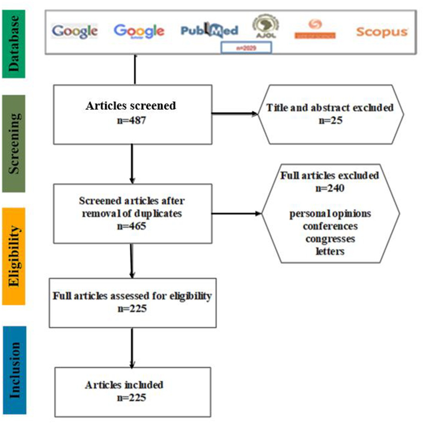
Figure 1. Selection of articles using different search engines and exclusion criteria.
3 Detection approaches
As stated above, LMO is an opportunistic pathogenic bacterium responsible for human listeriosis and often associated with contaminated foods (Andritsos et al., 2013). This species is ubiquitous in nature and able to survive in harsh environmental conditions, such as low temperature and pH (Sumrall et al., 2020). In humans, the diagnosis for listeriosis is established based on clinical symptoms and detection of the bacterium from bodily fluids such as blood, cerebrospinal fluid (CSF) and amniotic fluid (DiMaio, 2000). Various detection methods, including culture-dependent and culture-independent (serology, molecular biology) are used to monitor LMO in the food industry and clinical samples (Andritsos et al., 2013).
3.1 Culture-dependent approaches
Historically, it has been difficult to isolate LMO from food samples due to the presence of other bacterial species. To overcome this problem, a method was developed at an early stage by Gray (1957), consisting of storing the suspicious food at a low temperature for periods ranging from 1 week to 3 months or more to isolate LMO (Gray, 1957). This method, known as “cold enrichment,” was then used to isolate and characterise LMO from clinical samples by incubating them for prolonged periods at +4°C on agar plates until visible colonies were formed (Gasanov et al., 2005). This method has disadvantages, in that it generally does not allow for the isolation of damaged Listeria cells which are greatly outnumbered by competitors and will not grow or survive in harsh conditions (Gasanov et al., 2005). In France, different storage temperatures were set by Regulation 853/2004 for pre-packaged foodstuffs, including dairy products which were required to be stored at temperatures under 6°C. However, as mentioned above, storage at 4°C is not effective at preventing the growth of LMO. Subsequently, significant efforts were developed by researchers, focusing on enrichment media and the ideal protocols to improve the recovery of LMO cells damaged by competing microflora (Curiale and Lewus, 1994; Curtis and Lee, 1995). Several methods have been established by regulatory agencies to isolate LMO, including two widely used reference methods: the International Organization for Standardization (ISO) 11,290 method and the United States Food and Drug Administration (FDA) bacteriological and analytical methods (BAM; Fendri et al., 1989; Scotter et al., 2001; Kamana et al., 2014; Seyoum et al., 2015; Nwaiwu, 2016; Reda et al., 2016). Both methods require the enrichment of a sample in a selective broth, designed to slow the growth of competing organisms, before inoculation on selective agar and biochemical identification of colonies with the expected morphology (Fendri et al., 1989; Scotter et al., 2001; Kamana et al., 2014; Seyoum et al., 2015; Nwaiwu, 2016; Reda et al., 2016). In Africa, these reference methods are often used to isolate LMO from environment samples and food products.
3.2 Identification
To identify LMO isolates, several methods are used, including biochemical and phenotypic methods such as Gram staining, catalase testing, oxidase testing, motility testing, haemolysis testing, CAMP testing (Ikeh et al., 2010; Ernest et al., 2015; Seyoum et al., 2015; Tegegne et al., 2019), Api Listeria strip (Bille et al., 1992; Yehia et al., 2016; Osman et al., 2019; Drali et al., 2020), matrix assisted laser desorption ionisation-time of flight (MALDI-TOF) mass spectrometry (Fall et al., 2020; Togo et al., 2020), as well as molecular methods (Kaur et al., 2007; Kamana et al., 2014; Seyoum et al., 2015; Kawo and Bello, 2016; Nwaiwu, 2016; Reda et al., 2016; Fall et al., 2019; Drali et al., 2020; Togo et al., 2020; Sarr et al., 2021). All these identification methods, which are further detailed below, are used in Africa as they are less expensive.
3.2.1 Biochemical methods
Early identification methods based on biochemical and phenotypic markers are widely used. The esculinase reaction based on the detection of β-D-glucosidase activity is used to confirm that the isolated colonies using selective culture media are those of Listeria (Ikeh et al., 2010; Ernest et al., 2015; Seyoum et al., 2015; Tegegne et al., 2019). It is noteworthy that microorganisms from other genera (Enterococcus spp., Bacillus spp.) with a similar morphology can grow on selective plates and are also able to use esculin (Bille et al., 1992; Gasanov et al., 2005; Yehia et al., 2016; Osman et al., 2019; Drali et al., 2020).
The Christie-Atkins-Munch-Petersen (CAMP) test can be used to differentiate haemolytic species of the Listeria genus. In this instance, the suspected bacterium is grown horizontally between streaks of Staphylococous aureus and Rhodococcus equi on blood agar (Rocourt et al., 1985; Liu, 2006). LMO-induced haemolysis and, to a lesser extent, that induced by L. seeligeri is enhanced in the vicinity of S. aureus, whereas haemolysis by L. ivanovii is enhanced in the vicinity of R. equi. However, this test presents limitations as it sometimes fails to differentiate LMO and L. ivanovii in the vicinity of R.equi. The API Listeria strip (bioMérieux, Craponne, France) can be used to distinguish LMO and L. innocua based on the presence or the absence of arylamidase activity (DIM test; Bille et al., 1992; Kamana et al., 2014; Osman et al., 2019). Although these methods can successfully identify LMO, they can also yield ambiguous results (Shamloo et al., 2019).
3.2.2 Matrix assisted laser desorption ionisation-time of flight mass spectrometry
MALDI-TOF MS is a high-throughput soft ionisation technique based on the comparison of the protein fingerprint of microbial cells with a database of reference spectra through the use of various algorithms integrated in recently commercialised systems (Calderaro et al., 2014). This fast and accurate (Lagier et al., 2012, 2018) tool has been increasingly used in recent years and has revolutionised the identification of microorganisms in microbiology laboratories (Bizzini and Greub, 2010).
3.3 Culture-independent approaches
3.3.1 Immunological methods
These methods are based on LMO-specific antibodies and tests can be performed directly from the enrichment media without tedious sample preparation. They are widely applied in food testing due to their simplicity, sensitivity, accuracy and reproducibility. Two immunological methods are used, enzyme-linked immunosorbent assay (ELISA) and immuno-capture. ELISA allows the quantification of LMO based on the use of specific antibody-coated plates and a secondary antibody which enables a colorimetric reaction (Curiale et al., 1994). Immuno-capture also uses specific antibodies coated on magnetic beads to discriminate between LMO and the competing microflora (Jung et al., 2003).
3.3.2 Serological methods
Serological methods are mostly used for typing LMO strains linked to human infections and have been approved to differentiate lineages during an outbreak. Serological typing is based on monoclonal and polyclonal antibodies with the somatic O and flagellar H antigens of LMO (Seeliger and Höhne, 1979). Fifteen serotypes have been outlined based on the somatic antigen (O), whereas four serotypes have been defined based on the flagellar antigen (Seeliger and Höhne, 1979; Schönberg et al., 1996). At least 13 serotypes of LMO have been determined by combining the O and H antigens (1/2a, 1/2b, 1/2c, 3a, 3b, 3c, 4a, 4ab, 4b, 4c, 4d, 4e and 7) and serotypes 1/2a, 1/2b and 4b are the most common in human disease (Orsi et al., 2011).
Phage typing is also used to distinguish LMO strains, based on the specific interaction between a particular bacteriophage and its host strain, LMO, resulting in lysis of the host cell (Rocourt et al., 1985). A major drawback of the phage typing technique is that not all strains of LMO are typable (McLauchlin et al., 1986). Typing can also be achieved using the esterase typing method that measures the esterase activity of LMO strains on starch gels after electrophoresis (Harvey and Gilmour, 2001).
3.3.3 Molecular methods
The identification of LMO using molecular methods is now widely used, as these techniques are extremely sensitive, accurate and specific, although quite expensive. Most molecular methods are targeted towards virulence factor genes using either molecular typing (Cocolin et al., 1997) or gene detection (Bubert et al., 1999; Kaur et al., 2007). To confirm isolates and identify LMO in Africa using culture-independent methods, the most used methods are serotyping, PFGE, RAPD, RFLP, DNA sequencing, MLST, RT-PCR and Multiplex PCR. Molecular typing, consisting of DNA hybridisation-based methods and restriction enzyme analysis, aims at differentiating LMO from other Listeria species, as well as discriminating between different lineages of LMO. Molecular typing methods include DNA hybridisation and pulse field gel electrophoresis (PFGE; Bille and Rocourt, 1996). Gene detection is mostly achieved through single or multiplex PCR. The most targeted genes in the context of PCR are those of virulence factors, namely hlyA [listeriolysin O (LLO)], iap (Invasion-Associated Protein), inl (internalins), and prfA (regulatory protein for virulence cluster activation; Tang et al., 2011; Wang et al., 2015; Tang et al., 2017). Other DNA amplification-based methods include loop-mediated isothermal amplification (LAMP) and random amplified polymorphic DNA (RAPD; Harvey and Gilmour, 2001). Although most methods target virulence genes to identify LMO, it can also be identified using ribotyping (PCR-ribotyping), based on different ribosomal genes (Jacquet et al., 1995). Other typing methods target proteins such as multi-locus enzyme electrophoresis (MEE), based on the different electrostatic charge of proteins, thus reflecting the allelic variation of the genes encoding these amino acid sequences (Thomas et al., 2020). This reliable method is used by several WHO laboratories to detect Listeria serotypes, due to its high sensitivity and usability (Liu, 2006; Thomas et al., 2020).
Multilocus sequence typing (MLST) can also be used as it is the reference technique for discriminating between strains based on the sequencing of “housekeeping genes” encoding essential proteins of the bacterium (Ward et al., 2004; Smith et al., 2019). For instance, MLST with whole genome sequencing (WGS) showed that 91% of clinical isolates were sequence 6 (ST6) in South Africa, which determined that the outbreak in question was largely associated with LMO ST6. Most recently, next generation sequencing (NGS) can be applied to identify LMO in complex samples. For instance, 16S amplicon sequencing was used to determine the abundance of LMO in the breast milk of lactating women associated with severe acute malnutrition in Senegal (Sarr et al., 2021).
4 Characteristics and distribution of Listeria
It is well documented that Listeria species are widely distributed and commonly found in different environments. These species are spread through human and wildlife migration, the animal and food trade, as well as by wind and dust, which are all factors contributing to the global spread of LMO clones (Chenal-Francisque et al., 2011). Following outbreaks of listeriosis around the world, several contaminated samples have been sequenced, including clinical, animal, environmental and food samples. Thus, there are a total of 42,161 LMO genomes available on NCBI (last accessed January 2023).1 African genomes (Table S1) from South Africa (42 genomes), Senegal (11 genomes), Mali (eight genomes), Egypt (two genomes) and Algeria (one genome) were used to build a pangenome to assess the genomic variability of the African LMO strains (Figure 2).
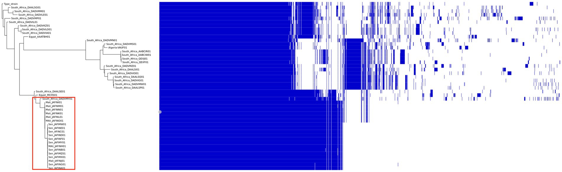
Figure 2. Pangenome analysis of African Listeria strains genome sequences.
We compared the genome sequences of Listeria monocytogenes isolated in Africa found in public databases. All genomes were re-annotated using the Prokka software, version 1.14.5 (Seemann, 2014). Comparisons between all selected genomes were done using Roary, a tool that rapidly builds large-scale pangenomes (Page et al., 2015), with a blast identity cut-off of 97% for the comparison between L. monocytogenes species. A maximum likelihood tree was constructed from the accessory genome elements (left). The presence (blue) and absence (white) of accessory genome elements is presented on the right. Figure 2 shows the dispersion of the pangenome of L. monocytogenes. The studied genomes exhibited a pangenome of 6,864 genes including a core genome of 2,207 genes (shared by all the analysed genomes). This analysis revealed the existence of three clusters in Africa, with South African strains distributed into two clusters and strains from Senegal and Mali clustered together (red box), suggesting the circulation of a single clone in these two west African countries. This clone might be derived from a South African strain which was part of the cluster as well.
4.1 Environmental distribution
LMO is ubiquitous in nature and widely distributed in the environment, including in dust, decaying vegetation and water, and can contaminate agricultural soils. To control its spread and to prevent contamination with this pathogen, studies have been conducted in several ecosystems in Nigeria and South Africa where LMO has been detected in agricultural soils, bodies of water (rivers, streams, ponds and wastewater; David and Odeyemi, 2007; Mawak et al., 2009; Sule et al., 2016; Iwu and Okoh, 2020), manure (cattle and poultry; Ogbonna et al., 2004; David and Odeyemi, 2007), and food processing environments (Adetunji and Adegoke, 2008; Stessl et al., 2020). These environments are potential sources of food, animal and human contamination (Table 1, Figure 3).
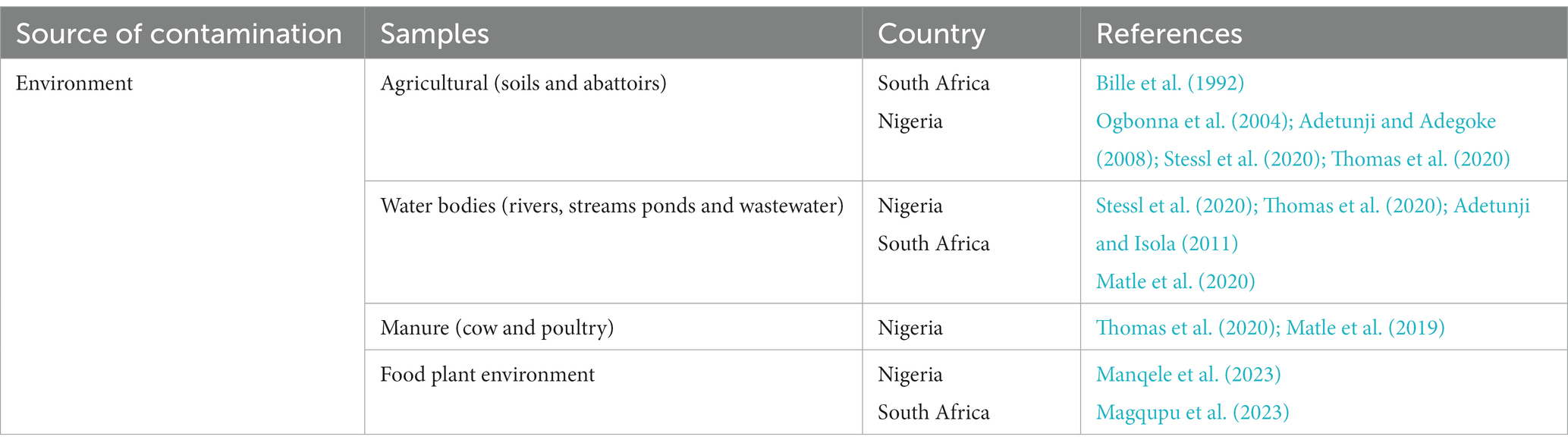
Table 1. Environmental distribution of LMO in Africa.

Figure 3. Distribution of Listeria monocytogenes in Africa.
4.2 Food distribution
4.2.1 Distribution of LMO in non-dairy products
Food is the main source of contamination and is responsible for outbreaks of listeriosis in humans and animals. The first description of LMO as a foodborne pathogen occurred in 1981 following an outbreak due to contaminated coleslaw in Canada (Schlech et al., 1983). The reported outbreak of listeriosis in South Africa in 2018 was associated with processed meat and led to other investigations (Thomas et al., 2020). To control food safety and prevent listeriosis infection or epidemics in African countries, studies have been conducted on several food products (ready-to-eat foods, cold cuts, dairy products, vegetables). LMO has been detected in meat products (beef, sheep and pork) in Nigeria (Ikeh et al., 2010; Adetunji and Isola, 2011), South Africa (Matle et al., 2019, 2020; Thomas et al., 2020; Magqupu et al., 2023; Manqele et al., 2023), Egypt (Yehia et al., 2016), Botswana (Morobe, 2009), Ethiopia (Gebretsadik et al., 2011), and Morocco (Cohen et al., 2008). It has also been detected in poultry in Nigeria (Okwori et al., 2011), Egypt (Yehia et al., 2016), Ethiopia (Gebretsadik et al., 2011) and South Africa (van Nierop et al., 2005) as well as in ready-to-eat foods in South Africa (Matle et al., 2019), Tunisia (Hmaïed et al., 2014), Morocco (Cohen et al., 2008) and Nigeria (Kawo and Bello, 2016), leafy vegetables in Nigeria (Mawak et al., 2009; Kawo and Bello, 2016; Nwaiwu, 2016; Miyebi et al., 2018) and Botswana (Morobe, 2009), fish (smoked and processed) in Nigeria (Chukwu et al., 2006) and Egypt (Yehia et al., 2016), and in ice cream and cakes in Ethiopia (Gebretsadik et al., 2011; Figure 3).
4.2.2 Distribution of LMO in dairy products
Milk (especially cow’s milk) is a common risk factor for contracting listeriosis. Weis et al. demonstrated excreted milk contamination by LMO in cows with mastitis (Weis and Seeliger, 1975) although the first occurrence was reported by Fleming et al. in 1988 (Fleming et al., 2010) with regards to the presence of LMO in 2% pasteurised milk in Massachusetts (Weis and Seeliger, 1975). This led to studies on milk and milk products to detect LMO to prevent and control its transmission. In this context, LMO has been detected from locally-fermented fresh milk in Nigeria (Kawo and Bello, 2016), traditionally fermented milk in Uganda (Mugampoza et al., 2011), raw milk (from cattle herds) in Nigeria (Yakubu et al., 2012), Botswana (Morobe, 2009), Ethiopia (Gebretsadik et al., 2011; Gume et al., 2023), Tanzania (Msalya, 2017), Uganda (Mugampoza et al., 2011) and Algeria (Hamdi et al., 2007), camel milk in Egypt (Yehia et al., 2016), yoghurt in Nigeria (Kawo and Bello, 2016), dairy products in Uganda (Mugampoza et al., 2011) and Algeria (Hamdi et al., 2007), and cheese (cottage and soft) in Botswana (Morobe, 2009), Ethiopia (Gebretsadik et al., 2011) and Nigeria (Adetunji et al., 2014; Table 2, Figure 3). A recently published review on the subject reported a global prevalence of 4.3% in dairy products in Africa with regional variations. West Africa presented a high prevalence with over 20% of positive dairy products while other regions (Southern, Northern, Eastern) had a prevalence under 6% (Oluwafemi et al., 2023).
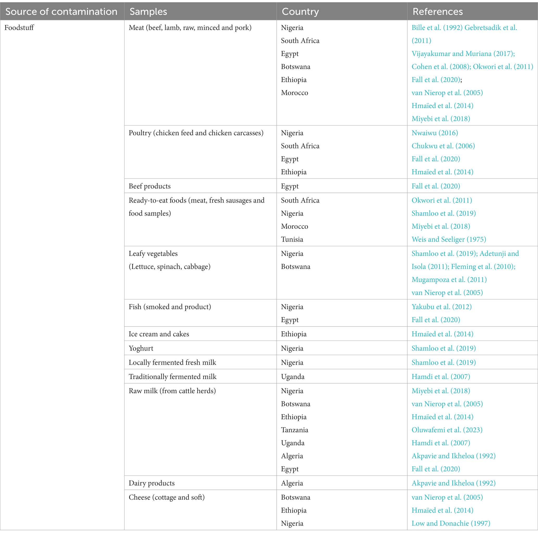
Table 2. Food distribution of LMO in Africa.
4.3 Animal distribution
Animal listeriosis caused by LMO has been reported worldwide, including in Africa (Akpavie and Ikheloa, 1992). This pathogen infects a wide variety of animal species, including mammals, birds, fish and shellfish (Low and Donachie, 1997; Dhama et al., 2015). However, the most commonly-infected animals are ruminants such as cattle, sheep and goats (Meredith and Schneider, 1984; Dhama et al., 2015). An epidemic in sheep herds was found subsequent to symptoms including depression, anorexia, diarrhoea, reduced milk production, fever and abortion in Ghana (Osei-Somuah et al., 2000) and Egypt (El-Beskawy et al., 2010). Subsequently, the first reported listeriosis epidemic in buffalo occurred in Nigeria (Chukwu et al., 2006) and cases have also been detected in laboratory animals and cattle (Akpavie and Ikheloa, 1992; Chukwu et al., 2006). These epidemics led to large-scale monitoring of LMO contamination in several African countries, resulting in its detection in ruminants (sheep, cattle and goats) in Nigeria (Lawan et al., 2013; Oyinloye et al., 2018), poultry in Nigeria (Ogbonna et al., 2004; Okwori et al., 2011) and Kenya (Njagi et al., 2004), fish in Nigeria (Ikeh et al., 2010; Adeshina and Adewale, 2015) and Egypt (Ahmed et al., 2013), shellfish in Egypt (Ahmed et al., 2013), and dogs in South Africa (Schroeder and van Rensburg, 1993; Table 3, Figure 3).
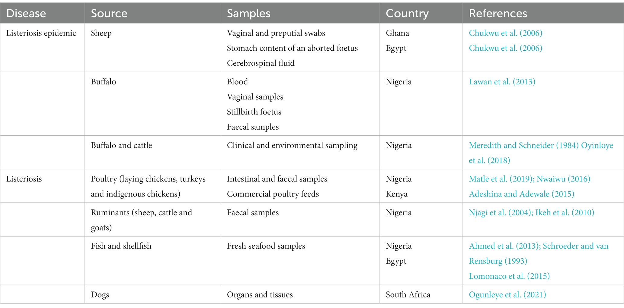
Table 3. Animal distribution of LMO in Africa.
4.4 Human distribution
The links between animal and human listeriosis are not fully understood. There may be a risk of zoonotic transmission of listeriosis through infected pathogens such as faeces, milk, birth fluids, placenta and the foetus (Dhama et al., 2015). As reported above, LMO is widely distributed in Africa in the environment including soil and water bodies as well as in livestock. As a major part of the African population lives in rural environment and lacks access to clean water, listeriosis is probably under-evaluated in this continent. This is should be taken into account when considering the data reported below.
4.4.1 Human listeriosis
Cases of human listeriosis have been reported around the world, including in Africa where the largest listeriosis outbreak was reported in South Africa in 2018 (Smith et al., 2019; Thomas et al., 2020). This disease, caused by LMO through contaminated food, can cause a non-invasive syndrome with febrile gastroenteritis in healthy people (Roberts and Wiedmann, 2003). LMO causes invasive syndromes such as bacteraemia, meningitis, encephalitis and focal abscesses of the central nervous system (CNS) in specific high-risk groups such as the elderly, immunocompromised individuals, and new-borns, and lethality can be as high as 30% (Lomonaco et al., 2015; Ogunleye et al., 2021). In pregnant women, it can lead to transplacental infection which results in miscarriages, premature delivery with serious illnesses such as early sepsis, late sepsis and after late delivery neonatal meningitis (Smíšková et al., 2010; Ogunleye et al., 2021).
Interestingly, the first reported case of listeriosis was an isolation of LMO from the cerebrospinal fluid (CSF) of a four-year-old immunocompromised child in Africa by Benallegue et al. (1968). Subsequently, cases of neonatal listeriosis have been reported in Algeria (Ramdani-Bouguessa and Rahal, 2000), Tunisia (Fendri et al., 1989) and South Africa (Dramowski et al., 2018). Researchers have conducted several studies in African countries following numerous reported infections with strong clinical symptoms. LMO was isolated from pregnant women in Tanzania (Ernest et al., 2015) and Ethiopia (Girma and Abebe, 2018), women who had experienced abortions in Nigeria (Shindang et al., 2013), Ethiopia (Ernest et al., 2015) and Senegal (Fall et al., 2019), an immunocompromised patient in South Africa (Opperman and Bamford, 2018), and in child patients (suffering from septicaemia and meningitis) in South Africa (Oppel et al., 2018), Egypt (Ahmed et al., 2013) and Algeria (Ramdani-Bouguessa and Rahal, 2000).
Despite this concern, human listeriosis, particularly pregnancy-associated listeriosis, is not reported as such although 46% of pregnant women lost their foetuses during the South African epidemic. Studies have reported variable prevalence among reported cases for pregnancy-associated listeriosis in Senegal, Ethiopia and Nigeria [8.04%; van Rensburg and Odendaal, 1992, 4.65%; Fall et al., 2019, 5.56%; Girma et al., 2021, respectively]. This low number of reported outbreaks might be due to the lack of investigation due to lack of resources on the African continent.
4.4.2 Non-symptomatic carriage of LMO in breast milk
Breast milk as a potential source of LMO infection has been overlooked and its prevalence in human milk has not been studied (de Noordhout et al., 2014). In 1988, Svabic-Vlahovic detected and reported in The Lancet the presence of LMO in the milk of a woman who transmitted it to her baby and to puppies (Svabić-Vlahović et al., 1988). Over 30 years later (2019), Togo and colleagues highlighted the presence of LMO in the breast milk of healthy Malian women in an area with a high prevalence of severe acute malnutrition (Togo et al., 2020). In 2021, we further demonstrated the presence in human breast milk in Senegal of a clone of LMO similar to the Malian clone circulating in West Africa, the abundance of which was associated with severe acute malnutrition (Sarr et al., 2021; Table 4).
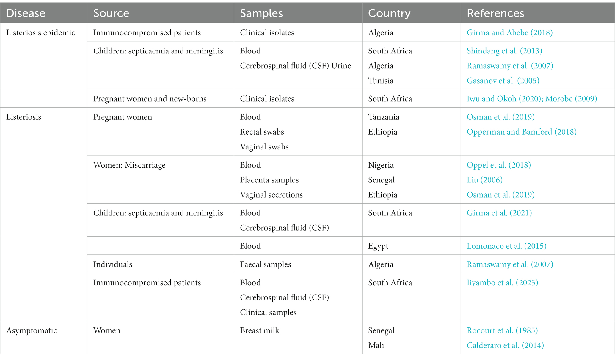
Table 4. Human distribution of LMO in Africa.
5 Treatment
5.1 Treatment of listeriosis
Listeriosis is currently exclusively treated through antibiotic therapy. The successful treatment of listeriosis lies in the administration of antibiotics with a high bactericidal activity during the early phase of the disease (Hof, 2004; Sharma et al., 2017). The most commonly-used treatment for listeriosis nowadays is ampicillin (an antibiotic of the β-lactam family; Hof, 2004; Conter et al., 2009; Bae et al., 2014; Tahoun et al., 2017; Keet and Rip, 2021). It is often used solely or in combination with an aminoglycoside active on Gram-stain positive bacteria (Hof, 2004) with gentamicin being the most commonly used (Charpentier et al., 1995; Bae et al., 2014). Gentamicin can also be combined with amoxicillin (Hof, 2004; Morvan et al., 2010; Keet and Rip, 2021). In pregnant women, amoxicillin or ampicillin are usually the first line of treatment either alone or in combination with gentamicin, followed by trimethoprim/sulfamethoxazole (Madjunkov et al., 2017). Trimethoprim (diaminopyrimidine family) combined with a sulphonamide-like drug is generally used for patients who are intolerant to (Charpentier et al., 1995; Chen et al., 2010; Morvan et al., 2010). It is often combined with sulfamethoxazole for better effectiveness (Chen et al., 2010). However, in a study carried out in Iraq on LMO isolates, it was shown that 98.1, 94.2 and 82.7% of the isolates were resistant to streptomycin, gentamicin and ampicillin, respectively. This resistance profile seems alarming as the aforementioned antibiotics are the drugs of choice for the treatment of listeriosis (Al-Mashhadany, 2019). In such cases, other antibiotics have been successfully used for the treatment of listeriosis such as vancomycin, erythromycin, tetracycline or chloramphenicol (Charpentier et al., 1995; Hof et al., 1997). It is noteworthy that lactic acid bacteria including Lactobacillus paracasei, Lactobacillus salivarius, and Streptococcus salivarius can also inhibit LMO, as demonstrated by several studies (Jiménez et al., 2008; Arroyo et al., 2010; Soleimani et al., 2010; Fernández et al., 2016; Sarr et al., 2021).
5.2 Antibiotic resistance of LMO
Acquired antibiotic resistance is a worldwide public health issue. Although there is a natural resistance, acquired resistance has increasingly emerged in foodborne and human strains. This resistance reportedly stems from the routine use of antibiotics in farms or livestock (Kayode and Okoh, 2023; Manyi-Loh et al., 2023). This acquired resistance requires a constant surveillance to provide adequate treatment for listeriosis and control its spread.
5.2.1 Natural resistance
A natural resistance to oxacillin, fosfomycin and fusidic acid has been described for LMO (Troxler and Graevenitz, 2000). The steady increase in antibiotic-resistant microbial pathogens has become a major public health issue worldwide (Shamloo et al., 2019). This trend was also observed for isolates of LMO (Komora et al., 2017). Antibiotic resistant strains of LMO were first identified in 1985 (Fleming et al., 2010) and later confirmed in subsequent studies (Poyart-Salmeron et al., 1990; Noll et al., 2018). Variability among strains regarding antibiotic resistance has been reported, although most resistant isolates are from food products (Conter et al., 2009; Morvan et al., 2010; Komora et al., 2017).
5.2.2 Acquired resistance
5.2.2.1 Resistance of foodborne LMO strains
The reported resistance of LMO to antibiotics has been determined in several food sources, including cow’s milk (Sharma et al., 2017; Tahoun et al., 2017; Shamloo et al., 2019), from which the first resistant strain was isolated in 1985 by Fleming et al. (2010). The reported resistance of foodborne LMO is concerning as some strains become susceptible to commonly-used antibiotics in the veterinary and human treatment of listeriosis (Keet and Rip, 2021).
5.2.2.2 Resistance of LMO strains isolated from raw milk
Milk is a common risk factor for contracting listeriosis. Although the first report of the presence of LMO in 2% pasteurised milk in Massachusetts was published by Fleming et al. in 1988 (Fleming et al., 2010), it had been previously shown by Weis (Weis and Seeliger, 1975) that LMO was a causative agent of mastitis in dairy cows which could lead to the contamination of excreted milk. A subsequent series of studies was conducted on LMO in milk and milk products (Shamloo et al., 2019). The most frequently-reported resistances in the literature for LMO in milk, apart from natural resistances, are those of beta-lactams (specifically penicillin G; Srinivasan et al., 2005; Jamali et al., 2013; Tahoun et al., 2017), rifampicin (Srinivasan et al., 2005; Tahoun et al., 2017), chloramphenicol (Srinivasan et al., 2005; Tahoun et al., 2017), and tetracycline (Srinivasan et al., 2005; Jamali et al., 2013; Tahoun et al., 2017; Terzi Gulel et al., 2020). The presence of multidrug-resistant LMO strains in raw milk has also been shown in different studies (Jamali et al., 2013; Tahoun et al., 2017).
5.2.2.3 Resistance of LMO strains isolated from raw meat
The antibiotic resistance of LMO in raw meat could compromise the effective treatment of listeriosis in humans, as LMO is becoming increasingly resistant to the antibiotics used to treat human listeriosis (Aras and Ardıç, 2015; Escolar et al., 2017). Raw meat isolates of LMO with antibiotic resistance have been obtained from sheep, beef, pigs, and camels, as well as from chicken and turkeys in different studies. The most frequently observed resistance from meat strains was that against tetracycline (Conter et al., 2009; Escolar et al., 2017; Chen et al., 2019; Keet and Rip, 2021). Other high levels of resistance have also been observed for ampicillin (Conter et al., 2009; Aras and Ardıç, 2015; Chen et al., 2019), oxacillin (Aras and Ardıç, 2015; Capita et al., 2019), and clindamycin (Kaur et al., 2007; Conter et al., 2009).
5.2.2.4 Other resistances of LMO strains isolated from humans
Although listeriosis generally responds to standard therapy, antibiotic-resistant strains have been reported. The study of the antimicrobial susceptibility of LMO in pregnant women, one of the highest risk groups, conducted in Ethiopia by Welekidan et al. (2019), showed a fairly high resistance to penicillin G (66.7%), clindamycin (66.7%), amoxicillin (50%) and vancomycin (50%; Welekidan et al., 2019). Unexpectedly high resistance (96.2) to clindamycin was also shown in another study on Listeria strains isolated from cancer patients with systemic listeriosis (Safdar and Armstrong, 2003). Resistance to cefuroxime (80.8%), cefotaxime (66.6%) and ceftriaxone (76.1%) was also observed in these same patients (Safdar and Armstrong, 2003). In addition, Polish isolates of LMO from invasive infections were resistant to tetracycline and minocycline and harboured the tet(M), tet(A) and tet(C) genes (Kuch et al., 2018). In a report describing the first human case of listeriosis meningo-encephalitis (a complication of gastrointestinal listeriosis), caused by the hypervirulent strain LM-ST-219, antibiotic susceptibility testing also detected resistance to clindamycin, and well as to erythromycin and oxacillin in these isolates (Sotgiu et al., 2018).
6 Scope and limitations
The review aimed at assessing the context of LMO infections across Africa, including sources of contamination, detection methods as well as clinical manifestations and treatment. Using six search engines with a specific query, we screened 225 articles which showed a continuous detection of LMO in different ecosystems (food, environment, hospital, animals). In fact, soil, water bodies and livestock seem to constitute a natural reservoir. Moreover, foodstuff mainly meat (raw and processed) and dairy products are also a source of contamination. Surprisingly, we highlighted in a study from our team in 2021 an asymptomatic carriage in the breast milk of healthy women from Mali and Senegal. This carriage should be further explored to understand its impact as a high relative abundance was associated with severe acute malnutrition in the children of the carriers.
Although cases of listeriosis are reported in Africa and more recently in South Africa, this subject seems under-investigated and under-reported. The high inter-study variability found while conducting this review mirrors the economic and cultural differences between countries. This is also reflected in the LMO surveillance practices between countries. Measures to prevent and control LMO contamination include sterilisation and decontamination of water, soil, and vegetation to prevent transmission to exposed individuals. In addition, the main sources of exposure to LMO are food processing plants, and sanitary controls are still needed to prevent them. It would be important to put in place sanitary procedures and quality controls to prevent new outbreaks of LMO contamination. The biggest challenge remains the implementation of health policies for the detection of LMO and to strengthen national commitments in favour of LMO surveillance.
Despite all the aspects regarding LMO conducted in this review, it presents several limitations. Due to the difference in the type of data reported in the different studies, we chose to cover all the aspects reported above and not focuses on numbers as they were highly variable and depended on the chosen variable in each study. Moreover, there were very few primary studies from other countries than South Africa and most studies presented low sample sizes.
7 Conclusion and perspectives
LMO is widespread in nature (in the environment, food, animals and humans) due to its ability to persist under extreme temperatures, high hydrostatic pressure, oxidative stress and high salt concentration (Bae et al., 2014). This pathogen is detected by several low-cost methods (including culture, biochemical and molecular methods) used in Africa but has been somewhat neglected in human breast milk. As breast milk is a medium of mother-to-child transfer, it is a potential source of LMO and deserves further investigation to shed more light upon the clone circulating in Africa, establish whether it is endemic, and investigate its relationship with malnutrition in larger studies in different localities or countries in Africa.
Author contributions
DR and MM: conceptualization. DR: methodology and funding acquisition. DR, MM, FF, SM, and MA: validation. MS, FD, and AP: formal analysis. MS: investigation. MS and MB: visualization. DR, MM, and MA: supervision. MS, MA, and MM: writing—original draft. MS, MA, MM, DR, SM, CS, and FF: writing—review and editing. All authors contributed to the article and approved the submitted version.
Funding
This study has received financial support from the French Government through the Agence Nationale pour la Recherche (ANR), including the “Programme d’Investissement d’Avenir” under the reference Méditerranée Infection 10-IAHU-03.
Conflict of interest
The authors declare that the research was conducted in the absence of any commercial or financial relationships that could be construed as a potential conflict of interest.
Publisher’s note
All claims expressed in this article are solely those of the authors and do not necessarily represent those of their affiliated organizations, or those of the publisher, the editors and the reviewers. Any product that may be evaluated in this article, or claim that may be made by its manufacturer, is not guaranteed or endorsed by the publisher.
Supplementary material
The Supplementary material for this article can be found online at: https://www.frontiersin.org/articles/10.3389/fmicb.2023.1213953/full#supplementary-material
Footnotes
References
Adeshina, I., and Adewale, Y. A. (2015). Preliminary studies on the prevalence of Listeria monocytogenes in Clarias gariepinus and Orechromis niloticus in Ibadan, Oyo state. J. Agricul. Res. Develop. 14:45. doi: 10.4314/jard.v14i1.5
Adetunji, V. O., and Adegoke, G. O. (2008). Formation of biofilm by strains of Listeria monocytogenes isolated from soft cheese “wara” and its processing environment. African J. Biotechnol.
Adetunji, V. O., Adesokan, H. K., and Agada, C. A., Isola TO (2014). Bacterial load and antimicrobial profile of Escherichia coli and Listeria spp. isolates from muscle tissues of slaughtered cattle at a major abattoir in Ibadan, South-Western Nigeria. J. Basic App. Sci. 10, 299–305. doi: 10.6000/1927-5129.2014.10.39
Adetunji, V. O., and Isola, T. O. (2011). Antibiotic resistance of Escherichia coli, Listeria and Salmonella isolates from retail meat tables in Ibadan municipal abattoir, Nigeria. Afr. J. Biotechnol. 10, 224–228. doi: 10.3923/pjn.2011.224.228
Ahmed, H. A., Hussein, M. A., and El-Ashram, A. M. (2013). Seafood a potential source of some zoonotic bacteria in Zagazig, Egypt, with the molecular detection of Listeria monocytogenes virulence genes. Vet. Ital. 49, 299–308. doi: 10.12834/VetIt.1305.05
Akpavie, S. O., and Ikheloa, J. O. (1992). An outbreak of listeriosis in cattle in Nigeria. Rev. Elev. Med. Vet. Pays Trop. 45, 263–264. doi: 10.19182/remvt.8914
Al-Mashhadany, D. A. (2019). Occurrence and antibiogram of Listeria monocytogenes isolates from retail meat shops at Erbil City, Kurdistan region, Iraq. Ital J Food Saf. 8:8451. doi: 10.4081/ijfs.2019.8451
Andritsos, N. D., Mataragas, M., Paramithiotis, S., and Drosinos, E. H. (2013). Quantifying Listeria monocytogenes prevalence and concentration in minced pork meat and estimating performance of three culture media from presence/absence microbiological testing using a deterministic and stochastic approach. Food Microbiol. 36, 395–405. doi: 10.1016/j.fm.2013.06.020
Aras, Z., and Ardıç, M. (2015). Occurrence and antibiotic susceptibility of Listeria species in Turkey meats. Korean J. Food Sci. Anim. Resour. 35, 669–673. doi: 10.5851/kosfa.2015.35.5.669
Arroyo, R., Martín, V., Maldonado, A., Jiménez, E., Fernández, L., and Rodríguez, J. M. (2010). Treatment of infectious mastitis during lactation: antibiotics versus oral administration of lactobacilli isolated from breast milk. Clin. Infect. Dis. 50, 1551–1558. doi: 10.1086/652763
Bae, D., Mezal, E. H., Smiley, R. D., Cheng, C. M., and Khan, A. A. (2014). The sub-species characterization and antimicrobial resistance of Listeria monocytogenes isolated from domestic and imported food products from 2004 to 2011. Food Res. Int. 64, 656–663. doi: 10.1016/j.foodres.2014.07.049
Benallegue, A., Benhassine, M., Grangaud, J. P., and Mazouni, M. (1968). Méningite à Listeria monocytogenes. Alger. Medicale 29:32.
Bille, J., Catimel, B., Bannerman, E., Jacquet, C., Yersin, M. N., Caniaux, I., et al. (1992). API Listeria, a new and promising one-day system to identify Listeria isolates. Appl. Environ. Microbiol. 58, 1857–1860. doi: 10.1128/aem.58.6.1857-1860.1992
Bille, J., and Rocourt, J. (1996). WHO international multicenter Listeria monocytogenes subtyping study--rationale and set-up of the study. Int. J. Food Microbiol. 32, 251–262. doi: 10.1016/S0168-1605(96)01140-3
Bizzini, A., and Greub, G. (2010). Matrix-assisted laser desorption ionization time-of-flight mass spectrometry, a revolution in clinical microbial identification. Clin. Microbiol. Infect. 16, 1614–1619. doi: 10.1111/j.1469-0691.2010.03311.x
Bubert, A., Hein, I., Rauch, M., Lehner, A., Yoon, B., Goebel, W., et al. (1999). Detection and differentiation ofListeria spp. by a single reaction based on multiplex PCR. Appl. Environ. Microbiol. 65, 4688–4692. doi: 10.1128/AEM.65.10.4688-4692.1999
Calderaro, A., Arcangeletti, M. C., Rodighiero, I., Buttrini, M., Gorrini, C., Motta, F., et al. (2014). Matrix-assisted laser desorption/ionization time-of-flight (MALDI-TOF) mass spectrometry applied to virus identification. Sci. Rep. 4:6803. doi: 10.1038/srep06803
Capita, R., Felices-Mercado, A., and García-Fernández, C. (2019). Alonso-Calleja C. Characterization of Listeria monocytogenes originating from the Spanish meat-processing chain. Foods 8:542. doi: 10.3390/foods8110542
Carlin, C. R., Liao, J., Weller, D. L., Guo, X., Orsi, R., and Wiedmann, M. (2021). Listeria cossartiae sp. nov., Listeria farberi sp. nov., Listeria immobilis sp. nov., Listeria portnoyi sp. nov. and Listeria rustica sp. nov., isolated from agricultural water and natural environments. Int. J. Syst. Evol. Microbiol. 71:004795. doi: 10.1099/ijsem.0.004795
Charlier, C., Perrodeau, É., Leclercq, A., Cazenave, B., Pilmis, B., Henry, B., et al. (2017). Clinical features and prognostic factors of listeriosis: the MONALISA national prospective cohort study. Lancet Infect. Dis. 17, 510–519. doi: 10.1016/S1473-3099(16)30521-7
Charpentier, E., Gerbaud, G., Jacquet, C., Rocourt, J., and Courvalin, P. (1995). Incidence of antibiotic resistance in Listeria species. J. Infect. Dis. 172, 277–281. doi: 10.1093/infdis/172.1.277
Chen, M., Cheng, J., Zhang, J., Chen, Y., Zeng, H., Xue, L., et al. (2019). Isolation, potential virulence, and population diversity of Listeria monocytogenes from meat and meat products in China. Front. Microbiol. 10:946. doi: 10.3389/fmicb.2019.00946
Chen, B. Y., Pyla, R., Kim, T. J., Silva, J. L., and Jung, Y. S. (2010). Antibiotic resistance in Listeria species isolated from catfish fillets and processing environment. Lett. Appl. Microbiol. 50, 626–632. doi: 10.1111/j.1472-765X.2010.02843.x
Chenal-Francisque, V., Lopez, J., Cantinelli, T., Caro, V., Tran, C., Leclercq, A., et al. (2011). Worldwide distribution of major clones of Listeria monocytogenes. Emerg. Infect. Dis. 17, 1110–1112. doi: 10.3201/eid/1706.101778
Chukwu, C. O., Ehizibolo, D. O., Mustapha, I. A., Chukwu, I. D., Olabode, O., and Ogbonna, C. I. (2006). Listeria species in retail smoked fish at Jos, Nigeria. Animal Production Res. Advan. 2:6349. doi: 10.4314/apra.v2i4.36349
Chukwu, C. O., Muhammad, M. J., Nwankpa, N. D., Chukwu, I. D., and Olabode, A. O. (2006). Listeric septicaemia and abotion in African buffalo (Syncerus caffer). Animal production research. Advances 2:339. doi: 10.4314/apra.v2i4.36339
Chukwu, C. O., Ogo, N. I., Antiabong, J. F., Muhammad, M. J., Ogbonna, C. I., and Chukwukere, S. C. (2006). Epidemiological evidence of listeriosis in guinea pigs fed with cabbage (Brassica oleracea) in Nigeria. Animal Production Res. Advan. 2:353. doi: 10.4314/apra.v2i4.36353
Cocolin, L., Manzano, M., Cantoni, C., and Comi, G. (1997). A PCR-microplate capture hybridization method to detect Listeria monocytogenes in blood. Mol. Cell. Probes 11, 453–455. doi: 10.1006/mcpr.1997.0133
Cohen, N., Filliol, I., Karraouan, B., Badri, S., Carle, I., Ennaji, H., et al. (2008). Microbial quality control of raw ground beef and fresh sausage in Casablanca (Morocco). J. Environ. Health 71, 51–63.
Conter, M., Paludi, D., Zanardi, E., Ghidini, S., Vergara, A., and Ianieri, A. (2009). Characterization of antimicrobial resistance of foodborne Listeria monocytogenes. Int. J. Food Microbiol. 128, 497–500. doi: 10.1016/j.ijfoodmicro.2008.10.018
Curiale, M. S., Lepper, W., and Robison, B. (1994). Enzyme-linked immunoassay for detection of Listeria monocytogenes in dairy products, seafoods, and meats: collaborative study. J. AOAC Int. 77, 1472–1489. doi: 10.1093/jaoac/77.6.1472
Curiale, M. S., and Lewus, C. (1994). Detection of Listeria monocytogenes in samples containing Listeria innocua. J. Food Prot. 57, 1048–1051. doi: 10.4315/0362-028X-57.12.1048
Curtis, G. D., and Lee, W. H. (1995). Culture media and methods for the isolation of Listeria monocytogenes. Int. J. Food Microbiol. 26, 1–13. doi: 10.1016/0168-1605(93)E0027-O
David, O. M., and Odeyemi, A. T. (2007). Antibiotic resistant pattern of environmental isolates of Listeria monocytogenes from ado-Ekiti, Nigeria. Afr. J. Biotechnol. 6, 2135–2139. doi: 10.5897/AJB2007.000-2332
de Noordhout, C. M., Devleesschauwer, B., Angulo, F. J., Verbeke, G., Haagsma, J., Kirk, M., et al. (2014). The global burden of listeriosis: a systematic review and meta-analysis. Lancet Infect. Dis. 14, 1073–1082. doi: 10.1016/S1473-3099(14)70870-9
Dhama, K., Karthik, K., Tiwari, R., Shabbir, M. Z., Barbuddhe, S., Malik, S. V. S., et al. (2015). Listeriosis in animals, its public health significance (food-borne zoonosis) and advances in diagnosis and control: a comprehensive review. Vet. Q. 35, 211–235. doi: 10.1080/01652176.2015.1063023
DiMaio, H. (2000). Listeria infection in women. Primary Care Update for OB/GYNS. 7, 40–45. doi: 10.1016/S1068-607X(99)00039-6
Drali, R., Deriet, A., Verhaegen, B., De Keersmaecker, S. C. J., Botteldoorn, N., Vanneste, K., et al. (2020). Whole-genome sequencing of Listeria monocytogenes serotype 4b isolated from ready-to-eat lentil salad in Algiers. New Microbes and New Infec. 33:100628. doi: 10.1016/j.nmni.2019.100628
Dramowski, A., Lloyd, L. G., Bekker, A., Holgate, S., Aucamp, M., Reddy, K., et al. (2018). Neonatal listeriosis during a countrywide epidemic in South Africa: A tertiary hospital’s experience. S. Afr. Med. J. 108, 818–827. doi: 10.7196/SAMJ.2018.v108i10.13207
El-Beskawy, M. A., Younis, E. E., Soumaya, E. A., and El-Sawalhy, A. A. (2010). Epidemiological studies on listeriosis in sheep. Bulletin of animal health and production in Africa [internet]. [cited 2020 Jan 12). Available from: https://www.ajol.info/index.php/bahpa/article/view/64215
Ernest, A. I., Ndaboine, E., Massinde, A., Kihunrwa, A., and Mshana, S. (2015). Maternal vaginorectal colonization by group B Streptococcus and Listeria monocytogenes and its risk factors among pregnant women attending tertiary hospital in Mwanza, Tanzania. Tanzan. J. Health Res. 17. doi: 10.4314/thrb.v17i2.4
Escolar, C., Gómez, D., Del Carmen Rota García, M., Conchello, P., and Herrera, A. (2017). Antimicrobial resistance profiles of Listeria monocytogenes and Listeria innocua isolated from ready-to-eat products of animal origin in Spain. Foodborne Pathog. Dis. 14, 357–363. doi: 10.1089/fpd.2016.2248
Fall, N. S., Sarr, M., Diagne, N., Bassène, H., Sokhna, C., Lagier, J.-C., et al. (2019). Listeria monocytogenes detected in vaginal self-samples of 2 women after spontaneous miscarriage, Senegal, West Africa. Eur. J. Clin. Microbiol. Infect. Dis. 1–2
Fall, N. S., Sarr, M., Diagne, N., Bassène, H., Sokhna, C., Lagier, J-C., et al. (2020). Listeria monocytogenes detected in vaginal self-samples of 2 women after spontaneous miscarriage, Senegal, West Africa. Eur. J. Clin. Microbiol. Infect. Dis. 39, 393–394. doi: 10.1007/s10096-019-03739-0
Fendri, C., Kechrid, A., Chebbi, F., Ben Hassen, A., and Ben, R. S. (1989). Listeriose en Tunisie: A Propos De Trois Cas. Med. Mal. Infect. 19, 470–471. doi: 10.1016/S0399-077X(89)80138-6
Fernández, L., Cárdenas, N., Arroyo, R., Manzano, S., Jiménez, E., Martín, V., et al. (2016). Prevention of infectious mastitis by Oral Administration of Lactobacillus salivarius PS2 during late pregnancy. Clin. Infect. Dis. 62, 568–573. doi: 10.1093/cid/civ974
Fleming, DW, Cochi, SL, MacDonald, KL, Brondum, J, Hayes, PS, Plikaytis, BD, et al. Massachusetts Medical Society. (2010) [cited 2021 Jun 1]. Pasteurized Milk as a vehicle of infection in an outbreak of Listeriosis. Available from: https://www.nejm.org/doi/10.1056/NEJM198502143120704
Gasanov, U., Hughes, D., and Hansbro, P. M. (2005). Methods for the isolation and identification of Listeria spp. and Listeria monocytogenes: a review. FEMS Microbiol. Rev. 29, 851–875. doi: 10.1016/j.femsre.2004.12.002
Gebretsadik, S., Kassa, T., Alemayehu, H., Huruy, K., and Kebede, N. (2011). Isolation and characterization of Listeria monocytogenes and other Listeria species in foods of animal origin in Addis Ababa, Ethiopia. J. Infect. Public Health 4, 22–29. doi: 10.1016/j.jiph.2010.10.002
Girma, Y., and Abebe, B. (2018). Isolation, identification and antimicrobial susceptibility of Listeria species from raw bovine milk in Debre-Birhan town, Ethiopia. J Zoonotic Dis Public Health. 2. doi: 10.1016/j.clnu.2015.05.002
Girma, L., Geteneh, A., Amenu, D., and Kassa, T. (2021). Isolation and characterization of Listeria monocytogenes among women attending Jimma University medical center. Southwest Ethiopia. BMC Infect Dis. 21:564. doi: 10.1186/s12879-021-06254-w
Gray, M. L. (1957). A rapid method for the detection of colonies of Listeria monocytogenes. Zentralbl Bakteriol Orig. 169, 373–377.
Gume, B., Berhanu, L., Kassa, T., Bediru, H., Fikre, A. G., Dadi, L. S., et al. (2023). Bacterial hazard identification and exposure assessment of raw milk consumption in Jimma zone, south West Ethiopia. BMC Microbiol. 23:166. doi: 10.1186/s12866-023-02910-0
Hamdi, T. M., Naïm, M., Martin, P., and Jacquet, C. (2007). Identification and molecular characterization of Listeria monocytogenes isolated in raw milk in the region of Algiers (Algeria). Int. J. Food Microbiol. 116, 190–193. doi: 10.1016/j.ijfoodmicro.2006.12.038
Harvey, J., and Gilmour, A. (2001). Characterization of recurrent and sporadic Listeria monocytogenes isolates from raw milk and nondairy foods by pulsed-field gel electrophoresis, monocin typing, plasmid profiling, and cadmium and antibiotic resistance determination. Appl. Environ. Microbiol. 67, 840–847. doi: 10.1128/AEM.67.2.840-847.2001
Hmaïed, F., Helel, S., François, J. M., Leclercq, A., Lecuit, M., Smaoui, H., et al. (2014). Prevalence, identification by a DNA microarray-based assay of human and food isolates Listeria spp. from Tunisia. Pathol. Biol. 62, 24–29. doi: 10.1016/j.patbio.2013.10.005
Hof, H. (2004). An update on the medical management of listeriosis. Expert. Opin. Pharmacother. 5, 1727–1735. doi: 10.1517/14656566.5.8.1727
Hof, H., Nichterlein, T., and Kretschmar, M. (1997). Management of listeriosis. Clin. Microbiol. Rev. 10, 345–357. doi: 10.1128/CMR.10.2.345
Iiyambo, O. J., Baba, V., Thomas, J., Sekwadi, P., and Naidoo, P. (2023). Presentation and outcomes of Listeria-affected pregnancies in Johannesburg tertiary hospitals: A 2-year review. Int. J. Gynecol. Obstet. 161, 989–996. doi: 10.1002/ijgo.14617
Ikeh, M. A. C., Obi, S. K. C., Ezeasor, D. N., Ezeonu, I. M., and Moneke, A. N. (2010). Incidence and pathogenicity profile of Listeria sp. isolated from food and environmental samples in Nsukka, Nigeria. Afr. J. Biotechnol. 9, 4776–4782. doi: 10.4314/ajb.v9i30
Iwu, C. D., and Okoh, A. I. (2020). Characterization of antibiogram fingerprints in Listeria monocytogenes recovered from irrigation water and agricultural soil samples. PLoS One 15:e0228956. doi: 10.1371/journal.pone.0228956
Jacquet, C., Catimel, B., Brosch, R., Buchrieser, C., Dehaumont, P., Goulet, V., et al. (1995). Investigations related to the epidemic strain involved in the French listeriosis outbreak in 1992. Appl. Environ. Microbiol. 61, 2242–2246. doi: 10.1128/aem.61.6.2242-2246.1995
Jamali, H., Radmehr, B., and Thong, K. L. (2013). Prevalence, characterisation, and antimicrobial resistance of Listeria species and Listeria monocytogenes isolates from raw milk in farm bulk tanks. Food Control 34, 121–125. doi: 10.1016/j.foodcont.2013.04.023
Jiménez, E., Fernández, L., Maldonado, A., Martín, R., Olivares, M., Xaus, J., et al. (2008). Oral Administration of Lactobacillus Strains Isolated from breast Milk as an alternative for the treatment of infectious mastitis during lactation. Appl. Environ. Microbiol. 74, 4650–4655. doi: 10.1128/AEM.02599-07
Jung, Y. S., Frank, J. F., and Brackett, R. E. (2003). Evaluation of antibodies for Immunomagnetic separation combined with flow cytometry detection of Listeria monocytogenes. J. Food Prot. 66, 1283–1287. doi: 10.4315/0362-028X-66.7.1283
Kamana, O., Ceuppens, S., Jacxsens, L., Kimonyo, A., and Uyttendaele, M. (2014). Microbiological quality and safety assessment of the Rwandan Milk and dairy chain. J. Food Prot. 77, 299–307. doi: 10.4315/0362-028X.JFP-13-230
Kaur, S., Malik, S. V. S., Vaidya, V. M., and Barbuddhe, S. B. (2007). Listeria monocytogenes in spontaneous abortions in humans and its detection by multiplex PCR. J. Appl. Microbiol. 103, 1889–1896. doi: 10.1111/j.1365-2672.2007.03414.x
Kawo, A. H., and Bello, A. M. (2016). Antimicrobial susceptibility profile of Listeria species isolated from some ready-to-eat foods sold in Kano, North-Western Nigeria. Bayero J. Pure App. Sci. 9, 217–222. doi: 10.4314/bajopas.v9i1.34
Kayode, A. J., and Okoh, A. I. (2023). Antimicrobial-resistant Listeria monocytogenes in ready-to-eat foods: implications for food safety and risk assessment. Foods. 12:1346. doi: 10.3390/foods12061346
Keet, R., and Rip, D. (2021). Listeria monocytogenes isolates from Western cape, South Africa exhibit resistance to multiple antibiotics and contradicts certain global resistance patterns. AIMS Microbiol. 7, 40–58. doi: 10.3934/microbiol.2021004
Komora, N., Bruschi, C., Magalhães, R., Ferreira, V., and Teixeira, P. (2017). Survival of Listeria monocytogenes with different antibiotic resistance patterns to food-associated stresses. Int. J. Food Microbiol. 245, 79–87. doi: 10.1016/j.ijfoodmicro.2017.01.013
Kuch, A., Goc, A., Belkiewicz, K., Filipello, V., Ronkiewicz, P., Gołębiewska, A., et al. (2018). Molecular diversity and antimicrobial susceptibility of Listeria monocytogenes isolates from invasive infections in Poland (1997–2013). Sci. Rep. 8:14562. doi: 10.1038/s41598-018-32574-0
Lagier, J. C., Armougom, F., Million, M., Hugon, P., Pagnier, I., Robert, C., et al. (2012). Microbial culturomics: paradigm shift in the human gut microbiome study. Clin. Microbiol. Infect. 18, 1185–1193. doi: 10.1111/1469-0691.12023
Lagier, J. C., Dubourg, G., Million, M., Cadoret, F., Bilen, M., Fenollar, F., et al. (2018). Culturing the human microbiota and culturomics. Nat. Rev. Microbiol. 16, 540–550. doi: 10.1038/s41579-018-0041-0
Lawan, F. A., Tijjani, A. N., Raufu, A. I., Ameh, J. A., Ngoshe, I. Y., and Auwal, M. S. (2013). Isolation and characterisation of Listeria species from ruminants in Maiduguri North–Eastern Nigeria. Afr. J. Biotechnol. 12. doi: 10.4314/ajb.v12i50
Liu, D. (2006). Identification, subtyping and virulence determination of Listeria monocytogenes, an important foodborne pathogen. J. Med. Microbiol. 55, 645–659. doi: 10.1099/jmm.0.46495-0
Lomonaco, S., Nucera, D., and Filipello, V. (2015). The evolution and epidemiology of Listeria monocytogenes in Europe and the United States. Infect. Genet. Evol. 35, 172–183. doi: 10.1016/j.meegid.2015.08.008
Low, J. C., and Donachie, W. (1997). A review of Listeria monocytogenes and listeriosis. Vet. J. 153, 9–29. doi: 10.1016/S1090-0233(97)80005-6
Madjunkov, M., Chaudhry, S., and Ito, S. (2017). Listeriosis during pregnancy. Arch. Gynecol. Obstet. 296, 143–152. doi: 10.1007/s00404-017-4401-1
Magqupu, S., Katiyatiya, C. L. F., Chikwanha, O. C., Strydom, P. E., and Mapiye, C. (2023). Quality and safety of pork sold in the informal urban street markets of the cape metropole. South Africa. Meat Sci. 204:109270. doi: 10.1016/j.meatsci.2023.109270
Manqele, A., Gcebe, N., Pierneef, R. E., Moerane, R., and Adesiyun, A. A. (2023). Identification of Listeria species and multilocus variable-number tandem repeat analysis (MLVA) typing of Listeria innocua and Listeria monocytogenes isolates from cattle farms and beef and beef-based products from retail outlets in Mpumalanga and north west provinces, South Africa. Pathogens. 12:147. doi: 10.3390/pathogens12010147
Manyi-Loh, C. E., Okoh, A. I., and Lues, R. (2023). Occurrence and multidrug resistance in strains of Listeria monocytogenes recovered from the anaerobic co-digestion sludge contained in a single stage steel biodigester: implications for antimicrobial stewardship. Microorganisms. 11:725. doi: 10.3390/microorganisms11030725
Mateus, T., Silva, J., Maia, R. L., and Teixeira, P. (2013). Vol. 2013, ISRN obstetrics and gynecology. Hindawi; [cited 2020 Dec 9]. p. e851712 Listeriosis during Pregnancy: A Public Health Concern. Available from: https://www.hindawi.com/journals/isrn/2013/851712/
Matle, I., Mafuna, T., Madoroba, E., Mbatha, K. R., Magwedere, K., and Pierneef, R. (2020). Population structure of non-ST6 Listeria monocytogenes isolated in the red meat and poultry value chain in South Africa. Microorganisms. 8:1152. doi: 10.3390/microorganisms8081152
Matle, I., Mbatha, K. R., Lentsoane, O., Magwedere, K., Morey, L., and Madoroba, E. (2019). Occurrence, serotypes, and characteristics of Listeria monocytogenes in meat and meat products in South Africa between 2014 and 2016. J. Food Saf. 39:e12629. doi: 10.1111/jfs.12629
Mawak, J. D., Dashen, M. M., Idolor, A. J., and Chukwu, O. O. C. (2009). Occurrence of Listeria Monocytogenes in irrigation water and vegetables at Jos, plateau state, Nigeria. Int. J. Tropical Agricul. Food Syst. 3, 279–282. doi: 10.4314/ijotafs.v3i4
McCollum, J. T., Cronquist, A. B., Silk, B. J., Jackson, K. A., O’Connor, K. A., Cosgrove, S., et al. (2013). Multistate outbreak of Listeriosis associated with cantaloupe. N. Engl. J. Med. 369, 944–953. doi: 10.1056/NEJMoa1215837
McLauchlin, J., Audurier, A., and Taylor, A. G. (1986). Aspects of the epidemiology of human Listeria monocytogenes infections in Britain 1967-1984; the use of serotyping and phage typing. J. Med. Microbiol. 22, 367–377. doi: 10.1099/00222615-22-4-367
Meredith, C. D., and Schneider, D. J. (1984). An outbreak of ovine listeriosis associated with poor flock management practices. J. S. Afr. Vet. Assoc. 55, 55–56.
Miyebi, J. I., Daniel, E. O., and Ezeanya, C. C. (2018). Listeria species as contaminants of lettuce and its resistant genes in Benin city, Nigeria. Nigerian J. Biotechnol. 35, 151–158. doi: 10.4314/njb.v35i2.18
Morobe, I. C. Prevalence, antimicrobial profiles, molecular serotyping and toxigenicity of" listeria monocytogenes" isolated from food in Gabarone, Botswana [PhD thesis]. (2009).
Morvan, A., Moubareck, C., Leclercq, A., Hervé-Bazin, M., Bremont, S., Lecuit, M., et al. (2010). Antimicrobial resistance of Listeria monocytogenes strains isolated from humans in France. Antimicrob. Agents Chemother. 54, 2728–2731. doi: 10.1128/AAC.01557-09
Msalya, G. (2017). Contamination levels and identification of Bacteria in Milk sampled from three regions of Tanzania: evidence from literature and laboratory analyses. Vet Med Int. 2017, 1–10. doi: 10.1155/2017/9096149
Mugampoza, D., Muyanja, C., Ogwok, P., Serunjogi, M. L., and Nasinyama, G. W. (2011). Occurrence of listeria monocytogenes in bulked raw milk and traditionally fermented dairy products in Uganda. Afr. J. Food Agric. Nutr. Dev. 11:916. doi: 10.4314/ajfand.v11i2.65916
Murray, E. G. D., Webb, R. A., and Swann, M. B. R. (1926). A disease of rabbits characterised by a large mononuclear leucocytosis, caused by a hitherto undescribed bacillus bacterium monocytogenes (n.sp.). J. Pathol. Bacteriol. 29, 407–439. doi: 10.1002/path.1700290409
Njagi, L. W., Mbuthia, P. G., Bebora, L. C., Nyaga, P. N., Minga, U., and Olsen, J. E. (2004). Carrier status for Listeria monocytogenes and other Listeria species in free range farm and market healthy indigenous chickens and ducks. East Afr. Med. J. 81, 529–533. doi: 10.4314/eamj.v81i10.9236
Noll, M., Kleta, S., and Al, D. S. (2018). Antibiotic susceptibility of 259 Listeria monocytogenes strains isolated from food, food-processing plants and human samples in Germany. J. Infect. Public Health 11, 572–577. doi: 10.1016/j.jiph.2017.12.007
Nwaiwu, O. (2016). Molecular serotype and evolutionary lineage of Listeria monocytogenes isolated from different Nigerian food items. Afr. J. Biotechnol. 15, 696–705. doi: 10.5897/AJB2015.15125
Ogbonna, C. I. C., Muhammad, M. J., Nwobu, O., Ogo, I. N., Makinde, A. A., Elem, H. E., et al. (2004). Listeria monocytogenes and other Listeria species in poul try faeces applied as manure on farm lands: environmental health and food safety. Nigerian. J. Biotechnol. 15, 52–59. doi: 10.4314/njb.v15i1
Ogunleye, O. O., Karimi, V., and Gujadhur, N. (2021). Listeria bacteremia presenting with cerebral abscess and endocarditis in an elderly patient with chronic immune thrombocytopenia. Cureus 13:e16601. doi: 10.7759/cureus.16601
Okwori, A. E. J., Otalike, P. E., Jamda, P. D., and Okwori, E. (2011). Bacteriological investigation of poultry feeds sold within Jos Metropolis. Journal of medical laboratory. Science 20, 21–26. doi: 10.4314/jmls.v20i1
Oluwafemi, Y. D., Igere, B. E., Ekundayo, T. C., and Ijabadeniyi, O. A. (2023). Prevalence of Listeria monocytogenes in milk in Africa: a generalized logistic mixed-effects and meta-regression modelling. Sci. Rep. 13:12646. doi: 10.1038/s41598-023-39955-0
Oppel, K. B., Holgate, S. L., and Finlayson, H. (2018). A retrospective review of Listeria monocytogenes infection at Tygerberg Children’s hospital, Cape Town, South Africa, from 2006 to 2016: is empirical ampicillin still indicated after the first month of life? S. Afr. Med. J. 108, 937–943. doi: 10.7196/SAMJ.2018.v108i11.13212
Opperman, C. J., and Bamford, C. (2018). Co-infection with Streptococcus pneumoniae and Listeria monocytogenes in an immunocompromised patient. S. Afr. Med. J. 108, 386–388. doi: 10.7196/SAMJ.2018.v108i5.12957
Orsi, R. H., den Bakker, H. C., and Wiedmann, M. (2011). Listeria monocytogenes lineages: genomics, evolution, ecology, and phenotypic characteristics. Int. J. Med. Microbiol. 301, 79–96. doi: 10.1016/j.ijmm.2010.05.002
Osei-Somuah, A., Aning, K. G., Nartey, P. K. W., and Abbam, J. K. (2000). Listeria abortions in sheep on the Accra plains: A case report. Ghana. J. Agric. Sci. 33, 87–91. doi: 10.4314/gjas.v33i1.1888
Osman, K. M., Kappell, A. D., Fox, E. M., Orabi, A., and Samir, A. Emerging public health challenges of Listeria monocytogenes isolated from different ecological niches in Egypt, Food, Humans, Animals and Environment: Egypt. (2019).
Oyinloye, M. A., David, O. M., and Famurewa, O. (2018). Pathogenic potential of Listeria monocytogenes isolated from cattle faeces in Adoekiti. Afr. J. Clin. Exp. Microbiol. 19:104. doi: 10.4314/ajcem.v19i2.5
Page, A. J., Cummins, C. A., Hunt, M., Wong, V. K., Reuter, S., Holden, M. T. G., et al. (2015). Roary: rapid large-scale prokaryote pan genome analysis. Bioinformatics 31, 3691–3693. doi: 10.1093/bioinformatics/btv421
Parte, A. C., Sardà Carbasse, J., Meier-Kolthoff, J. P., Reimer, L. C., and Göker, M. (2020). List of Prokaryotic names with Standing in Nomenclature (LPSN) moves to the DSMZ. Int. J. Syst. Evol. 70, 5607–5612. doi: 10.1099/ijsem.0.004332
Poyart-Salmeron, C., Carlier, C., Trieu-Cuot, P., Courtieu, A. L., and Courvalin, P. (1990). Transferable plasmid-mediated antibiotic resistance in Listeria monocytogenes. Lancet 335, 1422–1426. doi: 10.1016/0140-6736(90)91447-I
Ramaswamy, V., Cresence, V. M., Rejitha, J. S., Lekshmi, M. U., Dharsana, K. S., Prasad, S. P., et al. (2007). Listeria--review of epidemiology and pathogenesis. J. Microbiol. Immunol. Infect. 40, 4–13.
Ramdani-Bouguessa, N., and Rahal, K. (2000). Neonatal listeriosis in Algeria: the first two cases. Clin. Microbiol. Infect. 6, 108–111. doi: 10.1046/j.1469-0691.2000.00016-3.x
Reda, W. W., Abdel-Moein, K., Hegazi, A., Mohamed, Y., and Abdel-Razik, K. (2016). Listeria monocytogenes: an emerging food-borne pathogen and its public health implications. J. Infection in Developing Countries. 10, 149–154. doi: 10.3855/jidc.6616
Roberts, A. J., and Wiedmann, M. (2003). Pathogen, host and environmental factors contributing to the pathogenesis of listeriosis. Cell. Mol. Life Sci. 60, 904–918. doi: 10.1007/s00018-003-2225-6
Rocourt, J., Audurier, A., Courtieu, A. L., Durst, J., Ortel, S., Schrettenbrunner, A., et al. (1985). A multi-Centre study on the phage typing of Listeria monocytogenes. Zentralbl Bakteriol Mikrobiol Hyg A. 259, 489–497. doi: 10.1016/S0176-6724(85)80081-X
Safdar, A., and Armstrong, D. (2003). Listeriosis in patients at a Comprehensive Cancer Center, 1955–1997. Clin. Infect. Dis. 37, 359–364. doi: 10.1086/376631
Sarr, M., Tidjani Alou, M., Delerce, J., Khelaifia, S., Diagne, N., Diallo, A., et al. (2021). A Listeria monocytogenes clone in human breast milk associated with severe acute malnutrition in West Africa: A multicentric case-controlled study. PLoS Negl. Trop. Dis. 15:e0009555. doi: 10.1371/journal.pntd.0009555
Schlech, W. F., Lavigne, P. M., Bortolussi, R. A., Allen, A. C., Haldane, E. V., Wort, A. J., et al. (1983). Epidemic Listeriosis — evidence for transmission by food. N. Engl. J. Med. 308, 203–206. doi: 10.1056/NEJM198301273080407
Schönberg, A., Bannerman, E., Courtieu, A. L., Kiss, R., McLauchlin, J., Shah, S., et al. (1996). Serotyping of 80 strains from the WHO multicentre international typing study of Listeria monocytogenes. Int. J. Food Microbiol. 32, 279–287. doi: 10.1016/S0168-1605(96)01142-7
Schroeder, H., and van Rensburg, I. B. (1993). Generalised Listeria monocytogenes infection in a dog. J. S. Afr. Vet. Assoc. 64, 133–136.
Scotter, S. L., Langton, S., Lombard, B., Lahellec, C., Schulten, S., Nagelkerke, N., et al. (2001). Validation of ISO method 11290: part 2. Enumeration of Listeria monocytogenes in foods. Int. J. Food Microbiol. 70, 121–129. doi: 10.1016/S0168-1605(01)00530-X
Seeliger, H. P. R., and Höhne, K. (1979). “Chapter II serotyping of Listeria monocytogenes and related species” in Methods in microbiology. eds. T. Bergan and J. R. Norris (US: Academic Press), 31–49.
Seemann, T. (2014). Prokka: rapid prokaryotic genome annotation. Bioinformatics 30, 2068–2069. doi: 10.1093/bioinformatics/btu153
Seyoum, E. T., Woldetsadik, D. A., Mekonen, T. K., Gezahegn, H. A., and Gebreyes, W. A. (2015). Prevalence of Listeria monocytogenes in raw bovine milk and milk products from central highlands of Ethiopia. J. Infection in Developing Countries. 9, 1204–1209. doi: 10.3855/jidc.6211
Shamloo, E., Hosseini, H., Abdi Moghadam, Z., Halberg Larsen, M., Haslberger, A., and Alebouyeh, M. (2019). Importance of Listeria monocytogenes in food safety: a review of its prevalence, detection, and antibiotic resistance. Iran J Vet Res. 20, 241–254.
Sharma, S., Sharma, V., Dahiya, D. K., Khan, A., Mathur, M., and Sharma, A. (2017). Prevalence, virulence potential, and antibiotic susceptibility profile of Listeria monocytogenes isolated from bovine raw Milk samples obtained from Rajasthan. India. Foodborne Pathog Dis. 14, 132–140. doi: 10.1089/fpd.2016.2118
Shindang, J., Shindang, C. O., and Ekwempu, A. I. (2013). Incidence of Listeria monocytogenes and other Bacteria in spontaneous abortion cases in Jos. Nigerian J. Biotechnol. 25. doi: 10.4314/njb.v25i0
Smíšková, D., Karpíšková, R., Džupová, O., and Marešová, V. (2010). Listeriosis in pregnant women and newborns in Czech Republic. Klin. Mikrobiol. Infekc. Lek. 16, 211–214.
Smith, A. M., Tau, N. P., Smouse, S. L., Allam, M., Ismail, A., Ramalwa, N. R., et al. (2019). Outbreak of Listeria monocytogenes in South Africa, 2017–2018: laboratory activities and experiences associated with whole-genome sequencing analysis of isolates. Foodborne Pathog. Dis. 16, 524–530. doi: 10.1089/fpd.2018.2586
Soleimani, N. A., Kermanshahi, R. K., Yakhchali, B., and Sattari, T. N. (2010). Antagonistic activity of probiotic lactobacilli against Staphylococcus aureus isolated from bovine mastitis. AJMR. 4, 2169–2173. doi: 10.5897/AJMR.9000040
Sotgiu, G., Muresu, N., Dettori, M., Mura, E., Cossu, A., Dolores Masia, M., et al. (2018). A case of Listeria monocytogenes ST-219 Meningo-encephalitis. Int. J. Environ. Res. Public Health 16:10008. doi: 10.3390/ijerph16010008
Srinivasan, V., Nam, H. M., Nguyen, L. T., Tamilselvam, B., Murinda, S. E., and Oliver, S. P. (2005). Prevalence of antimicrobial resistance genes in Listeria monocytogenes isolated from dairy farms. Foodborne Pathog. Dis. 2, 201–211. doi: 10.1089/fpd.2005.2.201
Stessl, B., Szakmary-Brändle, K., Vorberg, U., Schoder, D., and Wagner, M. (2020). Temporal analysis of the Listeria monocytogenes population structure in floor drains during reconstruction and expansion of a meat processing plant. Int. J. Food Microbiol. 314:108360. doi: 10.1016/j.ijfoodmicro.2019.108360
Sule, I. O., Ahmed, R. N., Saliu, B. K., and Olayinka, K. J. (2016). Bacteriological and physicochemical analysis of waste water from fish ponds. Ethiopian J. Environ. Stud. Manag. 9:167. doi: 10.4314/ejesm.v9i2.5
Sumrall, E. T., Röhrig, C., Hupfeld, M., Selvakumar, L., Du, J., Dunne, M., et al. (2020). Glycotyping and specific separation of Listeria monocytogenes with a novel bacteriophage protein tool kit. Appl. Environ. Microbiol. 86:612. doi: 10.1128/AEM.00612-20
Svabić-Vlahović, M., Pantić, D., Pavićić, M., and Bryner, J. H. (1988). Transmission of Listeria monocytogenes from mother’s milk to her baby and to puppies. Lancet 2:1201. doi: 10.1016/S0140-6736(88)90276-0
Tahoun, A. B. M. B., Abou Elez, R. M. M., Abdelfatah, E. N., Elsohaby, I., El-Gedawy, A. A., and Elmoslemany, A. M. (2017). Listeria monocytogenes in raw milk, milking equipment and dairy workers: molecular characterization and antimicrobial resistance patterns. J Glob Antimicrob Resist. 10, 264–270. doi: 10.1016/j.jgar.2017.07.008
Tang, J. Y. H., Razali, N. A. S., Jalil, L. A., Mat-Sa’ad, S. H., Nakaguchi, Y., Nishibuchi, M., et al. (2017). Detection of Listeria Spp. and Listeria monocytogenes in vegetables by loop-mediated isothermal Amplifcation (LAMP) and multiplex polymerase chain reaction (PCR). J.Fundamental App. Sci. 9, 698–714. doi: 10.4314/jfas.v9i2s.43
Tang, M. J., Zhou, S., Zhang, X. Y., Pu, J. H., Ge, Q. L., Tang, X. J., et al. (2011). Rapid and sensitive detection of Listeria monocytogenes by loop-mediated isothermal amplification. Curr. Microbiol. 63, 511–516. doi: 10.1007/s00284-011-0013-3
Tegegne, H. A., Berhanu, A., Getachew, Y., Serda, B., Nölkes, D., Tilahun, S., et al. (2019). Microbiological safety and hygienic quality of camel meat at abattoir and retail houses in Jigjiga city. Ethiopia. J Infect Dev Ctries. 13, 188–194. doi: 10.3855/jidc.9686
Terzi Gulel, G., Gucukoglu, A., Cadirci, O., Saka, E., and Alisarli, M. (2020). Serotyping and antibiotic resistance of Listeria monocytogenes isolated from raw water buffalo milk and milk products. J. Food Sci. 85, 2889–2895. doi: 10.1111/1750-3841.15376
Thomas, J., Govender, N., McCarthy, K. M., Erasmus, L. K., Doyle, T. J., Allam, M., et al. (2020). Outbreak of Listeriosis in South Africa associated with processed meat. N. Engl. J. Med. 382, 632–643. doi: 10.1056/NEJMoa1907462
Togo, A. H., Dubourg, G., Camara, A., Konate, S., Delerce, J., Andrieu, C., et al. (2020). Listeria monocytogenes in human milk in Mali_ A potential health emergency. J. Inf. Secur. 80, 121–142. doi: 10.1016/j.jinf.2019.09.008
Troxler, R., and Graevenitz, A. (2000). von, Funke G, Wiedemann B, stock I. Natural antibiotic susceptibility of Listeria species: L. grayi, L. innocua, L. ivanovii, L. monocytogenes, L. seeligeri and L. welshimeri strains. Clin. Microbiol. Infect. 6, 525–535. doi: 10.1046/j.1469-0691.2000.00168.x
van Nierop, W., Dusé, A. G., Marais, E., Aithma, N., Thothobolo, N., Kassel, M., et al. (2005). Contamination of chicken carcasses in Gauteng, South Africa, by Salmonella, Listeria monocytogenes and Campylobacter. Int. J. Food Microbiol. 99, 1–6. doi: 10.1016/j.ijfoodmicro.2004.06.009
van Rensburg, H. J., and Odendaal, H. J. (1992). The prevalence of potential pathogenic micro-organisms in the endocervix of pregnant women at Tygerberg Hospital. S Afr Med. J 81, 156–157.
Vijayakumar, P. P., and Muriana, P. M. (2017). Inhibition of Listeria monocytogenes on ready-to-eat meats using Bacteriocin mixtures based on mode-of-action. Foods. 6:E22. doi: 10.3390/foods6030022
Wang, D., Wang, Y., Xiao, F., Guo, W., Zhang, Y., Wang, A., et al. (2015). A comparison of in-house Real-time LAMP assays with a commercial assay for the detection of pathogenic Bacteria. Molecules 20, 9487–9495. doi: 10.3390/molecules20069487
Ward, T. J., Gorski, L., Borucki, M. K., Mandrell, R. E., Hutchins, J., and Pupedis, K. (2004). Intraspecific phylogeny and lineage group identification based on the prfA virulence gene cluster of Listeria monocytogenes. J. Bacteriol. 186, 4994–5002. doi: 10.1128/JB.186.15.4994-5002.2004
Weis, J., and Seeliger, H. P. (1975). Incidence of Listeria monocytogenes in nature. Appl. Microbiol. 30, 29–32. doi: 10.1128/am.30.1.29-32.1975
Welekidan, L. N., Bahta, Y. W., Teklehaimanot, M. G., Abay, G. K., Wasihun, A. G., Dejene, T. A., et al. (2019). Prevalence and drug resistance pattern of Listeria monocytogenes among pregnant women in Tigray region, northern Ethiopia: a cross-sectional study. BMC. Res. Notes 12:538. doi: 10.1186/s13104-019-4566-8
Weller, D., Andrus, A., Wiedmann, M., and den Bakker, H. C. Y. (2015). Listeria booriae sp. nov. and Listeria newyorkensis sp. nov., from food processing environments in the USA. Int. J. Syst. Evol. Microbiol. 65, 286–292. doi: 10.1099/ijs.0.070839-0
World Health Organization. WHO estimates of the global burden of foodborne diseases: Foodborne disease burden epidemiology reference group 2007–2015. Geneva: World Health Organization. (2015).
Yakubu, Y., Salihu, M. D., Faleke, O. O., Abubakar, M. B., Junaidu, A. U., Magaji, A. A., et al. (2012). Prevalence and antibiotic susceptibility of Listeria monocytogenes in raw milk from cattle herds within Sokoto Metropolis, Nigeria. Sokoto J. Vet. Sci. 10, 13–17. doi: 10.4314/sokjvs.v10i2.3
Keywords: Listeria monocytogenes , environment, food, animal, human, treatment, Africa
Citation: Sarr M, Alou MT, Padane A, Diouf FS, Beye M, Sokhna C, Fenollar F, Mboup S, Raoult D and Million M (2023) A review of the literature of Listeria monocytogenes in Africa highlights breast milk as an overlooked human source. Front. Microbiol. 14:1213953. doi: 10.3389/fmicb.2023.1213953
Edited by:
Sukhadeo B. Barbuddhe, ICAR National Research Centre on Meat (NRCM), IndiaReviewed by:
Monirul Islam, University of Massachusetts Amherst, United StatesBeatrix Stessl, University of Veterinary Medicine Vienna, Austria
Chuck Czuprynski, University of Wisconsin-Madison, United States
Copyright © 2023 Sarr, Tidjani Alou, Padane, Diouf, Beye, Sokhna, Fenollar, Mboup, Raoult and Million. This is an open-access article distributed under the terms of the Creative Commons Attribution License (CC BY). The use, distribution or reproduction in other forums is permitted, provided the original author(s) and the copyright owner(s) are credited and that the original publication in this journal is cited, in accordance with accepted academic practice. No use, distribution or reproduction is permitted which does not comply with these terms.
*Correspondence: Maryam Tidjani Alou, tidjani_maryam@hotmail.com