 Weidong Wei1,2†
 Yinan Fan3†Jiaqi Song3Yedan Lou3
 Tao Zhang1,2Hua Yuan1,2
 Jieyuan Jin3Sinan Zhang4
 Xin Jin3*
- 1Shaoxing Maternity and Child Health Care Hospital, Shaoxing, Zhejiang, China
- 2Obstetrics and Gynecology Hospital of Shaoxing University, Shaoxing, Zhejiang, China
- 3Shaoxing University, Shaoxing, Zhejiang, China
- 4Department of Respiratory and Critical Care Medicine, Shanghai Changhai Hospital, The First Affiliated Hospital of Naval Military Medical University, Shanghai, China
Background: Fraser syndrome (FS) is an autosomal recessive inherited malformation disorder characterized by cryptophthalmos, syndactyly, and abnormalities of the respiratory and urogenital tracts. Variants in the FRAS1-related extracellular matrix 2 (FREM2) gene are the major genetic cause. However, clinical diagnosis remains challenging due to phenotypic heterogeneity.
Methods: A 24-week pregnant woman came to our hospital for genetic diagnosis. Ultrasound examination showed bilateral renal agenesis or dysplasia, absence of the bladder, and almost oligohydramnios. Trio whole-exome sequencing (Trio-WES) identified two novel compound heterozygous variants in the fetal FREM2 gene: a maternal, frameshift variant, c.5908_5909del, p.Leu1970ValfsTer33, and a paternal, nonsense variant, c.7881C>G,p.Tyr2627Ter.
Conclusion: We report a rare case of Fraser syndrome 2 caused by compound heterozygous mutations in the FREM2 gene. Our findings expanded the FREM2 genotypic spectrum and demonstrated the significance of Trio-WES in the prenatal diagnosis of recessive disorders.
1 Introduction
Fraser syndrome (FS) 2 (OMIM#617666) is an autosomal recessive genetic disorder characterized by cryptophthalmos, syndactyly, and abnormalities of the respiratory and genitourinary tracts (1). The clinical phenotype may also include a low-set hairline, hypoplastic alae nasi, small mouth, short thorax, abdominal distension, ambiguous genitalia, vaginal atresia, renal agenesis, renal hypoplasia, ureteral agenesis, direct connection of the ureter to the vagina, and bladder agenesis.
Fraser syndrome 2 is a very rare disorder with approximately 150 cases reported to date. The genetic background of this disease is usually associated with FRAS1, FREM2, and GRIP1. FRAS1 is a gene that is involved in skin epithelial morphogenesis during early development (2). FREM2 is located on chromosome 13, and GRIP1 is located on chromosome 12. The main function of the FREM2 gene is to participate in the formation and maintenance of the extracellular matrix. The extracellular matrix is a supporting structure that surrounds cells and significantly influences cell morphology, function, and interactions. The FREM2-encoded protein maintain normal tissue structure by interacting with other components in the extracellular matrix (3). These genes are crucial for cell adhesion and extracellular matrix structure during embryonic development, and variants can also lead to abnormal embryonic development.
Prenatal diagnosis of Fraser syndrome 2 can be performed by ultrasound examination; however, due to the great variety of possible malformations, the diagnosis will remain uncertain in most cases (4). The primary diagnostic criteria include cryptophthalmos, cutaneous syndactyly, genitourinary malformations, and laryngotracheal abnormalities. In response to this situation, we need to conduct further gene sequencing. Treatment and prognosis of FS usually involve dealing with the severity of cerebral, pulmonary, laryngeal, and renal malformations (5). Treating this disease requires multidisciplinary collaboration, including interventions such as total auricular reconstruction in otolaryngology and symblepharon release in ophthalmology.
In this study, we report a fetus with Fraser syndrome 2 caused by two variants in the FREM2 gene. Trio whole-exome sequencing (Trio-WES) revealed two novel variants not previously included in the ClinVar database: c.5908_5909del, p.Leu1970ValfsTer33 and c.7881C>G, p.Tyr2627Ter. Pedigree testing confirmed that the former variant originated from the mother, while the latter variant was inherited from the father. These findings demonstrate the importance of prenatal diagnosis. The genetic sites of these two variants were identified initially, and we hope this novel discovery will help geneticists gain further insights into Fraser syndrome 2.
2 Materials and methods
2.1 Sample collection
This study was approved by the Institutional Ethics Committee of Shaoxing Maternity and Child Health Care Hospital. An informed consent document for participation and publication was signed by the family members. With parental consent, we collected venous blood samples from both parents, and fetal DNA was extracted from the amniotic fluid.
2.2 Trio whole-exome sequencing
Genomic DNA was extracted from blood samples of the patient and both parents using a DNEasy Blood and Tissue Kit (Qiagen, Hilden, Germany), according to the manufacturer’s procedures. For Trio-WES, the genomic DNAs were enriched for coding exons using Agilent SureSelect Low Input Reagent Kit and sequenced on the Illumina HiSeq X Ten platform. The sequencing data captured 99.89% of coding regions across 37,837,472 base pairs across 20,858 genes. The average sequence depth was 144.446X, with 97.91% of targeted regions achieving a depth of ≥20X.
2.3 Data analysis
Quality assessment of raw sequencing data was performed using fastp software, and the low-quality reads, as well as those contaminated by adapters, were removed. The filtered data were aligned with the human hg19 reference genome using BWA software, and then the capture efficiency was evaluated. Then, the single-nucleotide variants (SNVs) and indels in the genome were analyzed using GATK software. Subsequently, the identified SNV and Indel were filtered using population databases, including 1,000 Genomes (1,000 human genome dataset), Genome Aggregation Database dataset (gnomAD) 2.1.1, and the Exome Aggregation Consortium dataset (ExAC). The pathogenicity of missense variants and splicing variants was predicted using the dbNSFP database. Reported variants were used in databases including OMIM, HGMD, and ClinVar. All variants were classified according to American College of Medical Genetics and Genomics (ACMG) genetic variant classification standards and guidelines. Finally, all potentially pathogenic sites were validated by Sanger sequencing.
Similar mitochondrial genome sequencing methods were used, and the obtained data were aligned with the mitochondrial reference genome NC_012920.1. The mitimpact29, HmtVar, and tRNA databases were used to predict the pathogenicity of missense variants and splicing variants. The ClinVar database was used to screen for reported variants.
3 Results
3.1 Clinical features
At 24 weeks of pregnancy, an ultrasound showed that the fetus had bilateral renal agenesis or dysplasia, with no visible bladder and almost no amniotic fluid. No renal echoes were visualized in the bilateral renal areas adjacent to the fetal spine, and the lying-down adrenal gland sign was observed (indicated by the white arrow). No bladder echo was seen between the left and right umbilical arteries on the transverse section of the fetal lower abdomen (Figure 1). Chromosomal testing confirmed a male fetus. The mother was a 28-year-old gravida 1, para 0, and the father was 31 years old. There was no documented history of consanguinity between the parents. This was their first pregnancy, and she conceived spontaneously.
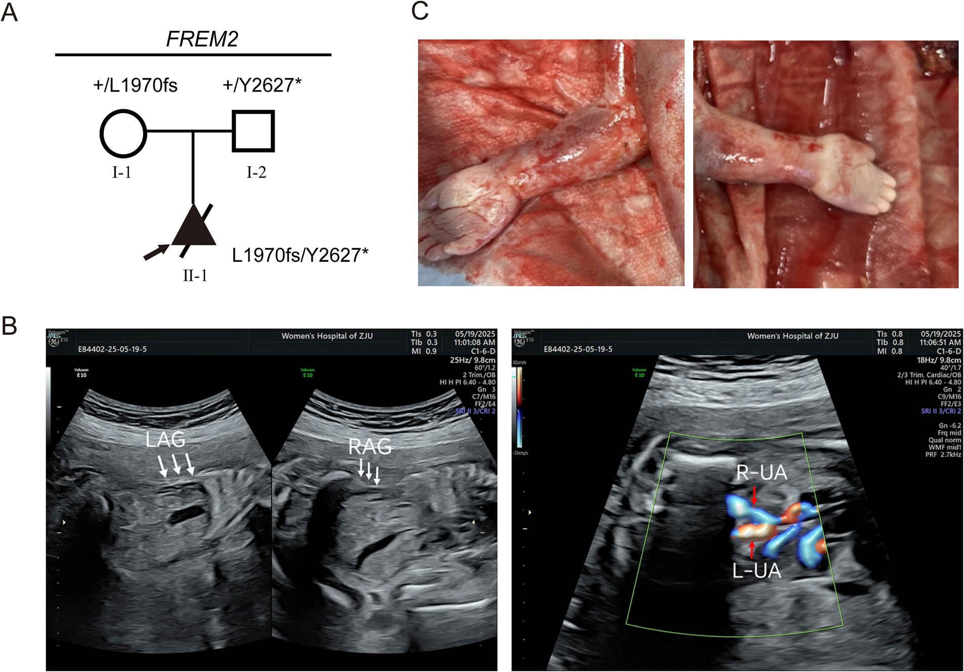
Figure 1. Clinical phenotypes of patients in the Fraser syndrome 2 pedigree associated with FREM2 variants. (A) Pedigree analysis of the presented family. The frameshift variant (c.5908_5909del, p.Leu1970ValfsTer33) occurred in a heterozygous state in the mother (I-1). The nonsense variant (c.7881C>G, p.Tyr2627Ter) occurred in a heterozygous state in the father (I-2). The compound heterozygous variants occurred in the fetus (II-1). Round symbols indicate female individuals; square symbols indicate male individuals; the triangle symbols indicate a fetus; arrow marks indicate the proband. The plus signs denote the reference allele. (B) No renal echoes were visualized in the bilateral renal areas adjacent to the fetal spine, and the lying-down adrenal gland sign was observed (indicated by the white arrow). LAG, left adrenal gland; RAG, right adrenal gland. No bladder echo was seen between the left and right umbilical arteries on the transverse section of the fetal lower abdomen. L-UA, left umbilical artery, R-UA, right umbilical artery. (C) Clinical photographs of the proband show cutaneous symbrachydactyly of the hands.
Parents have no relevant genetic history, and the fetal genetic inheritance was analyzed using STR testing.
3.2 Identification of a novel variant in FREM2 gene
Trio-WES was performed to detect the presence of any variant(s) in the related disease-causing genes. The sequencing was performed using capture high-throughput chip technology, and over 20,000 genes were detected from the human genome. The Sanger sequence was used to verify the variants. The results showed that two variants exist in the FREM2 gene, namely c.5908_5909del (PVS1 + PM2 + PM3) and c.7881C>G (PVS1 + PM2 + PM3), classified as pathogenic variants according to the American College of Medical Genetics and Genomics (ACMG) criteria. The former is a frameshift variant caused by the deletion of nucleotides 5,908 to 5,909 in the coding region, resulting in a change from leucine to valine at amino acid position 1970 and creating a new reading frame that terminates at codon 33 downstream (PVS1). This variant results in a premature termination of the coding protein sequence. The latter is a nonsense variant, which is a cytosine to guanine substitution at nucleotide position 7,881, resulting in a premature stop codon at amino acid position 2,627 (PVS1). This variant also causes the early termination of the coding protein sequence. Subsequently, DNA was extracted from the affected fetus and blood samples were collected from his parents, and the variants were verified by Sanger sequencing, which confirmed that the variant (FREM2 c.5908_5909del, p.Leu1970ValfsTer33) was inherited from the mother and the other variant (FREM2 c.7881C>G,p.Tyr2627Ter) was inherited from his father (PM3). This is the major cause of the fetus’s illness (Figures 2A,B). Neither of the two mutations has been reported in databases such as gnomAD (PM2).
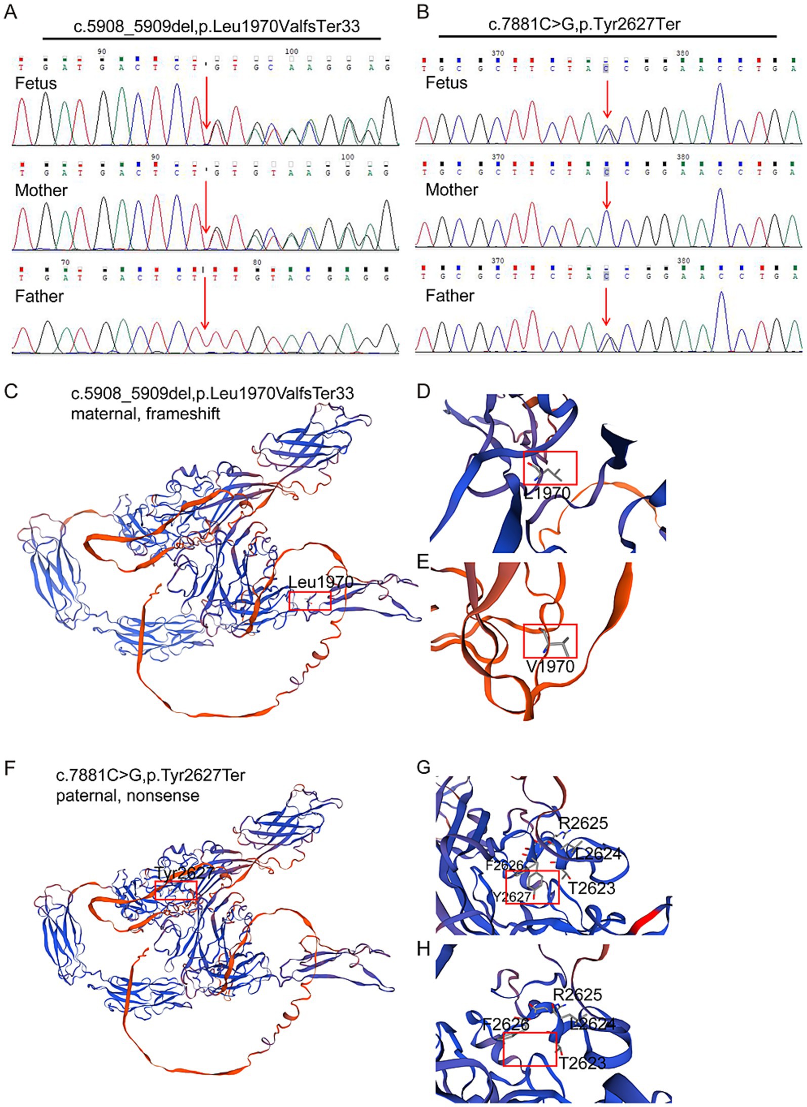
Figure 2. Characterization of identified novel mutations in FREM2. (A,B) Sanger sequencing verification of biallelic FREM2 variants in the pedigree. Variants are denoted in red font. The compound heterozygous variants of FREM2 were identified in the fetus. (C,D) Part of the three-dimensional structure of the wild-type FREM2; and (E) pathogenic variant type (c.5908_5909del; p.Leu1970ValfsTer33). (F,G) Part of the three-dimensional structure of the wild type FREM2; and (H) likely pathogenic variant type (c.7881C>G; p.Tyr2627Ter).
3.3 Structural analysis of FREM2 protein variants identified in the fetus
To investigate the impact of these variants on protein structure, the three-dimensional structures of the wild-type and mutant FREM2 proteins were analyzed. For the maternal frameshift variant c.5908_5909del (p.Leu1970ValfsTer33) (Figures 2C–E), the deletion leads to a shift in the reading frame, causing amino acid 1970 to change from leucine (Leu, shown in Figure 2D) to valine (Val, shown in Figure 2E), and subsequently causing premature termination. This results in a truncated protein structure, resulting from the loss of a C-terminal region beyond the new termination site. The alteration at position 1970 and the truncation disrupt the normal folding and spatial arrangement of the protein, particularly affecting the domain where Leu1970 is located, which may impair interactions with other molecules.
Regarding the paternal nonsense variant c.7881C>G (p.Tyr2627Ter) (Figures 2F–H), the substitution introduces a premature stop codon at amino acid 2,627. In the wild-type protein, the region around Tyr2627 (Figure 2F) has a specific conformation to neighboring residues such as R2625, F2626, L2624, and T2623. In the mutant protein (Figure 2H), the truncation removes Tyr2627 and the subsequent amino acids, leading to a loss of this structural segment and disrupting the overall protein architecture. Both variants result in significant structural abnormalities of the FREM2 protein, which is likely to compromise its normal function and contribute to the observed phenotypic abnormalities.
3.4 Predicted protein interaction network of FREM2
To explore the potential interacting proteins of FREM2 and gain insights into its functional network, we utilized the STRING database for protein–protein interaction (PPI) analysis (Figure 3). FREM2 (red node) was predicted to interact with a range of proteins. These include FRAS1, FREM1, and GRIP1, which are known to be associated with Fraser syndrome, suggesting a coordinated role in the biological processes underlying this disorder. Additionally, interactions were identified with proteins such as ITGA8, ITGB1, NPNT, PROSER1, TRPC4, FAU, and TCF12. The diverse array of interacting proteins implies that FREM2 may participate in multiple cellular pathways, including those related to cell adhesion, extracellular matrix organization, and signal transduction. This PPI network provides a foundation for further investigating the molecular mechanisms by which FREM2 variants lead to the phenotypic abnormalities observed in Fraser syndrome.

Figure 3. Human protein interactors as a candidate for FREM2 collected from the previously reported experimental methods. The network was created using STRING.
4 Discussion
Fraser syndrome 2 is a rare recessive genetic disorder with limited medical research. This article reports a case of Fraser syndrome 2 caused by compound heterozygous variants in the FREM2 gene, hoping to further enrich the understanding and research of this disease.
In this case, the fetus inherited different variants in the same gene (FREM2) from the parents. One is c.5908_5909del, p.Leu1970ValfsTer33 (a frameshift variant caused by the deletion of nucleotides 5,908 to 5,909 in the coding region, resulting in a change from leucine to valine at amino acid position 1970 and creating a new reading frame that terminates at codon 33 downstream). The other variant is c.7881C >G, p.Tyr2627Ter (a cytosine to guanine substitution at nucleotide position 7,881, resulting in a premature stop codon at amino acid position 2,627), which is a nonsense variant. According to the analysis of the pathogenic variant, both variants can lead to the premature termination of the coding protein sequence, resulting in truncated proteins. Many experiments have demonstrated that the introduction of a stop codon results in a truncated protein, leading to loss of biological function of protein (6). Therefore, in this case, the affected fetus is most likely suffering from Fraser syndrome 2 due to compound heterozygous variants that produce truncated proteins, disrupting the original structure of FREM2.
According to existing investigations, FREM2 is associated with immune checkpoints, and its variants may serve as prognostic markers in colorectal cancer patients (7). In another study, researchers focused on a common variant site (c.6499C>T) in three patients and concluded that loss-of-function variants in the FREM2 gene can disrupt eye morphology (8). Cryptophthalmos is a rare congenital disease that is also one of the characteristic features of Fraser syndrome 2. In recent years, cases have been discovered where variants in the FREM2 gene have led to premature aging and loss of normal function in fetuses (9). Some patients have received assistance based on NGS-based SNP haplotyping to select embryos that meet the requirements and have successfully given birth to a healthy baby through in vitro fertilization and embryo transfer (10).
To further explore the impacts of FREM2 gene mutation sites, we have collected and summarized pathogenic and likely pathogenic mutation sites of this gene from the ClinVar database, and creates a diagram (Figure 4). The updated spectrum of ClinVar-reported pathogenic variants in FREM2 (Figure 4) reveals notable clustering of truncating mutations within the large first exon, which encodes the vast N-terminal portion of the protein. This region encompasses critical structural elements, including the signal peptide and CSPG domains, which are essential for the protein’s secretion and its role in epithelial–mesenchymal interactions. The predominance of loss-of-function variants in this region suggests that complete disruption of the protein’s extracellular function is the primary disease mechanism for FS 2. During the retrieval process, we found that different mutation sites and mutation forms in the FREM2 gene lead to multiple outcomes. For example, both cases involve compound heterozygous mutations at two different sites of the same gene. However, in this case, the affected fetus mainly showed renal dysplasia, while in the other one, the mutations primarily led to dental and oral deformities (11). The most serious consequence would be the development of cryptophthalmos (8, 12).
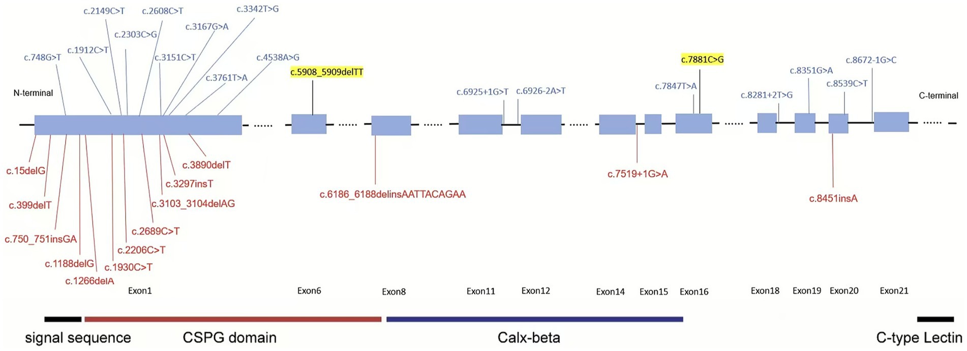
Figure 4. ClinVar-reported variants in the FREM2 gene. The figure displays pathogenic and likely pathogenic variants in the FREM2 gene documented in the ClinVar database, color-coded based on their classification: red for pathogenic (P) and blue for likely pathogenic (LP). Variants highlighted in yellow represent those identified and reported in the current study. The functional domains are highlighted using colored boxes.
From the perspective of protein structure, the two identified variants, c.5908_5909del (p.Leu1970ValfsTer33) and c.7881C>G (p.Tyr2627Ter), both cause severe truncation of the FREM2 protein. The maternal frameshift variant leads to a reading frame shift and premature termination, resulting in the loss of the C-terminal region and disruption of the normal folding and spatial arrangement, especially around the Leu1970 site. The paternal nonsense variant introduces a premature stop codon, removing Tyr2627 and subsequent amino acids, which destroys the overall protein architecture. As mentioned, truncated proteins caused by premature stop codons are generally considered to lose biological function (6), and such structural abnormalities in FREM2 likely impair its ability to perform normal cellular roles, which is a key factor in the development of Fraser syndrome 2 in the fetus.
Regarding the PPI network, FREM2 interacts with several proteins known to be associated with Fraser syndrome, such as FRAS1, FREM1, and GRIP1. This confirms the coordinated role of these proteins in the biological processes underlying the disorder. Additionally, interactions with proteins involved in cell adhesion, extracellular matrix organization, and signal transduction suggest that FREM2 is involved in multiple crucial cellular pathways during embryonic development. The variants in FREM2 not only affect its own structure and function but may also disrupt these interaction networks, further contributing to the complex phenotypic abnormalities observed in Fraser syndrome 2.
Existing studies have also implicated FREM2 in various biological processes and disease contexts. For example, FREM2 is associated with immune checkpoints and may serve as a prognostic marker in colorectal cancer patients (7). Loss-of-function variants in FREM2 can disrupt eye morphology (8), which is relevant to the cryptophthalmos characteristic of Fraser syndrome 2. Moreover, FREM2 variants have been linked to fetal developmental abnormalities, including premature aging and loss of normal function (9), and have implications for assisted reproductive technologies (10). The identification of two novel variants expands the FREM2 variant spectrum and also provides new insights into the genetic basis of Fraser syndrome 2, reinforcing the importance of high-throughput sequencing in uncovering the causes of rare genetic disorders.
In conclusion, this study reports two novel variants in the FREM2 gene associated with the risk of Fraser syndrome 2. The use of high-throughput sequencing technology provided valuable insights into the cause of variants and disease progression. This new discovery enhances our understanding of FS.
Data availability statement
The original contributions presented in the study are included in the article/supplementary material, further inquiries can be directed to the corresponding author.
Ethics statement
The studies involving humans were approved by the Institutional Ethics Committee of Shaoxing Maternity and Child Health Care Hospital. The studies were conducted in accordance with the local legislation and institutional requirements. Written informed consent for participation in this study was provided by the participants’ legal guardians/next of kin. Written informed consent was obtained from the individual(s), and minor(s)’ legal guardian/next of kin, for the publication of any potentially identifiable images or data included in this article.
Author contributions
WW: Formal analysis, Investigation, Writing – original draft, Writing – review & editing. YF: Conceptualization, Resources, Validation, Writing – original draft. JS: Data curation, Formal analysis, Methodology, Writing – original draft. YL: Conceptualization, Data curation, Validation, Writing – original draft. TZ: Data curation, Formal analysis, Funding acquisition, Investigation, Project administration, Resources, Writing – original draft, Writing – review & editing. HY: Investigation, Methodology, Validation, Visualization, Writing – review & editing. JJ: Investigation, Methodology, Writing – review & editing. SZ: Data curation, Investigation, Resources, Writing – original draft. XJ: Data curation, Formal analysis, Methodology, Resources, Software, Supervision, Validation, Visualization, Writing – original draft, Writing – review & editing.
Funding
The author(s) declare that financial support was received for the research and/or publication of this article. This work was supported by the Science Technology Department of Zhejiang Province, China (LTGY23H040004, LTGY23H040005, LTGY24H040005, and LTGY24H040006); the Health Commission of Zhejiang Province, China (WKJ-ZJ-2449 and 2023KY368); the Science Technology Department of Shaoxing, China (2022A14006, 2023A14034, and 2024A14004), the Health Commission of Shaoxing, China (2022KY035, 2022KY036, 2022KY038, 2022KY039, 2023SKY047, 2024SKY003, 2024SKY053, and 2024SKY052), the National Key R&D Program of China (2023YFC2705600) through its sub-project (2023YFC2705602), and Key Laboratory of Reproductive Health of Shaoxing City, Shaoxing Maternity and Child Health Care Hospital, Shaoxing, Zhejiang 312000, China (2023SSY004 and 2023SSY007)
Acknowledgments
We would like to thank our patients for agreeing to donate their personal data to our study and have these data being published.
Conflict of interest
The authors declare that the research was conducted in the absence of any commercial or financial relationships that could be construed as a potential conflict of interest.
Generative AI statement
The authors declare that no Gen AI was used in the creation of this manuscript.
Any alternative text (alt text) provided alongside figures in this article has been generated by Frontiers with the support of artificial intelligence and reasonable efforts have been made to ensure accuracy, including review by the authors wherever possible. If you identify any issues, please contact us.
Publisher’s note
All claims expressed in this article are solely those of the authors and do not necessarily represent those of their affiliated organizations, or those of the publisher, the editors and the reviewers. Any product that may be evaluated in this article, or claim that may be made by its manufacturer, is not guaranteed or endorsed by the publisher.
References
1. Slavotinek, AM, and Tifft, CJ. Fraser syndrome and cryptophthalmos: review of the diagnostic criteria and evidence for phenotypic modules in complex malformation syndromes. J Med Genet. (2002) 39:623–33. doi: 10.1136/jmg.39.9.623
2. McGregor, L, Makela, V, Darling, SM, Vrontou, S, Chalepakis, G, Roberts, C, et al. Fraser syndrome and mouse blebbed phenotype caused by mutations in FRAS1/Fras1 encoding a putative extracellular matrix protein. Nat Genet. (2003) 34:203–8. doi: 10.1038/ng1142
3. Jordan, VK, Beck, TF, Hernandez-Garcia, A, Kundert, PN, Kim, BJ, Jhangiani, SN, et al. The role of FREM2 and FRAS1 in the development of congenital diaphragmatic hernia. Hum Mol Genet. (2018) 27:2064–75. doi: 10.1093/hmg/ddy110
4. Berg, C, Geipel, A, Germer, U, Pertersen-Hansen, A, Koch-Dörfler, M, and Gembruch, U. Prenatal detection of Fraser syndrome without cryptophthalmos: case report and review of the literature. Ultrasound Obstet Gynecol. (2001) 18:76–80. doi: 10.1046/j.1469-0705.2001.00374.x
5. Dumitru, A, Costache, M, Lazaroiu, AM, Simion, G, Secara, D, Cirstoiu, M, et al. Fraser syndrome—a case report and review of literature. Maedica. (2016) 11:80–3.
6. Bartha, I, di Iulio, J, Venter, JC, and Telenti, A. Human gene essentiality. Nat Rev Genet. (2018) 19:51–62. doi: 10.1038/nrg.2017.75
7. Du, H, Wang, H, Kong, F, Wu, M, Chen, W, Lyu, J, et al. Identification and comprehensive analysis of FREM2 mutation as a potential prognostic biomarker in colorectal cancer. Front Mol Biosci. (2022) 9:839617. doi: 10.3389/fmolb.2022.839617
8. Zhang, X, Wang, D, Dongye, M, Zhu, Y, Chen, C, Wang, R, et al. Loss-of-function mutations in FREM2 disrupt eye morphogenesis. Exp Eye Res. (2019) 181:302–12. doi: 10.1016/j.exer.2019.02.013
9. Ikeda, S, Akamatsu, C, Ijuin, A, Nagashima, A, Sasaki, M, Mochizuki, A, et al. Prenatal diagnosis of Fraser syndrome caused by novel variants of FREM2. Hum Genome Var. (2020) 7:32. doi: 10.1038/s41439-020-00119-5
10. Zhou, Y, Yang, X, Liu, Z, Zhang, Y, Chen, H, Zhang, Y, et al. Excluding embryos with two novel mutations in FREM2 gene by the next-generation sequencing-based single nucleotide polymorphism haplotyping. Aging. (2021) 13:24786–94. doi: 10.18632/aging.203715
11. Kantaputra, PN, Wangtiraumnuay, N, Ngamphiw, C, Olsen, B, Intachai, W, Tucker, AS, et al. Cryptophthalmos, dental anomalies, oral vestibule defect, and a novel FREM2 mutation. J Hum Genet. (2022) 67:115–8. doi: 10.1038/s10038-021-00972-4
Keywords: Fraser syndrome 2, FREM2 , compound heterozygous, prenatal diagnosis, Trio-WES
Citation: Wei W, Fan Y, Song J, Lou Y, Zhang T, Yuan H, Jin J, Zhang S and Jin X (2025) Case Report: A case of Fraser syndrome 2 in a Chinese fetus caused by novel compound heterozygous variants in the FREM2 gene. Front. Med. 12:1705708. doi: 10.3389/fmed.2025.1705708
Edited by:
Mustafa Salih, King Saud University, Saudi ArabiaCopyright © 2025 Wei, Fan, Song, Lou, Zhang, Yuan, Jin, Zhang and Jin. This is an open-access article distributed under the terms of the Creative Commons Attribution License (CC BY). The use, distribution or reproduction in other forums is permitted, provided the original author(s) and the copyright owner(s) are credited and that the original publication in this journal is cited, in accordance with accepted academic practice. No use, distribution or reproduction is permitted which does not comply with these terms.
*Correspondence: Xin Jin, TWVycnlfSmluQG91dGxvb2suY29t
†These authors have contributed equally to this work