 Xiaoyan Wu1†Wenfeng Peng2†Xu Zhang1†
 Tao Tang1Ling Deng1Yuxia Xu1
 Xiaoyun Liu1
 Fang Wang1Wujian Peng3
 Jianrong Huang3*Xiaoni Zhong4*
- 1Department of Molecular Diagnostics, State Key Laboratory of Oncology in South China, Guangdong Provincial Clinical Research Center for Cancer, Sun Yat-sen University Cancer Center, Guangzhou, China
- 2Department of Pathology, Shenzhen Second People's Hospital, Shenzhen University 1st Affiliated Hospital, Shenzhen University School of Medicine, Shenzhen, China
- 3Department of Nephrology, The Third People's Hospital of Shenzhen, The Second Affiliated Hospital of Southern University of Science and Technology, Shenzhen, China
- 4Department of Pathology, Shenzhen People's Hospital, The Second Affiliated Hospital of Jinan University, The First Affiliated Hospital of Southern University of Science and Technology, Shenzhen, China
A corrigendum on
Clinicopathological and molecular characterization of astroblastoma
by Wu, X., Peng, W., Zhang, X., Tang, T., Deng, L., Xu, Y., Liu, X., Wang, F., Peng, W., Huang, J., and Zhong, X. (2025). Front. Mol. Neurosci. 18:1483833. doi: 10.3389/fnmol.2025.1483833
In the published article, the word “astrocytoma” was erroneously used in place of the correct word “astroblastoma” throughout the article including in the title, keywords section, citation section, main body text, and the captions of Tables 2, 3 and Figures 1, 2. As astroblastoma is a rare and distinct CNS tumor that has been clearly established as an independent entity in the WHO classification system, the astroblastoma discussed in this research is therefore distinct from astrocytoma and should therefore be presented accurately. All mentions of astrocytoma have now been replaced with astroblastoma in the corrected article where appropriate. The corrected versions of Tables 2, 3 and Figures 1, 2, along with their revised captions, appear below.
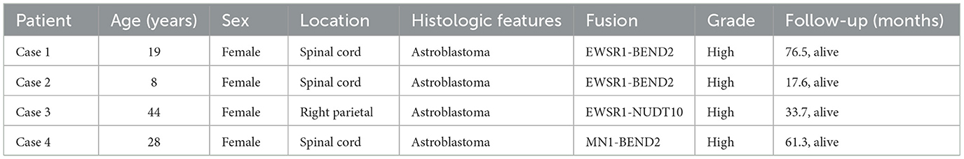
Table 2. Clinical information on patients with astroblastoma in this study.
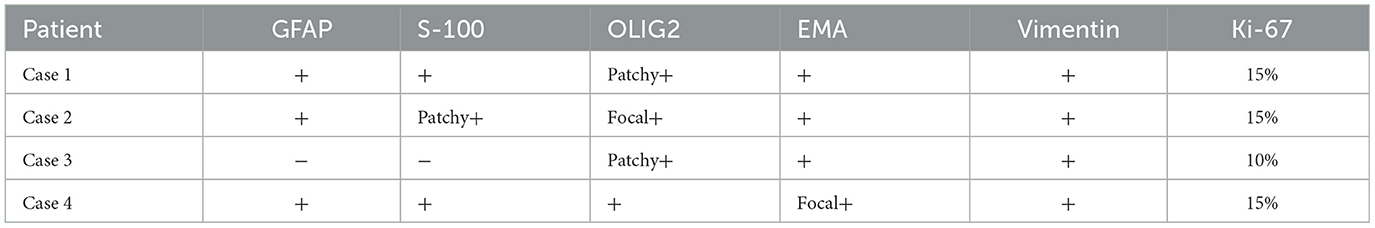
Table 3. IHC characteristics of astroblastoma cases.
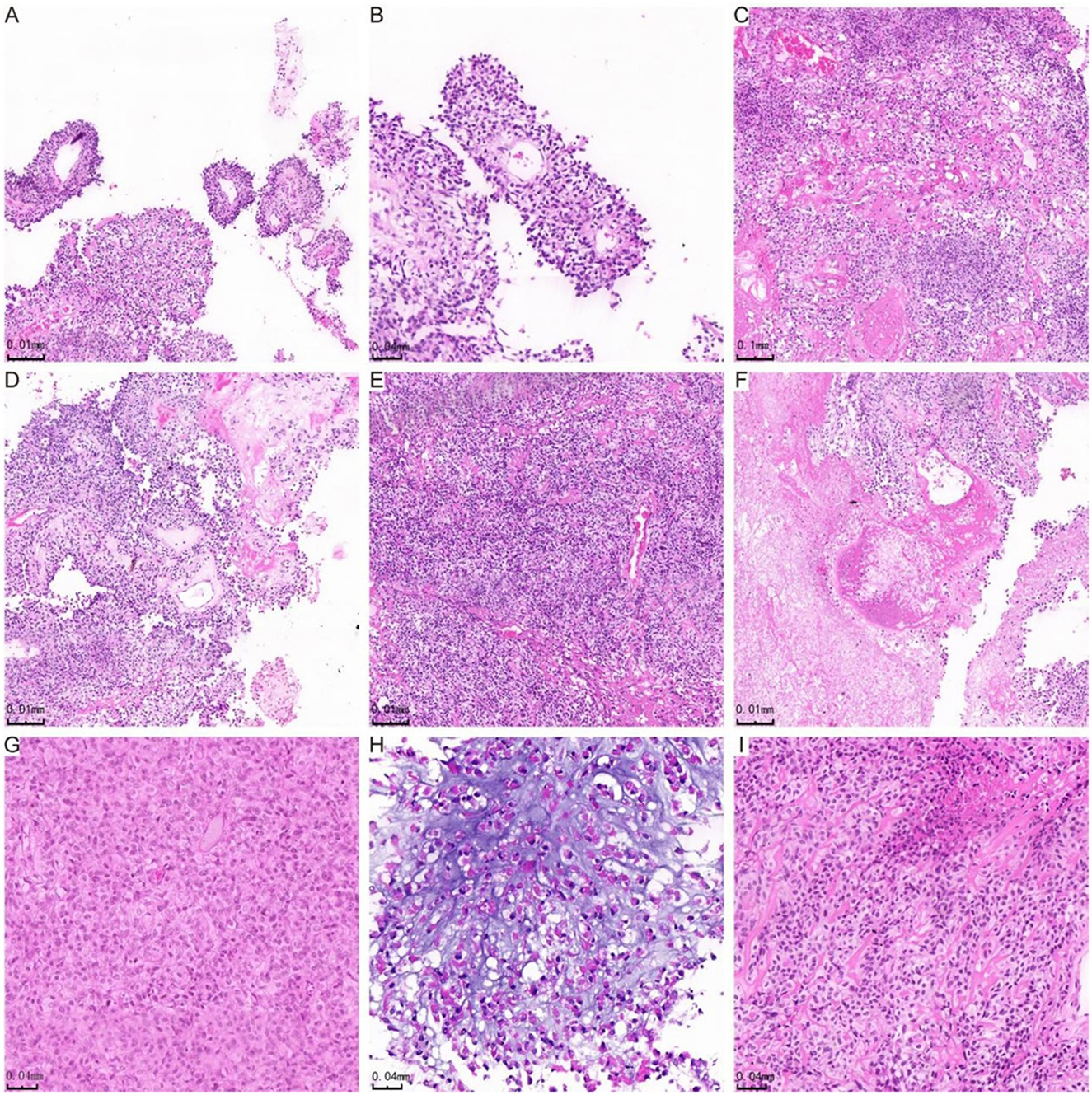
Figure 1. H&E staining showing histological manifestations of astroblastoma. (A) The tumor cells are organized in false rosette-like clusters surrounding blood vessels and exhibit a papillary growth pattern. (B) Shows the tumor cells arranged in a radial pattern. (C) The cells of the astroblastoma are positioned within a hardened matrix. (D) Highlights flake and trabecular structures, along with abundant vascularity and perivascularity. (E) Demonstrates prominent hyaline degeneration and a high density of tumor cells. (F) Indicates areas of necrosis, while (G) reveals undifferentiated cells characterized by abundant eosinophilic cytoplasm. (H) Star-shaped microcystic structures are observed in the astroblastoma, and (I) illustrates cells in astroblastomas exhibiting an epithelioid morphology.
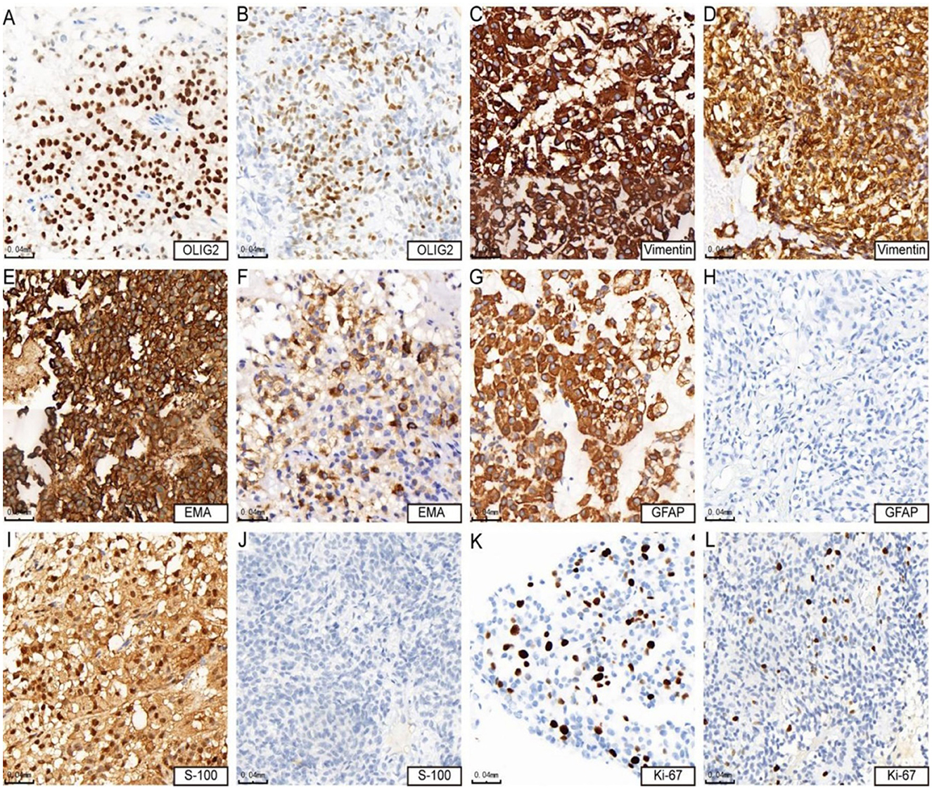
Figure 2. Immunohistochemical features of astroblastoma. (A, B) Show positive expression of OLIG2 (cases 3 and 4). (C, D) Demonstrate strong positive expression of vimentin (cases 3 and 4). (E) indicates strong positive expression of EMA (case 4), while (F) Shows positive expression of EMA (case 3). (G) Reveals strong positive expression of GFAP (case 4), whereas (H) shows negative expression of GFAP (case 3). (I) Presents positive expression of S-100 (case 4), in contrast to (J), which indicates negative expression of S-100 (case 3). Lastly, (K) shows that the KI-67 hotspot area is approximately 15% + (case 4), while (L) indicates that the KI-67 hotspot area is about 10% + (case 3).
The authors apologize for this error and state that this does not change the scientific conclusions of the article in any way. The original article has been updated.
Publisher's note
All claims expressed in this article are solely those of the authors and do not necessarily represent those of their affiliated organizations, or those of the publisher, the editors and the reviewers. Any product that may be evaluated in this article, or claim that may be made by its manufacturer, is not guaranteed or endorsed by the publisher.
Keywords: astroblastoma, clinicopathological, molecular characterization, next-generation sequencing, EWSR1-NUDT10 gene fusion
Citation: Wu X, Peng W, Zhang X, Tang T, Deng L, Xu Y, Liu X, Wang F, Peng W, Huang J and Zhong X (2025) Corrigendum: Clinicopathological and molecular characterization of astroblastoma. Front. Mol. Neurosci. 18:1580624. doi: 10.3389/fnmol.2025.1580624
Received: 20 February 2025; Accepted: 20 May 2025;
Published: 09 June 2025.
Edited and reviewed by: Jean-Marc Taymans, Institut National de la Santé et de la Recherche Médicale (INSERM), France
Copyright © 2025 Wu, Peng, Zhang, Tang, Deng, Xu, Liu, Wang, Peng, Huang and Zhong. This is an open-access article distributed under the terms of the Creative Commons Attribution License (CC BY). The use, distribution or reproduction in other forums is permitted, provided the original author(s) and the copyright owner(s) are credited and that the original publication in this journal is cited, in accordance with accepted academic practice. No use, distribution or reproduction is permitted which does not comply with these terms.
*Correspondence: Jianrong Huang, amlhbnJvbmcyMDA2QDE2My5jb20=; Xiaoni Zhong, eGlhb25pMTk5MTEyM0AxNjMuY29t
†These authors have contributed equally to this work and share first authorship