

- 1 Division of Exercise Physiology, School of Medicine, West Virginia University, Morgantown, WV, USA
- 2 Laboratory of Cardiovascular Science, Intramural Research Program, National Institute on Aging, National Institutes of Health, Baltimore, MD, USA
Age is the dominant risk factor for cardiovascular diseases. Understanding the coupling between the left ventricle (LV) and arterial system, termed arterial–ventricular coupling (EA/ELV), provides important mechanistic insights into the complex cardiovascular system and its changes with aging in the absence and presence of disease. EA/ELV can be indexed by the ratio of effective arterial elastance (EA; a measure of the net arterial load exerted on the LV) to left ventricular end-systolic elastance (ELV; a load-independent measure of left ventricular chamber performance). Age-associated alterations in arterial structure and function, including diameter, wall thickness, wall stiffness, and endothelial dysfunction, contribute to a gradual increase in resting EA with age. Remarkably there is a corresponding increase in resting ELV with age, due to alterations to LV remodeling (loss in myocyte number, increased collagen) and function. These age-adaptations at rest likely occur, at least, in response to the age-associated increase in EA and ensure that EA/ELV is closely maintained within a narrow range, allowing for optimal energetic efficiency at the expense of mechanical efficacy. This optimal coupling at rest is also maintained when aging is accompanied by the presence of hypertension, and obesity, despite further increases in EA and ELV in these conditions. In contrast, in heart failure patients with either reduced or preserved ejection fraction, EA/ELV at rest is impaired. During dynamic exercise, EA/ELV decreases, due to an acute mismatch between the arterial and ventricular systems as ELV increases disproportionate compared to EA (≈200 vs. 40%), to ensure that sufficient cardiac performance is achieved to meet the increased energetic requirements of the body. However, with advancing age the reduction in EA/ELV during acute maximal exercise is blunted, due to a blunted increase ELV. This impaired EA/ELV is further amplified in the presence of disease, and may explain, in part, the reduced cardiovascular functional capacity with age and disease. Thus, although increased stiffness of the arteries itself has important physiological and clinical relevance, such changes also have major implications on the heart, and vice versa, and the manner in the way they interact has important ramifications on cardiovascular function both at rest and during exercise. Examination of the alterations in arterial–ventricular coupling with aging and disease can yield mechanistic insights into the pathophysiology of these conditions and increase the effectiveness of current therapeutic interventions.
Introduction
The population in the Western world is aging; by 2030, there will be 71 million individuals in the United States over 65 years of age, representing ≈20% of the U.S. population. Aging significantly increases cardiovascular morbidity even in the absence of other risk factors (e.g., hypertension, obesity, diabetes, hypercholesterolemia). Thus, the risk of death from heart disease is ≈60-fold greater in individuals in the eighth decade compared to individuals in the 4th decade of life. Not only does clinically overt cardiovascular disease increase dramatically with aging, but so do subclinical or occult diseases, such as silent coronary atherosclerosis. Therefore, the aging of the U.S. population is one of the major public health challenges that we face in the twenty-first century.
The cardiovascular system is modulated to provide sufficient pressure and flow to the tissues at rest and during exercise. Understanding the performance (pressure and flow output) of the left ventricle (LV) requires not only examining the properties of the LV itself (power and stroke capacity of the heart), but also investigating the modulating effects of the arterial system on left ventricular performance. These modulating effects of the vasculature include the capacitance and inertial properties of the aorta, along with the resistance capacity of the microcirculation. The interaction of the LV with the arterial system, termed arterial–ventricular coupling (EA/ELV), is a central determinant of cardiovascular performance and cardiac energetics. EA/ELV can be indexed by the ratio of effective arterial elastance (EA) to left ventricular end-systolic elastance (ELV) and is best displayed in the pressure–volume plane (Figure 1). This review will describe the concept of arterial ventricular coupling and how aging, in the absence and presence of cardiovascular disease, affects the coupling both at rest and during exercise, and its physiological consequences. Further, we will discuss potential therapeutic interventions to restore the coupling between the heart and arteries.
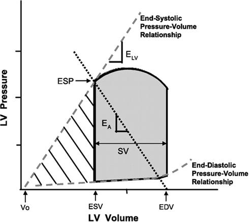
Figure 1. Ventricular pressure–volume diagram from which effective arterial elastance (EA) and left ventricular (LV) end-systolic elastance (ELV) are derived. EA represents the negative slope of the line joining the end-diastolic volume (EDV) and the end-systolic pressure (ESP) points. ELV represents the slope of the ESP–volume relationship passing through the volume intercept (V0). The shaded area represents the cardiac stroke work, and the hatched area represents the potential energy. LV ESP is the LV pressure at the end of systole. EDV is the LV volume at the end of diastole. End-systolic volume (ESV) is the LV volume at the end of systole. Stroke volume (SV) is the volume of blood ejected by the LV with each beat and is obtained from subtracting ESV from EDV. From Chantler et al. (2008a) with permission.
Arterial–Ventricular Coupling
The gold standard to assess the arterial afterload that opposes left ventricular ejection independent of left ventricular function is through aortic input impedance derived from the Fourier analysis of aortic pressure and flow (Murgo et al., 1980). However, aortic input impedance is described in the frequency domain, whereas measures of left ventricular contraction are best described in the time domain; consequently making direct comparisons between arterial and left ventricular function difficult. The pioneering work from Sunagawa et al. (1983) conceived a measure of arterial load (EA) that could be directly compared to a measure of left ventricular contraction (ELV) in the same units (elastance; change in pressure for a given change in volume). Rather than specific arterial properties, EA simplifies the arterial load into an integrative index that incorporates the principal elements of arterial load, including peripheral vascular resistance (which is determined, in large part, by the small arteries), total arterial compliance (which is determined, in large part, by the central elastic arteries), characteristic impedance, and systolic and diastolic time intervals (Sunagawa et al., 1983). Indeed EA is directly related to heart rate and peripheral resistance, and is inversely related to compliance (Chemla et al., 2003). EA can, therefore, be considered a measure of the net arterial load that is imposed on the LV. Further, EA measured invasively as end-systolic pressure/stroke volume closely approximates the arterial load obtained from aortic input impedance and arterial compliance data based on a three-element Windkessel model (Kelly et al., 1992b). Invasively EA is determined from pressure–volume loops as the negative slope of the line joining the end-diastolic volume and end-systolic pressure points (Figure 1).
The contractile function of the LV can also be expressed from the slope of the end-systolic pressure–volume relationship (ELV; Figure 1), which can be obtained from a series of pressure–volume loops recorded while the preload of the heart is altered. ELV reflects a relatively (within normal physiological limits) load-independent measure of left ventricular contraction (chamber stiffness at end systole). An increase in contractility is depicted by an increase in the slope and a shift in the end-systolic pressure–volume relationship to the left, which allows the ventricle to generate more pressure for a given left ventricular volume. However, in addition to the inotropic state, ELV is also influenced by the geometric (structural remodeling) and biochemical properties (i.e., stiffness or compliance of myocytes, composition of muscle, fibrosis, collagen, etc., in the LV wall) that underlie end-systolic stiffness (Borlaug and Kass, 2008). Whereby a “stiffer” LV due to remodeling leads to a higher ELV. Thus, caution should be exercised in interpreting the significance of an elevated ELV, particularly when other measures of left ventricular systolic function are normal. ELV should, therefore, be considered an integrated measure of left ventricular chamber performance that can be related to an integrated measure of arterial load (i.e., EA). Importantly, EA shares common units with ELV, and their ratio EA/ELV is a measure of the interaction between the heart and the arterial system and provides information about how blood pressure and flow change due to different loading conditions. See Chantler et al. (2008a) for a more detailed review of the methods to measure EA and ELV.
At rest, in healthy individuals the properties of the heart and arteries are closely matched so that near maximal cardiac work, power, and chamber efficiency are achieved (Little and Cheng, 1991; De Tombe et al., 1993). Values obtained in isolated canine hearts show the efficiency and stroke work of the heart to be optimal over an EA/ELV ratio ranging 0.3–1.3 (Figure 2). Whereas, in healthy humans the optimal range of EA/ELV to cardiac efficiency and stroke work are generally ranging from 0.7 to 1.0 (Asanoi et al., 1992; Najjar et al., 2004, Redfield et al., 2005). During exercise, an acute mismatch between the arterial and ventricular systems occurs due to a disproportionate increase in ELV vs. EA (Najjar et al., 2004). As a result EA/ELV decreases to ensure that sufficient cardiac performance is achieved to meet the increased energetic requirements of the body. In the next section, we will review the alterations in EA/ELV at rest and during exercise due to aging and illustrate how the age–disease interaction accentuates the changes in EA/ELV. We will also discuss therapeutic interventions aimed to restore the relationship between EA and ELV, including the physiological meaning behind the adaptations.
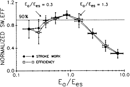
Figure 2. Average normalized SW and SW/MVO2 (efficiency; EFF) as a function of arterial–ventricular coupling ratio. Both parameters reach optimal values between EA/Ees (Ees = ELV) ratios spanning 0.3–1.3. From De Tombe et al. (1993) with permission.
Healthy Aging and Arterial Ventricular Coupling
At Rest
Numerous studies have documented a gradual increase in EA with advancing age (Cohen-Solal et al., 1996; Chen et al., 1998, Redfield et al., 2005). Two small clinical cohort studies calculated EA from invasive methods of flow and pressure and reported a significant rise in EA (44–73%, p < 001) from ≈20 to 70 years of age (Cohen-Solal et al., 1996; Chen et al., 1998). Although the strength of these data lies in the invasive assessment of EA, the small sample size (Cohen-Solal et al., 1996; Chen et al., 1998), and the inclusion of patients with coronary artery disease, and individuals on chronic medications (Chen et al., 1998), provides limited insight into the influence of healthy aging on EA. Non-invasive studies of EA have also reported an increase in EA with age. In a larger epidemiological study consisting of 623 individuals from Olmsted County, Minnesota study, Redfield et al. (2005) observed an age-associated increase in EAI (EA normalized for body surface area) in both men (r = 0.18, p < 0.01) and women (r = 0.13, p = 0.02). Inclusion of individuals with existing cardiovascular disease further increased the age-associated change in EAI in both men (r = 0.28, p < 0.001) and women (r = 0.28, p < 0.001; Figure 3A).
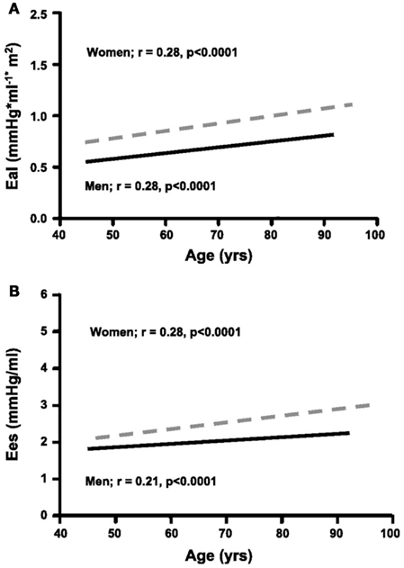
Figure 3. The association between age and EA indexed to body surface area (EAI) (A) and ELV (Ees = ELV) (B) in men (solid line) and women (dashed line) at rest. Pearson correlation coefficients and probability values for each association are shown. With advancing age, both EAI and Ees increase. However, the increase in Ees with age is significantly greater in women than men. Furthermore, EAI and Ees are higher in women vs. men at all ages. Modified from Redfield et al. (2005) with permission.
The specific mechanisms for the increased EA with age reflect the age-associated changes in the arterial properties of individuals. EA is a lumped parameter incorporating mean resistance and pulsatile properties of the arterial load, and is therefore influenced by changes in arterial compliance, wave reflection, and characteristic impedance (Kelly et al., 1992a). During the past two decades, we have characterized the effects of aging on multiple aspects of arterial structure and function in a single study population in the Baltimore Longitudinal Study on Aging (BLSA) who are rigorously screened to exclude both clinical and occult cardiovascular disease. Although at rest, peripheral resistance is the dominant factor affecting EA (Chemla et al., 2003) the age-associated actions of peripheral resistance are heterogeneous to the extent that only an age-associated increase in resting peripheral resistance is noted in healthy women but not men (Fleg et al., 1995). Indeed, the increase in EA with age noted by Redfield et al. (2005) is not attributed to an increase in peripheral resistance as no relationship between age and peripheral resistance existed. EA is also altered by arterial stiffening and blood pressure pulsatility increases with age (Franklin et al., 1997) thus raising the end-systolic pressure required to eject blood thereby increasing EA. Findings from the BLSA indicate that healthy aging is associated with an increase in aortic root diameter, an increase in carotid wall intimal media thickness, and an overall stiffening of the large elastic arteries (Najjar et al., 2005). Redfield et al. (2005) proposed the increase in EA with age is attributable to an increase in arterial stiffness, manifested by an increase in pulse pressure (a surrogate measure of arterial stiffening). Thus, the age-associated increase in EA is largely attributed to an increase arterial stiffness.
The heart seems to respond to an increase in EA with a corresponding increase in ELV with age (Cohen-Solal et al., 1996; Chen et al., 1998, Redfield et al., 2005). Cohen-Solal et al. (1996) noted a 28% higher ELV in men ≈60 vs. 30 years of age. In the Olmsted County, Minnesota study, Redfield et al. (2005) noted a 10% (r = 0.16, p < 0.001) and 15% (r = 23, p < 0.0001) increase in ELV in men and women without cardiovascular disease, respectively (Figure 3B). These results would suggest that ELV increases to compensate for the increase in EA ensuring that their ratio, EA/ELV, is matched for maximal efficiency at rest. Indeed, significant relationships exist between EA and ELV ranging from correlations of 0.50 to 0.73 (Chen et al., 1998; Borlaug et al., 2009) suggesting that a change in EA accounts for a ≈25% change in ELV (Figure 4). This small percentage is not surprising given that both components have multiple different determinants. Nevertheless, older individuals with increased arterial stiffness (EA) are more likely to have an increased left ventricular stiffness (ELV), i.e., a stiffer heart coupled to a stiffer vascular system. A consequence of the age-associated increase in ELV, along with an increase in EA, essentially permits the EA/ELV ratio to remain relatively matched (coupled; Cohen-Solal et al., 1996; Chen et al., 1998, Redfield et al., 2005). However, Redfield et al. (2005) did note a slight sex difference in EA/ELV with age whereby healthy women demonstrated a slight decline in EA/ELV with age, reflecting a disproportionate increase in ELV compared with EA. This suggests a greater impact of aging on ventricular vs. arterial properties in women compared to men, as indicated in Figure 4.
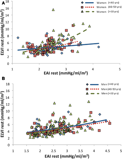
Figure 4. Relationship between arterial (EAI) and left ventricular (ELVI) elastance indexed to body surface area in healthy men (A) and women (B) from the Baltimore longitudinal Study of Aging. Note the linear relationship between EAI and ELVI irrespective of age and gender (Chantler and Lakatta, unpublished data).
Given that ELV is as a load-independent index of left ventricular contractility (Sagawa, 1978), one might suggest that an increase in ELV with age (and more so in women) reflects enhanced contractility. This is unlikely given that other measures of left ventricular function do not increase with age (Lakatta, 1993). Thus, what does the increase in ELV likely reflect? ELV is also influenced by the structural/geometric and biochemical properties (i.e., stiffness or compliance of myocytes, composition of muscle, fibrosis, collagen, etc., in the heart wall) that underlie left ventricular end-systolic stiffness (Borlaug and Kass, 2008; Chantler et al., 2008a). Advancing age is also associated with alterations in left ventricular structure. Most notably, there is a reduction in myocyte number, and there is an increase in left ventricular wall thickness and collagen deposition in the heart (Olivetti et al., 1995; Lakatta, 2003). Thus, the increase in ELV with age could represent differences in left ventricular geometry and/or structure between young and older individuals. For example, concentric remodeling leads to a higher ELV (Borlaug et al., 2009). However, the increase in ELV noted in Redfield et al. (2005) are unlikely to be due to differences in LV chamber size, as similar results are obtained when ELV is normalized to end-diastolic volume (a crude estimate of chamber size). An increase in the amount (focal increases) and a change in the physical properties of collagen (purportedly due to non-enzymatic cross-linking) also occur within the myocardium with aging (Lakatta and Levy, 2003). Thus, the increase in ELV with age is likely attributed to a combination of left ventricular remodeling but more so due to a loss of myocyte number and an increase in myocardial collagen content, thereby increasing myocardial stiffness.
During Exercise
During exercise EA has been shown to increase (Najjar et al., 2004; Otsuki et al., 2006), decline (Asanoi et al., 1992), or remain unchanged (Cohen-Solal et al., 1998). The response of EA during exercise is dependent on the changes in its components and has important consequences on the Frank–Starling mechanism (Chantler et al., 2011). EA is linearly related to heart rate and peripheral resistance, and inversely related to compliance (Segers et al., 2002; Chemla et al., 2003, Otsuki et al., 2006; Chantler et al., 2011). Both resistance and compliance usually decrease during exercise (reflecting less resistance to blood flow in the microcirculation, but increased stiffness of the conduit arteries) and the relative contribution of the pulsatile component (compliance) to EA increases, so that by 80% of peak exercise the resistive and the pulsatile components provide nearly equal contributions to EA (Otsuki et al., 2006). With advancing age, the ability to increase heart rate, and lower resistance during exercise is blunted. In addition the reduction in compliance (due to a greater increase in pulse pressure) is also limited during exercise (Lakatta, 1993; Fleg et al., 1995). Although the blunted maximum cardiovascular responses with age are, in part, due to older individuals achieving a lower maximal workload, similar cardiovascular deficits are also evident at submaximal workloads (Lakatta, 1993; Fleg et al., 1995). The only study to directly examine the age-associated change in EA/ELV in the absence of cardiovascular disease reported that EA, in general, increases during exercise and that EA does not differ between young and older subjects (Najjar et al., 2004). Perhaps the blunted changes in resistance, compliance, and heart rate are compensated for by the greater increase in blood pressure during exercise in older vs. younger healthy individuals (Ogawa et al., 1992). Indeed, some of the components of the changes in EA seem to be related to each other (Chantler et al., 2012). That is, greater preservation of compliance during exercise is associated with a greater reduction in resistance (and a smaller increase in mean and pulse pressure). This suggests that the tandem changes in resistance and compliance appear to be linked, and raises the possibility of a crosstalk between central and peripheral arteries. Further, the change in EA during exercise is also linked to a specific pattern of change in ventricular volumes and function. The change in EA is inversely related to the recruitment of end-diastolic volume, and the enhancement of stroke volume and cardiac output with exercise. Indeed individuals expressing a large increase in EA during exercise demonstrate a blunted utilization of the Frank–Starling mechanism, irrespective of age, sex, and body size (Chantler et al., 2012). The cardiovascular system responds to exercise by increasing cardiac output predominately through an increase in heart rate, and in the upright position stroke volume is increased. The perfusion of the tissues depends both on the ability of the LV to produce flow and sustain perfusion pressure. Thus, the speed and force of left ventricular contraction increases during exercise and this is reflected by an increase in ELV during exercise (Little and Cheng, 1993). Unlike EA, aging impairs the increase in ELV during exercise. Najjar et al. (2004) reports that the blunted increase in ELV with aging begins to appear at 50% of maximal workload and that at maximal exercise older men and women (>60 years) have ≈40–55% smaller ELV compared to men and women <40 years of age. A consequence of the blunted increase in ELV during exercise in older healthy individuals is a corresponding blunted reduction in EA/ELV (Najjar et al., 2004). Furthermore, the altered coupling noted at peak exercise with age is also associated with a blunted increase in ejection fraction. Indeed, EA/ELV is inversely related to ejection fraction [EA/ELV ≈ (1/ejection fraction) − 1; Cohen-Solal et al., 1994]. The advantage of EA/ELV over ejection fraction is that examining the components of EA/ELV permits evaluation of whether alterations in EA/ELV are due to alterations in arterial properties, left ventricular properties, or both. Consequently, advancing age is associated with a smaller EA/ELV (and ejection fraction) reserve (EAI/ELVI peak − EA/ELV rest) and ELV reserve (ELV peak − ELV rest; Najjar et al., 2004). Unlike at rest when an increase in ELV could represent enhanced left ventricular contraction, decreased left ventricular mass (or concentric remodeling), or increased myocardial stiffness (due to alterations in biochemical properties), an increase in ELV during exercise likely reflects an increase in inotropy. This would suggest a decrease in left ventricular contraction during exercise with increasing age.
Aging–CV Disease Interactions Affect Arterial Ventricular Coupling
Unfortunately in today’s society, for the most part, aging is highly linked to the occurrence of cardiovascular diseases. Further, cardiovascular disease risk factors (obesity, hypercholesterolemia, diabetes, and hypertension) often co-vary in number or severity with increasing age. Although measuring the age-associated changes in cardiovascular structure and function in a “healthy” aging population is important to provide insights into the normal aging process, the generalizability of these findings are sometimes limited. The age and disease interaction has important consequences on arterial ventricular coupling both at rest and during exercise as outlined below.
Hypertension
The prevalence of hypertension markedly increases with advancing age, such that the relative risk of acquiring hypertension is ≈90% of individuals over 40 years of age (Lloyd-Jones et al., 2009; Figure 5). Age-associated changes in arterial and left ventricular structure and function are accelerated in the presence of hypertension. Hypertensive patients exhibit greater carotid wall thickness (Arnett et al., 1996), central arterial wall stiffness (Amar et al., 2001), and reflected waves (Nichols et al., 1992) than normotensive subjects, even after adjusting for age. Furthermore, hypertension is associated with left ventricular remodeling and fibrosis (Mayet and Hughes, 2003). As such, EA and ELV are reported to be increased, between 15–60 and 16–95%, respectively, in hypertensive patients compared with normotensive controls (Cohen-Solal et al., 1994; Saba et al., 1999, Lam et al., 2007). As with aging, however, the coupling ratio (EA/ELV) remains matched (coupled) between normotensives and hypertensives (Cohen-Solal et al., 1994; Saba et al., 1999). We found similar results (matched EA/ELV due to tandem increase in EA and ELV) when comparing men with predominantly systolic hypertension to normotensive men. In contrast, women with predominantly systolic hypertension, have a 23% lower resting EA/ELV than normotensive women, a finding that persisted even after adjusting for age (Chantler et al., 2008b). The lower EA/ELV in women with systolic hypertension is due to a disproportionate increase in ELV compared with EA (45 vs. 16%), suggesting an adaptation by these women to limit the impact of systolic hypertension on the vasculature or, alternatively, a more pronounced impact of systolic hypertension on ventricular vs. arterial elastance.
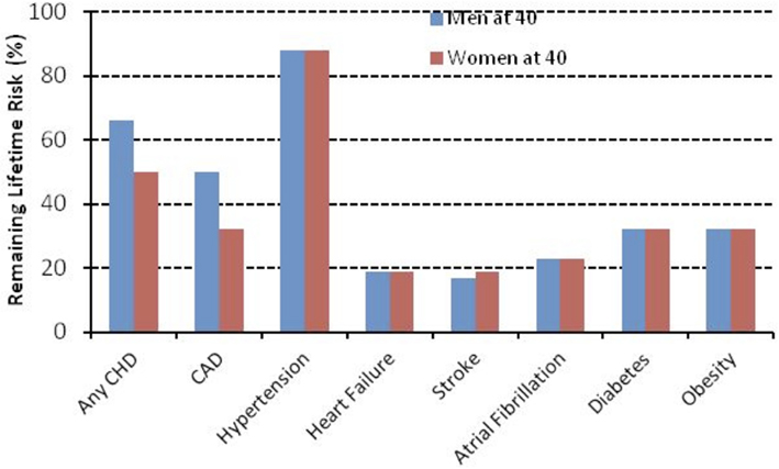
Figure 5. Remaining lifetime risk for cardiovascular disease and other diseases among men and women free of disease at 40 years of age from the Framingham Study. Modified from Lloyd-Jones et al. (2009).
There are a limited number of studies that have examined the coupling response during dynamic exercise in hypertensive individuals. Borlaug et al. (2010) showed that hypertensive individuals express similar changes in EA, ELV and EA/ELV at submaximal and maximal exercise compared to normotensive individuals matched by age and sex. In contrast, Chantler et al. (2008b) reported that the effects of predominantly systolic hypertension on the changes in EA/ELV are similar between normotensive men and systolic hypertensive men and women at 50% of maximal workload and at peak upright bicycle exercise. In men, this is because EA and ELV are proportionally higher at peak exercise in systolic hypertensive compared with normotensive individuals. In women, this is because EA and ELV are similar at peak exercise between systolic hypertensive and normotensive women. Thus, EA/ELV reserve is similar between systolic hypertensive and normotensive men, but 61% lower in systolic hypertensive vs. normotensive women because of the uncoupling in EA/ELV at rest in these systolic hypertensive women.
Obesity
About one-third of U.S. adults (33.8%) are obese and the prevalence of overweight/obesity increases with advancing age. Obesity-related conditions include heart disease, stroke, type 2 diabetes, and certain types of cancer, some of the leading causes of death (Flegal et al., 2010). The physiological changes that occur with obesity are similar to those in hypertension, and often hypertension and obesity co-exist in a given individual complicating interpretations due to obesity alone (Stamler et al., 1978, 1991). Indeed, Zebekakis et al. (2005) identified that obese individuals (14% on antihypertensive medications) have increased carotid, brachial, and femoral diameter with age, which remained significant after adjusting for differences in BP, medications, and smoking status. Further, increasing body mass index is associated with a reduced arterial distensibility (Zebekakis et al., 2005) and increased arterial stiffness (Toto-Moukouo et al., 1986; Zebekakis et al., 2005). In individuals with uncomplicated obesity (without the presence of cardiovascular disease), an increase in body mass index (45 ± 5 vs. 21 ± 2) is associated with an increase in left ventricular mass (irrespective of normalized to height or body surface area), and end-diastolic volume (unadjusted or indexed to height; Rider et al., 2011). Although such alterations in arterial and left ventricular structure/function increase resting EA and ELV in obese vs. non-obese controls, their coupling (EA/ELV), however, remains matched, irrespective of body size. Rider et al. (2011) noted a similar resting ejection fraction, which is inversely related to EA/ELV, between individuals of various body mass index categories. However, whether the increase in EA and ELV noted in obese individuals represents the pathologic effects of excess adipose tissue on cardiovascular function, or the normal physiological relationship between body size and cardiovascular function is important to decipher. In order to have meaningful clinical and scientific comparisons, differences in body size must be accounted for (Chantler et al., 2005; Chantler and Lakatta, 2009). Indeed, in the unadjusted form, resting EA and ELV are slightly reduced (4 and 3%, respectively) in obese vs. healthy non-obese controls (Chirinos et al., 2009). In contrast, when EA and ELV are scaled either ratiometrically or allometrically to body surface area, EA and ELV are between 16–20 and 18–19% higher (p < 0.001) in obese vs. controls. Irrespective of the scaling technique employed there was no relationship between obesity and EA/ELV (Chirinos et al., 2009). Unfortunately, the changes in EA and ELV, and subsequently EA/ELV have not been reported during dynamic exercise in obese vs. non-obese individuals.
Heart Failure
Heart failure (HF) is a syndrome that is characterized by an inability of the heart to pump a sufficient amount of blood to meet the demands of the metabolizing tissues, or can do so only at the expense of elevated filling pressures (Adams et al., 2006). The inability to meet the tissues’ demands is attributed to the ability, or lack thereof, for the LV to fill (diastolic properties) or to eject (systolic properties) blood. Although there is considerable confusion as to the pathological mechanisms that describes individuals with systolic (HF with reduced ejection fraction), or diastolic (HF with preserved ejection fraction) the coupling between the heart and arteries does seem to be impacted differently by the type of HF.
HF with a Reduced EF
Heart failure patients with systolic dysfunction are characterized by a diminished resting ejection fraction and left ventricular contractility (Asanoi et al., 1989). Patients with HF and a reduced ejection faction have a downward and rightward shift of the end-systolic pressure–volume relationship, reflecting a reduced ELV (range 0.6–2.6 mmHg·ml−1·m−2; Asanoi et al., 1989). Patients with HF and a reduced ejection faction have an augmented EA (range 1.7–3.7 mmHg·ml−1·m−2) due to a decrease in stroke volume and increases in heart rate and peripheral resistance (Asanoi et al., 1989). The increase in EA and decrease in ELV result in a three to fourfold increase in EA/ELV (range 1.3–4.3; Asanoi et al., 1989; Sasayama and Asanoi, 1991). This suboptimal coupling reflects diminished cardiovascular performance and efficiency of the failing heart.
During exercise, the traditional reduction in EA/ELV due to a substantial increase in ELV vs. any change in EA are virtually absent in HF patients with a reduced ejection fraction (Cohen-Solal et al., 1998). Thus the limited capacity of systolic HF patients to augment their cardiovascular function during times of stress (such as exercise) involve marked deficits in both left ventricular and arterial elastance reserves.
HF with a Preserved EF
Heart failure patients with a preserved ejection fraction (≥50%) represent ≈40% of patients with HF (Owan et al., 2006). HF with a preserved ejection fraction is more prevalent with advancing age, in women, and in individuals with systolic hypertension (Klapholz et al., 2004). Recent interest has refocused attention on examining the coupling between the heart (systolic and diastolic properties) and arterial system in order to gain further insights into the pathophysiological mechanisms that underlies HF with a preserved ejection fraction. Some of the pathophysiological adaptations that occur in HF with a preserved ejection fraction can be attributed to normal adaptations evident with aging. Indeed the relative risk (controlled for gender, race, medical history, and admission characteristics) of developing HF with a preserved ejection fraction is ≈10–27% between 65 and 90 years of age (Masoudi et al., 2003). However, aging alone does not account for all the physiological changes noted in HF with a preserved ejection fraction. For example, compared to age-matched normotensive controls, HF patients with a preserved ejection fraction typically have an EA that is ≈40% higher and an ELV that is approximately twofold greater, and thus a lower EA/ELV is noted in HF patients with a preserved ejection fraction vs. healthy controls (Kawaguchi et al., 2003). However, when comparing HF patients with a preserved ejection fraction with age, and hypertensive matched controls without HF EA/ELV is similar because of a tandem rise in both EA (≈40%) and ELV (≈50%) in HF patients with a preserved ejection fraction (Kawaguchi et al., 2003). In larger epidemiological studies in whom EA/ELV is examined non-invasively, HF patients with a preserved ejection fraction have matched increases in EA and ELV (and thus similar EA/ELV) compared to non-hypertensive controls without HF (Lam et al., 2007; Borlaug et al., 2009). Further, no differences in EA and ELV, or EA/ELV are found between individuals with hypertension without HF and HF patients with a preserved ejection fraction (Lam et al., 2007; Borlaug et al., 2009). Also the increase in ELV in HF patients with a preserved ejection fraction compared to non-hypertensive controls is not due to differences in left ventricular remodeling but more likely due to an increased passive myocardial stiffening (Borlaug et al., 2009). However, the vast majority of HF patients with a preserved ejection fraction in these epidemiological studies are hypertensive (>95%), which may contribute to the matched increases in EA and ELV. Indeed, some patients with HF patients with a preserved ejection fraction who are normotensive have values of EA and ELV that are similar to those of healthy normotensive controls (Maurer et al., 2005). These findings highlight the difficulty in understanding the pathophysiological mechanisms that are evident in a disease that is comprised of a very heterogeneous group of individuals.
Recent studies have highlighted the importance of altered arterial–ventricular interactions during exercise. HF patients with a preserved ejection fraction who have increased EA and ELV at rest exhibited a marked hypertensive response and elevated diastolic pressures to sustained handgrip exercise (Kawaguchi et al., 2003). Further, during upright cycle ergometry exercise at maximal effort, HF patients with a preserved ejection fraction have a threefold smaller increase in ELV and a reduced ability to lower their peripheral resistance and increase their heart rate during exercise compared with hypertensive controls with left ventricular hypertrophy (Borlaug et al., 2006). Some of the peak cardiovascular deficits noted between HF patients with a preserved ejection fraction and hypertensive controls could be related to the fact that the HF patients with a preserved ejection fraction are unable to attain the same level of exercise workload compared to controls. In other words, are the peak cardiovascular deficits a mechanism or consequence of exercise limitation? However, similar deficits are also noted between HF patients with a preserved ejection fraction and hypertensive controls at matched submaximal workloads (Borlaug et al., 2010). Thus, HF patients with a preserved ejection fraction are characterized by a diminished increase in ELV and a smaller reduction in EA/ELV compared to controls without hypertension, and to those with hypertension but without HF, both at submaximal exercise workloads (20 W) and at maximum exercise (Borlaug et al., 2010). In addition, the depressed reserve responses correlated with reduced exercise capacity and greater subjective symptoms at low-level workloads. Since female sex, systolic hypertension, and older age are risk factors for HF with a preserved ejection fraction (Klapholz et al., 2004), and as the pathophysiology of HF with a preserved ejection fraction involves a limited cardiovascular reserve (Kitzman et al., 1991), the diminished EA/ELV reserve observed in older systolic hypertensive women without HF (Chantler et al., 2008b) suggests that they may be exhibiting signs of subclinical (Stage B) HF. This raises the possibility that they may be on a trajectory to progressive exercise intolerance and perhaps functional limitations.
In summary, the pathophysiological mechanisms that contribute to HF with a preserved ejection fraction are due to the accumulation of multiple cardiovascular impairments that are expressed during exercise, reflecting impaired inotropic, chronotropic, lusitropic, and vasodilatory responses that impair EA/ELV (Kawaguchi et al., 2003; Borlaug et al., 2010).
Consequences of Alterations in EA/ELV with Aging, Hypertension, or Heart Failure
In a young, healthy individual the coupling between the arteries and heart are well matched to: (1) maintain an optimal transfer of blood from heart to periphery without excessive changes in blood pressure and; (2) provide optimal cardiovascular flow reserve without compromising arterial pressures (Kass, 2005). However, the increased stiffness noted in both the arteries and heart with age, which is further exacerbated in the presence of disease (hypertension, HF, etc.) reflects a coupling disease (Kass, 2005). That is the stiff arteries and LV interact to limit the cardiovascular response to stress (reduced performance) and generates clinical symptoms (Kass, 2005).
As illustrated in Figure 6, a stiffer heart-arterial system increases load-sensitivity even if the coupling ratio is normal. That is, an increased resting EA and ELV means that systolic pressures are much more sensitive to changes in left ventricular volume. This is clearly observed in young vs. old individuals, whereby a decreased preload in younger individual results in a modest drop in systolic pressure but in older individuals there is a much greater change in systolic pressure (Chen et al., 1998). This exaggerated systolic pressure response is also evident in hypertensives and HF patients with a preserved ejection fraction compared to controls (Kawaguchi et al., 2003). Consequently, the stroke work (myocardial demand) required to perform this task is increased and can potently influence systolic and diastolic function, including coronary flow (i.e., greater dependence upon systolic pressure for coronary flow; Kawaguchi et al., 2003). Thus, older individuals are working at a higher setpoint regarding changes in pressure for a given change in loading conditions and this disadvantage is further exaggerated in hypertensives and HF patients with a preserved ejection fraction.
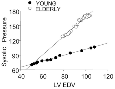
Figure 6. Relationship between changes in systolic pressure to alterations in ventricular diastolic volume (induced by balloon obstruction of inferior vena caval inflow) in a young and in an elderly patient. There is much greater sensitivity of systolic pressure to volume changes in the elderly patient, indicated by the steeper slope. Modified from Chen et al. (1998) with permission.
In addition to enhanced load-sensitivity, the global (systolic and diastolic) reserve capacity becomes blunted with arterial and left ventricular stiffening. Increased ELV at rest translates into less effective changes in ELV during exercise (or stress) thereby limiting cardiovascular performance (Borlaug et al., 2010). For example, individuals who start at a higher ELV have a limited capacity to further increase stroke volume, and the limited stroke volume response is further exacerbated when a stiff heart is connected to a stiff artery (Chen et al., 1998; Kawaguchi et al., 2003). It is therefore not surprising that a stiffer LV and arterial system are linked to a reduced aerobic capacity (Borlaug et al., 2010). Further, in an isolated canine heart model, Kass et al. (1989) reported, larger reductions in ELV after a myocardial infarction in hearts with a higher resting ELV. This greater mechanical vulnerability to an ischemic insult may help to explain why older individuals may experience worse outcomes following a myocardial infarction (Maggioni et al., 1993).
Another a major consequence of arterial stiffening is an increased pulse pressure, which also increases cyclic changes of arterial flow. As such, the microcirculation receives larger pulsatile pressures which can damage the vascular beds and in turn, cause damage to the end organs (such as the kidney and brain). Indeed increased arterial stiffness is independently associated with dementia (Hanon et al., 2005). Increased arterial stiffness can also lead to endothelial dysfunction and an abnormal vasodilation response to stress.
Therapeutic Interventions
Interventions related to improving arterial ventricular coupling span from pharmacological to lifestyle (exercise and diet) approaches. We will briefly highlight some important interventions in this area. The abnormalities in combined arterial and ventricular stiffening leading to a mismatch in their interaction, as highlighted above, has important physiological consequences. Numerous approaches have been taken to reverse the age and disease associated changes. By reducing the increase in arterial and left ventricular systolic stiffness with age or disease we may improve arterial–ventricular interactions and thus cardiovascular performance by being more efficient blood pressure regulation for a given volume of blood. For example, acute administration of sodium nitroprusside, a balanced vasodilator, acutely reduces EA (10%) and increases ELV (47%) at rest in older (70 ± 8 years) individuals (Chantler et al., 2011). Further, at peak exercise, sodium nitroprusside leads to an increase in ELV (68%) without a change in EA and consequently the normal reduction in EA/ELV during exercise is enhanced (36%). Importantly, sodium nitroprusside acutely attenuates the age-associated deficits in EA/ELV and ELV during exercise (Chantler et al., 2011). Similarly, acute intravenous verapamil (calcium-channel blocker) in healthy older (70 ± 10 years) volunteers reduces resting arterial–ventricular stiffening and EA during exercise (though a decline in heart rate, pulsatile, and resistive arterial load), and improves (13%) aerobic exercise performance (Chen et al., 1999). These results in older persons highlight that, at least acutely, the abnormalities in arterial–ventricular interactions can be partly restored. Of note, verapamil also improved arterial ventricular interactions and exercise capacity in HF patients with a preserved ejection fraction and hypertrophic cardiomyopathy (Brown et al., 1985; Setaro et al., 1990). However, whether the acute effects of sodium nitroprusside or verapamil identified can be maintained with chronic drug administration is unknown.
Hypertensive patients on optimal brachial and central blood pressure antihypertensive therapies shifts arterial–ventricular coupling from blood flow maximization to left ventricular mechanical efficiency optimization (Osranek et al., 2008). Further, the effects of antihypertensive monotherapy on EA/ELV examined in 10,670 patients over a 6-month period indicated that angiotensin converting enzyme inhibitors (ACEI), angiotensin II receptor blockers (AIIRA), and dihydropyridine calcium antagonists decrease EA/ELV, whereas diuretics, α-blockers, both β-blocker groups (with and without intrinsic sympathomimetic activity), and non-dihydropyridines significantly increase EA/ELV compared to baseline measurements (Figure 7; Iakovou et al., 2004). Thus various antihypertensive drugs have a differential effect on EA/ELV with ACEI, AIIRA, and dihydropyridine calcium antagonists have the most favorable effect on this index, likely through their actions on causing vasodilatation and by actually inhibiting the vasoconstrictive results of neurohormonal activation (Iakovou et al., 2004). However, the effects of these drugs on the components of EA/ELV are not reported.
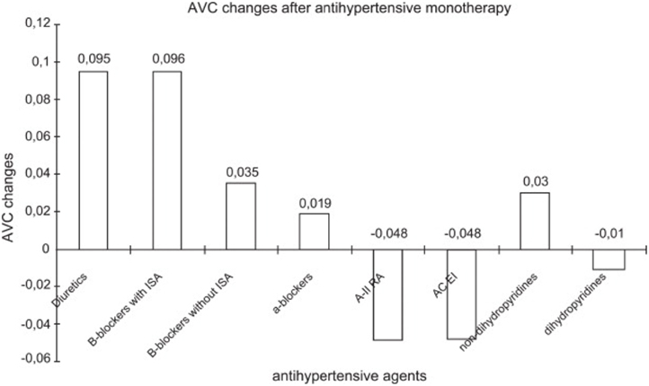
Figure 7. Arterial–ventricular coupling (AVC) percentage changes after antihypertensive monotherapy in 10,670 patients over 6 months of treatment. Abbreviations: ACE inhibitors, angiotensin converting enzyme inhibitors; AIIRA, angiotensin II receptor antagonists; ISA, intrinsic sympathomimetic activity. From Iakovou et al. (2004) with permission.
Other therapies shown to improve EA/ELV are exercise training. In healthy older men, 24–32 weeks of aerobic endurance exercise training does not alter baseline ejection fraction (inverse of EA/ELV) or left ventricular contractility (systolic blood pressure/end-systolic volume), but increases peak ejection fraction, suggesting that EA/ELV would have further decreased during exercise, due to a greater peak left ventricular contractility (Schulman et al., 1996). In the same study, eight master athletes stopped their endurance training for 12 weeks, which tended to decrease peak ejection fraction and LV contractility (Schulman et al., 1996). In patients with coronary artery disease, 12 months of aerobic endurance exercise training did not alter resting EA/ELV, or ELV, but produced a slight 13% reduction in EA at rest (Rinder et al., 1999). Further, exercise training led to a 37% increase in ELV and a 23% decrease in EA/ELV during handgrip exercise performed at 30% of maximal voluntary contraction. However, the change in EA during handgrip exercise remained unaltered after the exercise training. The results of this study suggest that long-term endurance exercise training induces significant cardiovascular adaptations both in the basal state and during an afterload stress in patients with coronary artery disease.
One year of progressive and vigorous endurance training in sedentary healthy older (70 ± 3 years) individuals resulted in slight reductions in EA at rest (14%) and peak exercise (20%). This coincided with an improvement in compliance at rest (Fujimoto et al., 2010). However, the exercise training failed to reverse cardiac stiffening. One possible reason for the lack of changes in left ventricular stiffness with exercise training is the development of cross-linked advanced glycation end products in the left ventricular wall along with a loss in the number and the volume of cardiac myocytes that occur with older age and which are pathologically irreversible once formed (Aronson, 2003). Thus any improvement in left ventricular function could have been constrained by cross-linked collagen. A phase II drug, alagebrium, is a cross-link breaker and is known to improve left ventricular and arterial stiffness in animals (Kass, 2003), and a clinical trial is currently underway to examine the effects of exercise and alagebrium combined on cardiovascular stiffening in the elderly (NIH clinicaltrials.gov identifier NCT01014572). However, alagebrium administered to patients with HF and systolic dysfunction does not improve exercise tolerance or cardiac function (Hartog et al., 2011). Whether HF patients with a preserved ejection fraction, in whom the largest effect of alagebrium could be expected, would benefit from an advanced glycation end products breaker therapy remains unknown.
Conclusion
Although increased stiffness of the arteries itself has important physiological and clinical relevance, such changes also have major implications on the heart, and vice versa, and the manner in the way they interact has important ramifications on cardiovascular function both at rest and during exercise. Examination of the alterations in arterial–ventricular coupling with aging and disease can yield mechanistic insights into the pathophysiology of these conditions and increase the effectiveness of current therapeutic interventions. Future studies should identify agents to chronically reverse increases in EA and ELV that occur with age and disease. Furthermore, longitudinal studies are needed to evaluate whether alterations in EA/ELV, EA, and ELV provide any prognostic information for adverse outcomes, such as HF.
Conflict of Interest Statement
The authors declare that the research was conducted in the absence of any commercial or financial relationships that could be construed as a potential conflict of interest.
Acknowledgments
This research was supported in part by the Intramural Research Program of the NIH, National Institute on Aging.
References
Adams, K. F., Lindenfeld, J., and Arnold, J. M. O. (2006). Executive summary: HFSA 2006 comprehensive heart failure practice guideline. J. Card. Fail. 12, 10–38.
Amar, J., Ruidavets, J. B., Chamontin, B., Drouet, L., and Ferrieres, J. (2001). Arterial stiffness and cardiovascular risk factors in a population-based study. J. Hypertens. 19, 381–387.
Arnett, D. K., Tyroler, H. A., Burke, G., Hutchinson, R., Howard, G., and Heiss, G. (1996). Hypertension and subclinical carotid artery atherosclerosis in blacks and whites. The Atherosclerosis Risk in Communities Study. ARIC Investigators. Arch. Intern. Med. 156, 1983–1989.
Aronson, D. (2003). Cross-linking of glycated collagen in the pathogenesis of arterial and myocardial stiffening of aging and diabetes. J. Hypertens. 21, 3–12.
Asanoi, H., Kameyama, T., Ishizaka, S., Miyagi, K., and Sasayama, S. (1992). Ventriculoarterial coupling during exercise in normal human subjects. Int. J. Cardiol. 36, 177–186.
Asanoi, H., Sasayama, S., and Kameyama, T. (1989). Ventriculoarterial coupling in normal and failing heart in humans. Circ. Res. 65, 483–493.
Borlaug, B. A., and Kass, D. A. (2008). Ventricular-vascular interaction in heart failure. Heart Fail. Clin. 4, 23–36.
Borlaug, B. A., Lam, C. S., Roger, V. L., Rodeheffer, R. J., and Redfield, M. M. (2009). Contractility and ventricular systolic stiffening in hypertensive heart disease insights into the pathogenesis of heart failure with preserved ejection fraction. J. Am. Coll. Cardiol. 54, 410–418.
Borlaug, B. A., Melenovsky, V., Russell, S. D., Kessler, K., Pacak, K., Becker, L. C., and Kass, D. A. (2006). Impaired chronotropic and vasodilator reserves limit exercise capacity in patients with heart failure and a preserved ejection fraction. Circulation 114, 2138–2147.
Borlaug, B. A., Olson, T. P., Lam, C. S. P., Flood, K. S., Lerman, A., Johnson, B. D., and Redfield, M. M. (2010). Global cardiovascular reserve dysfunction in heart failure with preserved ejection fraction. J. Am. Coll. Cardiol. 56, 845–854.
Brown, F., Campbell, W., Mitchell, P. J., and Randall, K. (1985). Dopamine autoreceptors and the effects of drugs on locomotion and dopamine synthesis. Br. J. Pharmacol. 84, 853–860.
Chantler, P. D., Clements, R. E., Sharp, L., George, K. P., Tan, L. B., and Goldspink, D. F. (2005). The influence of body size on measurements of overall cardiac function. Am. J. Physiol. Heart Circ. Physiol. 289, H2059–H2065.
Chantler, P. D., and Lakatta, E. G. (2009). Role of body size on cardiovascular function can we see the meat through the fat? Hypertension 54, 459–461.
Chantler, P. D., Lakatta, E. G., and Najjar, S. S. (2008a). Arterial-ventricular coupling: mechanistic insights into cardiovascular performance at rest and during exercise. J. Appl. Physiol. 105, 1342–1351.
Chantler, P. D., Melenovsky, V., Schulman, S. P., Gerstenblith, G., Becker, L. C., Ferrucci, L., Fleg, J. L., Lakatta, E. G., and Najjar, S. S. (2008b). The sex-specific impact of systolic hypertension and systolic blood pressure on arterial-ventricular coupling at rest and during exercise. Am. J. Physiol. Heart Circ. Physiol. 295, H145–H153.
Chantler, P. D., Nussbacher, A., Gerstenblith, G., Schulman, S. P., Becker, L. C., Ferrucci, L., Fleg, J. L., Lakatta, E. G., and Najjar, S. S. (2011). Abnormalities in arterial-ventricular coupling in older healthy persons are attenuated by sodium nitroprusside. Am. J. Physiol. Heart Circ. Physiol. 300, H1914–H1922.
Chantler, P. D., Melenovsky, V., Schulman, S. P., Gerstenblith, G., Becker, L. C., Ferrucci, L., Fleg, J. L., Lakatta, E. G., and Najjar, S. S. (2012). Use of the Frank-Starling mechanism during exercise is linked to exercise-induced changes in arterial load. Am. J. Physiol. Heart Circ. Physiol. 302, H349–H358.
Chemla, D., Antony, I., Lecarpentier, Y., and Nitenberg, A. (2003). Contribution of systemic vascular resistance and total arterial compliance to effective arterial elastance in humans. Am. J. Physiol. Heart Circ. Physiol. 285, H614–H620.
Chen, C. H., Nakayama, M., Nevo, E., Fetics, B. J., Maughan, W. L., and Kass, D. A. (1998). Coupled systolic-ventricular and vascular stiffening with age: implications for pressure regulation and cardiac reserve in the elderly. J. Am. Coll. Cardiol. 32, 1221–1227.
Chen, C.-H., Nakayama, M., Talbot, M., Nevo, E., Fetics, B., Gerstenblith, G., Becker, L. C., and Kass, D. A. (1999). Verapamil acutely reduces ventricular-vascular stiffening and improves aerobic exercise performance in elderly individuals. J. Am. Coll. Cardiol. 33, 1602–1609.
Chirinos, J. A., Rietzschel, E. R., De Buyzere, M. L., De Bacquer, D., Gillebert, T. C., Gupta, A. K., Segers, P., Asklepios Investigators. (2009). Arterial load and ventricular-arterial coupling. Hypertension 54, 558–566.
Cohen-Solal, A., Caviezel, B., Himbert, D., and Gourgon, R. (1994). Left ventricular-arterial coupling in systemic hypertension: analysis by means of arterial effective and left ventricular elastances. J. Hypertens. 12, 591–600.
Cohen-Solal, A., Caviezel, B., Laperche, T., and Gourgon, R. (1996). Effects of aging on left ventricular-arterial coupling in man: assessment by means of arterial effective and left ventricular elastances. J. Hum. Hypertens. 10, 111–116.
Cohen-Solal, A., Faraggi, M., Czitrom, D., Le Guludec, D., Delahaye, N., and Gourgon, R. (1998). Left ventricular-arterial system coupling at peak exercise in dilated nonischemic cardiomyopathy. Chest 113, 870–877.
De Tombe, P. P., Jones, S., Burkhoff, D., Hunter, W. C., and Kass, D. A. (1993). Ventricular stroke work and efficiency both remain nearly optimal despite altered vascular loading. Am. J. Physiol. 264, H1817–H1824.
Fleg, J. L., O’Connor, F., Gerstenblith, G., Becker, L. C., Clulow, J., Schulman, S. P., and Lakatta, E. G. (1995). Impact of age on the cardiovascular response to dynamic upright exercise in healthy men and women. J. Appl. Physiol. 78, 890–900.
Flegal, K. M., Carroll, M. D., Ogden, C. L., and Curtin, L. R. (2010). Prevalence and trends in obesity among US adults, 1999-2008. JAMA 303, 235–241.
Franklin, S. S., Gustin, W. IV, Wong, N. D., Larson, M. G., Weber, M. A., Kannel, W. B., and Levy, D. (1997). Hemodynamic patterns of age-related changes in blood pressure: the Framingham Heart Study. Circulation 96, 308–315.
Fujimoto, N., Prasad, A., Hastings, J. L., Arbab-Zadeh, A., Bhella, P. S., Shibata, S., Palmer, D., and Levine, B. D. (2010). Cardiovascular effects of 1 year of progressive and vigorous exercise training in previously sedentary individuals older than 65 years of age. Circulation 122, 1797–1805.
Hanon, O., Haulon, S., Lenoir, H., Seux, M.-L., Rigaud, A.-S., Safar, M., Girerd, X., and Forette, F. (2005). Relationship between arterial stiffness and cognitive function in elderly subjects with complaints of memory loss. Stroke 36, 2193–2197.
Hartog, J. W. L., Willemsen, S., van Veldhuisen, D. J., Posma, J. L., van Wijk, L. M., Hummel, Y. M., Hillege, H. L., and Voors, A. A. (2011). Effects of alagebrium, an advanced glycation endproduct breaker, on exercise tolerance and cardiac function in patients with chronic heart failure. Eur. J. Heart Fail. 13, 899–908.
Iakovou, I., Karpanou, E. A., Vyssoulis, G. P., Toutouzas, P. K., and Cokkinos, D. V. (2004). Assessment of arterial ventricular coupling changes in patients under therapy with various antihypertensive agents by a non-invasive echocardiographic method. Int. J. Cardiol. 96, 355–360.
Kass, D. A. (2003). Getting better without AGE: new insights into the diabetic heart. Circ. Res. 92, 704–706.
Kass, D. A. (2005). Ventricular arterial stiffening: integrating the pathophysiology. Hypertension 46, 185–193.
Kass, D. A., Marino, P., Maughan, W. L., and Sagawa, K. (1989). Determinants of end-systolic pressure-volume relations during acute regional ischemia in situ. Circulation 80, 1783–1794.
Kawaguchi, M., Hay, I., Fetics, B., and Kass, D. A. (2003). Combined ventricular systolic and arterial stiffening in patients with heart failure and preserved ejection fraction: implications for systolic and diastolic reserve limitations. Circulation 107, 714–720.
Kelly, R., Tunin, R., and Kass, D. (1992a). Effect of reduced aortic compliance on cardiac efficiency and contractile function of in situ canine left ventricle. Circ. Res. 71, 490–502.
Kelly, R. P., Ting, C. T., Yang, T. M., Liu, C. P., Maughan, W. L., Chang, M. S., and Kass, D. A. (1992b). Effective arterial elastance as index of arterial vascular load in humans. Circulation 86, 513–521.
Kitzman, D. W., Higginbotham, M. B., Cobb, F. R., Sheikh, K. H., and Sullivan, M. J. (1991). Exercise intolerance in patients with heart failure and preserved left ventricular systolic function: failure of the Frank-Starling mechanism. J. Am. Coll. Cardiol. 17, 1065–1072.
Klapholz, M., Maurer, M., Lowe, A. M., Messineo, F., Meisner, J. S., Mitchell, J., Kalman, J., Phillips, R. A., and Steingart, R. (2004). Hospitalization for heart failure in the presence of a normal left ventricular ejection fraction: results of the New York heart failure registry. J. Am. Coll. Cardiol. 43, 1432–1438.
Lakatta, E. G. (1993). Cardiovascular regulatory mechanisms in advanced age. Physiol. Rev. 73, 413–467.
Lakatta, E. G. (2003). Arterial and cardiac aging: major shareholders in cardiovascular disease enterprises. Part III: cellular and molecular clues to heart and arterial aging. Circulation 107, 490–497.
Lakatta, E. G., and Levy, D. (2003). Arterial and cardiac aging: major shareholders in cardiovascular disease enterprises. Part II: the aging heart in health: links to heart disease. Circulation 107, 346–354.
Lam, C. S., Roger, V. L., Rodeheffer, R. J., Bursi, F., Borlaug, B. A., Ommen, S. R., Kass, D. A., and Redfield, M. M. (2007). Cardiac structure and ventricular-vascular function in persons with heart failure and preserved ejection fraction from Olmsted County, Minnesota. Circulation 115, 1982–1990.
Little, W. C., and Cheng, C. P. (1991). Left ventricular-arterial coupling in conscious dogs. Am. J. Physiol. 261, H70–H76.
Little, W. C., and Cheng, C. P. (1993). Effect of exercise on left ventricular-arterial coupling assessed in the pressure-volume plane. Am. J. Physiol. 264, H1629–H1633.
Lloyd-Jones, D., Adams, R. J., Brown, T. M., Carnethon, M., Dai, S., De Simone, G., Ferguson, T. B., Ford, E., Furie, K., Gillespie, C., Go, A., Greenlund, K., Haase, N., Hailpern, S., Ho, P. M., Howard, V., Kissela, B., Kittner, S., Lackland, D., Lisabeth, L., Marelli, A., McDermott, M. M., Meigs, J., Mozaffarian, D., Mussolino, M., Nichol, G., Roger, V., Rosamond, W., Sacco, R., Sorlie, P., Stafford, R., Thom, T., Wasserthiel-Smoller, S., Wong, N. D., Wylie-Rosett, J., American Heart Association Statistics Committee and Stroke Statistics Subcommittee. (2009). Heart disease and stroke statistics – 2010 update. A report from the American Heart Association. Circulation 121, e1–e170.
Maggioni, A. P., Maseri, A., Fresco, C., Franzosi, M. G., Mauri, F., Santoro, E., and Tognoni, G. (1993). Age-related increase in mortality among patients with first myocardial infarctions treated with thrombolysis. The Investigators of the Gruppo Italiano per lo Studio della Sopravvivenza nell’Infarto Miocardico (GISSI-2). N. Engl. J. Med. 329, 1442–1448.
Masoudi, F. A., Havranek, E. P., Smith, G., Fish, R. H., Steiner, J. F., Ordin, D. L., and Krumholz, H. M. (2003). Gender, age, and heart failure with preserved left ventricular systolic function. J. Am. Coll. Cardiol. 41, 217–223.
Maurer, M. S., King, D. L., El-Khoury Rumbarger, L., Packer, M., and Burkhoff, D. (2005). Left heart failure with a normal ejection fraction: identification of different pathophysiologic mechanisms. J. Card. Fail. 11, 177–187.
Mayet, J., and Hughes, A. (2003). Cardiac and vascular pathophysiology in hypertension. Heart 89, 1104–1109.
Murgo, J. P., Westerhof, N., Giolma, J. P., and Altobelli, S. A. (1980). Aortic input impedance in normal man: relationship to pressure wave forms. Circulation 62, 105–116.
Najjar, S. S., Schulman, S. P., Gerstenblith, G., Fleg, J. L., Kass, D. A., O’Connor, F., Becker, L. C., and Lakatta, E. G. (2004). Age and gender affect ventricular-vascular coupling during aerobic exercise. J. Am. Coll. Cardiol. 44, 611–617.
Najjar, S. S., Scuteri, A., and Lakatta, E. G. (2005). Arterial aging: is it an immutable cardiovascular risk factor? Hypertension 46, 454–462.
Nichols, W. W., Nicolini, F. A., and Pepine, C. J. (1992). Determinants of isolated systolic hypertension in the elderly. J. Hypertens. 10, 573.
Ogawa, T., Spina, R. J., Martin, W. H. III, Kohrt, W. M., Schechtman, K. B., Holloszy, J. O., and Ehsani, A. A. (1992). Effects of aging, sex, and physical training on cardiovascular responses to exercise. Circulation 86, 494–503.
Olivetti, G., Giordano, G., Corradi, D., Melissari, M., Lagrasta, C., Gambert, S. R., and Anversa, P. (1995). Gender differences and aging: effects on the human heart. J. Am. Coll. Cardiol. 26, 1068–1079.
Osranek, M., Eisenach, J. H., Khandheria, B. K., Chandrasekaran, K., Seward, J. B., and Belohlavek, M. (2008). Arterioventricular coupling and ventricular efficiency after antihypertensive therapy. Hypertension 51, 275–281.
Otsuki, T., Maeda, S., Iemitsu, M., Saito, Y., Tanimura, Y., Ajisaka, R., and Miyauchi, T. (2006). Contribution of systemic arterial compliance and systemic vascular resistance to effective arterial elastance changes during exercise in humans. Acta Physiol. (Oxf.) 188, 15–20.
Owan, T. E., Hodge, D. O., Herges, R. M., Jacobsen, S. J., Roger, V. L., and Redfield, M. M. (2006). Trends in prevalence and outcome of heart failure with preserved ejection fraction. N. Engl. J. Med. 355, 251–259.
Redfield, M. M., Jacobsen, S. J., Borlaug, B. A., Rodeheffer, R. J., and Kass, D. A. (2005). Age- and gender-related ventricular-vascular stiffening: a community-based study. Circulation 112, 2254–2262.
Rider, O. J., Petersen, S. E., Francis, J. M., Ali, M. K., Hudsmith, L. E., Robinson, M. R., Clarke, K., and Neubauer, S. (2011). Ventricular hypertrophy and cavity dilatation in relation to body mass index in women with uncomplicated obesity. Heart 97, 203–208.
Rinder, M. R., Miller, T. R., and Ehsani, A. A. (1999). Effects of endurance exercise training on left ventricular systolic performance and ventriculoarterial coupling in patients with coronary artery disease. Am. Heart J. 138, 169–174.
Saba, P. S., Ganau, A., Devereux, R. B., Pini, R., Pickering, T. G., and Roman, M. J. (1999). Impact of arterial elastance as a measure of vascular load on left ventricular geometry in hypertension. J. Hypertens. 17, 1007–1015.
Sasayama, S., and Asanoi, H. (1991). Coupling between the heart and arterial system in heart failure. Am. J. Med. 90, 14S–18S.
Schulman, S. P., Fleg, J. L., Goldberg, A. P., Busby-Whitehead, J., Hagberg, J. M., O’Connor, F. C., Gerstenblith, G., Becker, L. C., Katzel, L. I., Lakatta, L. E., and Lakatta, E. G. (1996). Continuum of cardiovascular performance across a broad range of fitness levels in healthy older men. Circulation 94, 359–367.
Segers, P., Stergiopulos, N., and Westerhof, N. (2002). Relation of effective arterial elastance to arterial system properties. Am. J. Physiol. Heart Circ. Physiol. 282, H1041–H1046.
Setaro, J. F., Zaret, B. L., Schulman, D. S., Black, H. R., and Soufer, R. (1990). Usefulness of verapamil for congestive heart failure associated with abnormal left ventricular diastolic filling and normal left ventricular systolic performance. Am. J. Cardiol. 66, 981–986.
Stamler, R., Ford, C. E., and Stamler, J. (1991). Why do lean hypertensives have higher mortality rates than other hypertensives? Findings of the Hypertension Detection and Follow-up Program. Hypertension 17, 553–564.
Stamler, R., Stamler, J., Riedlinger, W. F., Algera, G., and Roberts, R. H. (1978). Weight and blood pressure. Findings in hypertension screening of 1 million Americans. JAMA 240, 1607–1610.
Sunagawa, K., Maughan, W. L., Burkhoff, D., and Sagawa, K. (1983). Left ventricular interaction with arterial load studied in isolated canine ventricle. Am. J. Physiol. 245, H773–H780.
Toto-Moukouo, J. J., Achimastos, A., Asmar, R. G., Hugues, C. J., and Safar, M. E. (1986). Pulse wave velocity in patients with obesity and hypertension. Am. Heart J. 112, 136–140.
Keywords: left ventricular function, arterial system, exercise, aging, disease
Citation: Chantler PD and Lakatta EG (2012) Arterial–ventricular coupling with aging and disease. Front. Physio. 3:90. doi: 10.3389/fphys.2012.00090
Received: 11 January 2012; Accepted: 25 March 2012;
Published online: 07 May 2012.
Edited by:
Ana Paula Dantas, Institut d’Investigacions Biomediques August Pi i Sunyer, SpainCopyright: © 2012 Chantler and Lakatta. This is an open-access article distributed under the terms of the Creative Commons Attribution Non Commercial License, which permits non-commercial use, distribution, and reproduction in other forums, provided the original authors and source are credited.
*Correspondence: Paul D. Chantler, Division of Exercise Physiology, School of Medicine, Robert C. Byrd Health Sciences Center, West Virginia University, P.O. Box 9227, Morgantown, WV 26506, USA. e-mail:cGNoYW50bGVyQGhzYy53dnUuZWR1