AC
anderson camargo moreira
Federal University of Santa Catarina
Florianopolis, Brazil
44
Total downloads
2,870
Total views and downloads
Submit your idea
You will be redirected to our submission process.
Manuscript Submission Deadline 21 February 2026
This Research Topic is currently accepting articles.
The research regarding bone tissues comprises the development of new techniques ranging from medication administration, used to prevent and remediate bone issues, to the use of scaffolds to support neo-bone regeneration. Different techniques can be combined not just to treat bone injuries, but also to accelerate the healing process, providing patients a shorter and more effective recovery. Regarding scaffolds, the level of biocompatibility of their raw material and the use of pharmaceuticals adhered to its components, which can stimulate bone formation, have been meticulously studied, however, geometrical features such as microstructural architecture, pore interconnectivity, pore size, and surface roughness are also important parameters to evaluate their performance as a bone supporter.
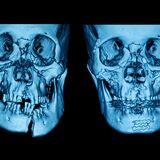
The study of neo-bone formations is, nowadays, supported by the use of imaging techniques. Currently, X-ray microtomography (microCT) is the most employed one, since it can provide 3D images of mineralized bone and scaffolds/implant issues. The performance of new scaffold geometries, in which the 3D connectivity is also crucial, has also been assessed with microCT.
Mineralized bones are the first tissue ever imaged via an X-ray imaging technique while still functional and inside the human body. It was a remarkable achievement more than one century ago because no surgery process was indeed required. The level of information taken from bone images has been escalating as new analysis methods are also increasing. The process of bone regeneration can be evaluated via the analysis of some parameters such as; volume fraction (BV/TV), which consists of the ratio between mineralized bone and volume of the analyzed tissue, trabecular thickness (Tb.Th), and trabecular separation (Tb.Sp), among others, all of them determined via image analysis. Another important parameter that has been correlated with bone diseases is the interconnectivity index. Even though the above-mentioned parameters can be estimated via 2D images, to accurately analyze the bone architecture and connectivity, high-resolution 3D images are a fine option. Even the contact area between scaffolds (and/or implants) and the bone is more effective via 3D analysis.
A strong tool that helps bone and scaffold image analysers are based on Machine Learning methods. Besides acting in the binarization process of bone structures from surrounding tissues and scaffolds, enabling the creation of accurate 3D models for surgical planning or prosthetics designs, Machine Learning contributes significantly to medical research, diagnostics, and treatment planning. Some models even analyze patterns in imaging data to assess bone density and quality.
The main goal of this research topic is to explore the use of 3D images applied to the study of bones and scaffolds/implant issues and available tools used in the process, such as Machine Learning methods. This topic research intends to highlight the advantages of using 3D image analysis over conventional 2D image analysis.
This Research Topic accepts the following article types, unless otherwise specified in the Research Topic description:
Articles that are accepted for publication by our external editors following rigorous peer review incur a publishing fee charged to Authors, institutions, or funders.
Keywords: Bone, scaffold, 3D image, image analysis, machine learning.
Important note: All contributions to this Research Topic must be within the scope of the section and journal to which they are submitted, as defined in their mission statements. Frontiers reserves the right to guide an out-of-scope manuscript to a more suitable section or journal at any stage of peer review.
Manuscripts can be submitted to this Research Topic via the main journal or any other participating journal.
Submit your idea
You will be redirected to our submission process.
